
Medical expert of the article

New publications
Osteoarthritis: Causes and Risk Factors
Last updated: 12.03.2026

All iLive content is medically reviewed or fact checked to ensure as much factual accuracy as possible.
We have strict sourcing guidelines and only link to reputable media sites, academic research institutions and, whenever possible, medically peer reviewed studies. Note that the numbers in parentheses ([1], [2], etc.) are clickable links to these studies.
If you feel that any of our content is inaccurate, out-of-date, or otherwise questionable, please select it and press Ctrl + Enter.
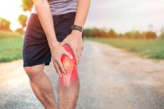
Osteoarthritis is a heterogeneous disease of the entire joint, not simply a wear and tear of cartilage due to age. The pathological process involves articular cartilage, subchondral bone, synovial membrane, ligaments, capsule, periarticular muscles, and adipose tissue. Current reviews emphasize that obesity and joint trauma remain the leading modifiable factors, while the disease mechanisms themselves include inflammatory, metabolic, and post-traumatic processes. [1]
The global burden of the disease is rapidly growing. The Global Burden of Disease study estimated that approximately 595 million people, equivalent to 7.6% of the world's population, lived with osteoarthritis in 2020, and the total number of cases could approach 1 billion by 2050. The knee joint remains the most common site. [2]
It's also important that osteoarthritis is no longer perceived as a disease exclusively of the elderly. According to large reviews, the proportion of early onset is increasingly noticeable, often associated with trauma, excess fat, decreased muscle mass, high mechanical stress, or congenital joint shape. This changes the approach: prevention should begin not after age 65, but much earlier. [3]
From a clinical perspective, osteoarthritis is significant for more than just pain. It limits walking, standing, climbing stairs, everyday activities, hand grip, sleep quality, and work performance. The National Arthritis Institute notes that as the disease progresses, even simple activities such as rising from a chair, holding a pot, or walking across a parking lot become difficult. [4]
Another fundamental point: the degree of pain does not always strictly correspond to the picture on an X-ray. The severity of symptoms is influenced by synovitis, bone changes, muscle weakness, biomechanical disturbances, and central sensitization mechanisms. Therefore, a modern article on osteoarthritis cannot be limited to the words "there is wear" or "there are osteophytes." [5]
Table 1. Main groups of risk factors for osteoarthritis
| Group | What's included | Practical meaning |
|---|---|---|
| Non-modifiable | age, female gender after 50 years, family predisposition, joint shape features | help explain the underlying vulnerability of the joint |
| Metabolic | obesity, central obesity, hyperglycemia, dyslipidemia, metabolic syndrome | increase the risk not only through stress, but also through inflammatory-metabolic mechanisms |
| Post-traumatic | ligament rupture, meniscus injury, joint surgery, chronic instability | often accelerate the early development of the disease |
| Biomechanical | varus, valgus, muscle weakness, abnormal axis, pathological shape of the hip joint | create a local overload on a certain part of the joint |
| Professional and behavioral | heavy physical labor, lifting heavy objects, prolonged standing, kneeling, squatting, and some high-impact sports | These are the factors that can most often be influenced preventively. |
Sources for the table. [6]
How Osteoarthritis Develops: A Modern Understanding of Pathogenesis
The classic old model described the disease as a slow wear and tear of cartilage. The modern model is much more complex. Osteoarthritis is now viewed as an imbalance between microdamage and joint tissue repair. When stress, age-related changes, injury, or metabolic influences exceed repair capacity, a chronic process of remodeling and degradation begins. [7]
The composition of the extracellular matrix in cartilage changes, chondrocyte function is disrupted, and biologically active matrix fragments appear, which themselves support the inflammatory response. Simultaneously, remodeling occurs in the subchondral bone, and the synovial membrane begins to produce inflammatory mediators. This explains why pain, swelling, morning stiffness, and limited function are associated with more than just cartilage. [8]
Low-grade inflammation occupies a special place. This is not the inflammation typical of rheumatoid arthritis, but it is active enough to support pain, synovitis, and the progression of structural changes. Modern reviews identify at least bone, synovial, and cartilaginous variants of molecular dominance, which explains the clinical heterogeneity of the disease. [9]
Pain in osteoarthritis is also multifactorial. It can originate from synovial fluid, bone microremodeling, capsular stretching, muscle spasm, and changes in pain processing in the nervous system. This is why some patients experience severe pain with relatively mild radiographic changes, while others, conversely, experience pronounced radiographic changes with tolerable symptoms. Studies on central sensitization confirm this clinical-radiographic dissociation. [10]
A modern practical conclusion from pathogenesis is crucial: osteoarthritis cannot be treated with a single pill or injection. If the disease is accompanied by weight gain, muscle weakness, limb axis abnormalities, synovitis, and pain sensitization, then intervention must be multifaceted. This is why international recommendations base treatment on physical activity, patient education, and weight management. [11]
Table 2. The main pathogenetic variants of osteoarthritis
| Option | What dominates | What does this usually mean clinically? |
|---|---|---|
| Post-traumatic | damage to ligaments, meniscus, cartilage, kinematic disorders | earlier onset, often in people under 50 years of age |
| Metabolic | obesity, insulin resistance, adipokines, low-grade systemic inflammation | combination with abdominal obesity, diabetes, dyslipidemia |
| Biomechanical | varus, valgus, pathological axis, abnormal joint shape, muscle imbalance | overload of a certain part of the joint and faster local wear |
| Age | cellular aging, decreased repair, accumulation of microdamage | gradual progression, often in combination with other factors |
| Pain sensitized | enhancing pain processing at the peripheral and central levels | the pain may be more severe than expected from the images |
Sources for the table. [12]
Causes and risk factors: what really increases the likelihood of disease
Age, gender and hormonal background
Age remains the most consistent risk factor, but it is not the only cause. The National Arthritis Institute clearly states that osteoarthritis is more common with age, but it also develops in young people—usually after an injury, with abnormal joint structure, or due to a genetic predisposition. [13]
Women are more likely to develop the disease than men, especially after age 50. According to the CDC and NIAMS, female gender is a significant risk factor after this age. For many women, the disease becomes noticeable after menopause, supporting the role of hormonal and tissue changes, not just mechanical stress. [14]
In practice, this means that a woman with emerging knee, wrist, or hip pain after menopause requires a more careful evaluation, even if the radiographic changes are still moderate. In real-life clinics, it is precisely in this group that the early stage is often underestimated, with complaints attributed solely to age. [15]
Heredity and genetic predisposition
Osteoarthritis has a strong hereditary component. Modern family and twin studies indicate that heritability is approximately 39%-65% for hand and knee lesions and approximately 60% for the hip joint. This does not mean genetic inevitability, but rather indicates different underlying vulnerabilities of joint tissues in different individuals. [16]
GDF5 remains one of the most studied genetic signals. Reviews and meta-analyses in recent years confirm the association of variants of this gene with osteoarthritis of the knee, hip, and hand joints. GDF5 is involved in joint development and the regulation of tissue repair, so its role is biologically plausible and not a random statistical finding. [17]
This is important for two reasons. First, family history does play a role in risk assessment. Second, genetic predisposition does not negate the value of prevention: weight control, injury prevention, and maintaining muscle strength are especially important for individuals with a genetic predisposition. [18]
Obesity, body composition and metabolic syndrome
Obesity is not simply a factor in overloading the knees and hip joints. Large reviews and the Nature Reviews Disease Primers guideline emphasize that obesity is one of the two main modifiable risk factors, with its effects partially mediated by inflammatory and metabolic pathways. Therefore, a link is observed not only with weight-bearing joints but also with the joints of the hand. [19]
In a UK Biobank study, people with metabolic syndrome had a 15% higher risk of osteoarthritis, and central obesity was associated with a 58% increased risk. Hyperglycemia also increased the risk, and the combination of metabolic syndrome and elevated C-reactive protein levels further strengthened the adverse association. [20]
New reviews from 2025 further emphasize not only body weight per se, but also body composition: a high percentage of fat tissue with low muscle mass is associated with a more severe course of the disease and poorer physical function. This is an important shift in thinking: we need to focus not only on body weight, but also on sarcopenic obesity, waist circumference, and the patient's physical fitness. [21]
Injuries, surgeries and post-traumatic osteoarthritis
A history of knee injury is one of the strongest risk factors. Meta-analyses show that after a knee injury, the risk of osteoarthritis increases approximately 4-6 times, and for some types of injuries, the pooled odds ratio exceeds 4. This is one of the key pathways for the development of early osteoarthritis in people of working age. [22]
Of particular importance are anterior cruciate ligament tears, meniscal injuries, and chronic instability. A 2024 systematic review comparing meniscectomy and meniscal repair found a higher rate of osteoarthritis progression after meniscectomy than after meniscal preservation. This supports the principle that, whenever possible, tissue preservation is better than removal. [23]
Femoroacetabular impingement syndrome is of great importance for the hip joint. A prospective study with 10-year follow-up showed that this clinical variant was associated with a nearly seven-fold increase in the likelihood of radiographic coxarthrosis, and the absolute risk was very high. This is particularly important for middle-aged patients with groin pain and limited hip internal rotation. [24]
Biomechanics, muscle weakness, work and physical activity
Muscle weakness is not just a consequence of disease, but sometimes even a predictor. An updated meta-analysis involving 46,819 people found that knee extensor weakness was associated with the development of symptomatic and radiographic knee osteoarthritis in both women and men. [25]
Occupational mechanical stress is also significant. Systematic reviews show that heavy physical labor, heavy lifting, squatting, kneeling, knee bending, climbing, and prolonged standing are associated with a higher risk of lower extremity osteoarthritis compared to sedentary work. [26]
However, not all physical activity is harmful. Patient and clinical literature emphasizes that ordinary regular activity and exercise do not cause osteoarthritis. The risk is primarily associated with injury, very high repetitive loads, and poorly controlled biomechanics. New studies describe a U-shaped logic: moderate activity is beneficial, while extremely high and traumatic loads can increase the risk. [27]
Table 3. Which risk factors are particularly important for different joints?
| Joint | Particularly significant factors |
|---|---|
| Knee | obesity, trauma, meniscus injury, muscle weakness, varus, heavy work with squats and kneeling |
| Hip | abnormal joint shape, femoroacetabular impingement syndrome, dysplastic features, obesity, heavy mechanical work |
| Brush | female gender, age, heredity, metabolic factors, repetitive manual loads |
| Spine and other joints | age, systemic metabolic influences, previous injuries, posture and local load characteristics |
Sources for the table. [28]
How does osteoarthritis manifest itself and what are the typical signs?
Symptoms typically begin gradually and initially affect one or a few joints. The most common symptoms include pain with joint use, brief stiffness after rest or in the morning, decreased range of motion, swelling after exercise, and a feeling of instability. According to NIAMS, morning stiffness in typical osteoarthritis typically lasts less than 30 minutes. [29]
The location of symptoms depends on the joint. In the hands, bony thickening and changes in the shape of the interphalangeal joints are typical. In the knee, pain when walking, a crunching sound, and a feeling of buckling are typical. In the hip joint, pain is felt in the groin, inner thigh, or buttock with limited range of motion. In the neck and lower back, pain and stiffness are typical, sometimes with secondary spinal stenosis. [30]
As the condition progresses, the person begins to limit activity, which leads to further muscle weakness and additional stress on the joint. This creates a "vicious cycle": pain reduces movement, reduced movement weakens the muscles, and weakness increases pain and instability. This is why modern treatment almost never recommends long-term immobilization as a strategy. [31]
It's important to remember that pain severity may differ from imaging data. In some patients, severe pain is explained by more pronounced pain sensitization or active synovitis, not just the volume of osteophytes and the degree of joint space narrowing. Therefore, in real-world clinical practice, patient complaints should not be discounted by the words "the image doesn't look that bad." [32]
Table 4. Typical osteoarthritis and signs requiring alertness
| Sign | More typical for osteoarthritis | Requires exclusion of other pathology |
|---|---|---|
| Pain | increases with load | very severe pain at rest with rapid increase |
| Stiffness in the morning | up to 30 minutes | prolonged and severe stiffness |
| Joint | without pronounced fever | hot, sharply swollen joint |
| Dynamics | slow gradual progression | rapid deterioration, rapid deformation |
| Context | without systemic symptoms | recent injury, suspected infection or tumor |
Sources for the table. [33]
When you urgently need to think about more than just osteoarthritis
Current NICE guidelines recommend making a typical diagnosis clinically in people aged 45 years and older if they have joint pain under load and no morning stiffness, or if it lasts no more than 30 minutes. However, this algorithm is only suitable for the typical presentation. [34]
If a patient has a recent injury, a sharp increase in deformity, prolonged morning stiffness, a hot, swollen joint, or a suspected infection or malignancy, these are considered atypical signs. In such situations, a more comprehensive assessment is required rather than simply repeating the "age-related osteoarthritis" template. [35]
In practice, this is especially important in three groups of patients. The first are young or middle-aged individuals following a sports injury. The second are patients with pronounced inflammatory symptoms, where other arthritis must be ruled out. The third are individuals with hip pain and limited hip rotation, in whom femoroacetabular impingement and rapidly progressing coxarthrosis may be missed. [36]
Patients with pain clearly disproportionate to the radiographic findings, sleep disturbances, severe fatigue, anxiety, or signs of generalized pain require special attention. In these patients, pain sensitization, along with joint damage, may play a significant role, and this situation requires more careful management rather than automatic enhancement of pain relief. [37]
Diagnostics: What's Really Needed and What's Often Done in Vain
For typical osteoarthritis, the basis for diagnosis remains clinical assessment: age, pain pattern, duration of stiffness, joint examination, range of motion, deformity, limb axis, muscle strength, and history of previous injuries. NICE guidelines explicitly recommend diagnosis without routine imaging in the typical presentation. [38]
Routine imaging is not always necessary. NICE specifically emphasizes that imaging should not be routinely used for making a typical diagnosis, for routine monitoring, or for selecting non-surgical treatment. The point is that images often add little to the already obvious clinical picture and can delay the initiation of appropriate management. [39]
Imaging becomes useful when atypical symptoms are present or another pathology needs to be ruled out. It then helps search for alternative or additional diagnoses, such as inflammatory arthritis, the consequences of a recent injury, a tumor, or another cause of pain. This is especially important in cases of rapid deterioration, a hot joint, and unusually prolonged stiffness. [40]
Today, proper diagnosis involves not only determining whether osteoarthritis is present, but also its type. For one patient, the leading factor may be a meniscal injury, for another, central obesity and hyperglycemia, for a third, a pathological hip joint, and for a fourth, decades of heavy knee work. Without this, it is impossible to develop truly targeted prevention of progression. [41]
Therefore, the modern diagnostic formula is: clinical diagnosis plus risk factor phenotyping. This approach is most consistent with current reviews from 2025, which emphasize the need to consider metabolic, biomechanical, and psychosocial factors simultaneously. [42]
Table 5. Practical diagnostic scheme
| Stage | What is being assessed | When it's especially important |
|---|---|---|
| Anamnesis | load-related pain, short-term stiffness, injuries, occupation, family history | Always |
| Inspection | deformity, pain, range of motion, muscle strength, limb axis | Always |
| Visualization | not routinely, but in case of an atypical picture or suspicion of another pathology | in case of red flags and doubts |
| Risk factor assessment | weight, waist circumference, metabolic syndrome, previous surgeries, joint shape characteristics | for the selection of prevention and management |
| Assessment of pain and function | walking, getting up, stairs, sleep, impact on daily life | to decide on treatment tactics |
Sources for the table. [43]
What really helps reduce the risk of progression?
The most consistent international guidelines agree on one thing: the mainstay of management remains exercise therapy, patient education, and weight control if overweight or obese. NICE lists exercise and weight management as core areas, while the American College of Rheumatology and the Arthritis Foundation have made strong recommendations in favor of exercise, weight loss, self-management programs, and some supportive measures. [44]
Exercises should be individually tailored. NICE recommends offering therapeutic exercise to all patients with osteoarthritis, including local muscle strengthening and general aerobic training, and also considering supervised exercise. It is important to explain in advance that pain may temporarily increase when starting exercise, but regular exercise usually reduces pain and improves function in the long term. [45]
Weight loss is particularly important for knee and hip osteoarthritis, but it is also beneficial in more general areas. NICE indicates that any weight loss helps, and that a loss of approximately 10% of body weight is likely more beneficial than a 5% loss. This is consistent with population data, where a high body mass index is associated with approximately 20.4% of the global osteoarthritis burden. [46]
Medications are considered adjunctive rather than the primary strategy. The ACR recommends topical nonsteroidal anti-inflammatory drugs for knee osteoarthritis, as well as oral nonsteroidal anti-inflammatory drugs and intra-articular glucocorticoids for the knee in certain situations. NICE, however, advises against the routine use of paracetamol or weak opioids, and does not recommend intra-articular hyaluronic acid injections for routine management. [47]
If symptoms significantly reduce quality of life and comprehensive non-surgical measures prove insufficient or unacceptable, NICE recommends considering referral for hip, knee, or shoulder osteoarthritis. The decision should be based on the impact of symptoms on life, not just the imaging results. [48]
Table 6. What to do to reduce the risk of progression
| Direction | What really works |
|---|---|
| Movement | Regular, individually tailored exercise, muscle strengthening, aerobic activity |
| Weight | weight loss in case of excess weight, waist circumference control |
| Metabolic health | control of glucose, lipids, blood pressure |
| Injury prevention | Knee and hip protection in sports, proper rehabilitation after injuries |
| Biomechanics | correction of movement technique, work on muscle strength and stability |
| Profession and everyday life | reducing chronic overload, frequent squats, prolonged kneeling and heavy lifting |
Sources for the table. [49]
Forecast
Osteoarthritis is a chronic disease, but its course is highly variable. In some people, progression occurs slowly over decades, while in others, it accelerates significantly due to a combination of obesity, injury, instability, abnormal joint shape, and muscle weakness. Therefore, not only age but also the entire range of risk factors is more important for prognosis. [50]
From a population perspective, the disease will become increasingly common. A projected increase to nearly 1 billion cases by 2050 means that prevention, early recognition, and correction of modifiable factors will be increasingly important for both individual patients and healthcare systems. [51]
The good news is that osteoarthritis doesn't necessarily mean permanent loss of mobility. Even in advanced cases, a significant proportion of patients can reduce pain, improve function, and delay severe stages by addressing exercise, body weight, metabolic status, movement technique, and the consequences of old injuries. [52]
FAQ
Can osteoarthritis be considered simply age-related wear and tear? No. Modern reviews emphasize that it is a disease of the entire joint with inflammatory, metabolic, and post-traumatic mechanisms, and not simply a result of aging. [53]
Is it true that regular exercise damages joints? No. NICE and ACR guidelines consider therapeutic exercise as the foundation of treatment, not a harmful factor. The main risks are injuries and chronic, uncontrolled overuse. [54]
Should everyone have an X-ray or MRI? No. With a typical presentation, the diagnosis is often made clinically. Imaging is needed for atypical signs or suspected other pathologies. [55]
Can osteoarthritis begin before age 50? Yes. It is especially often associated with joint trauma, abnormal hip shape, meniscus surgery, obesity, or intense mechanical overload. [56]
Why does it sometimes hurt so much, but the images aren't so bad? Because the pain is determined not only by cartilage and osteophytes, but also by synovitis, bone changes, muscle weakness, and pain sensitization. [57]
Does losing weight help? Yes. NICE states that any weight loss is beneficial, and that a loss of about 10% of body weight is usually more beneficial than a 5% loss if you are overweight or obese. [58]
Are all meniscus surgeries equally safe in the long term? No. Current reviews show that meniscus removal is associated with a higher risk of osteoarthritis progression than meniscus preservation and reconstruction when possible. [59]
When to consider joint replacement. When pain, stiffness, decreased function, or deformity significantly impair quality of life, and non-surgical measures are no longer helpful or appropriate. [60]
Key points from experts
Flavia Cicuttini, Professor, Head of the Musculoskeletal Diseases Unit at Monash University and Head of Rheumatology at the Alfred Hospital. The key message of her current research direction and review in Nature Reviews Disease Primers is that osteoarthritis is a disease of the entire joint, and the two main modifiable risk factors are obesity and joint trauma. [61]
Margrit Kloppenburg, MD, PhD, is a professor of rheumatology and a researcher at Osteoarthritis Research Society International. Her research focuses on the inflammatory, metabolic, and genetic mechanisms of disease development and progression. The practical implication of this thesis is that osteoarthritis does not have a single pathology, and treatment must take into account the patient's phenotype. [62]
Nigel Arden, Professor of Rheumatic Diseases and Consultant Rheumatologist at the University of Oxford, focuses on the epidemiology of osteoarthritis, risk factors, and clinical trials. His key practical finding is that disease outcome is largely determined by how early modifiable risk factors are addressed. [63]
Virginia Byers Kraus, MD, PhD, Mary Bernheim Distinguished Professor of Medicine, Duke University. Her work focuses on biomarkers, early diagnosis, and prognosis of osteoarthritis; Duke reports predictive markers that can identify the risk of knee osteoarthritis years before clinical symptoms. This highlights the reality of an early window for intervention. [64]
Conclusion
Osteoarthritis isn't just about age or cartilage. It's a multifaceted disease in which age, gender, genetics, obesity, metabolic syndrome, injury, muscle weakness, work, and biomechanics intersect. The most powerful modifiable targets today are body weight, injury consequences, load patterns, and muscle function. These factors should be the primary focus of a modern article on the causes and risk factors of osteoarthritis. [65]