Medical expert of the article

New publications
Quantitative electroencephalography
Last reviewed: 04.07.2025

All iLive content is medically reviewed or fact checked to ensure as much factual accuracy as possible.
We have strict sourcing guidelines and only link to reputable media sites, academic research institutions and, whenever possible, medically peer reviewed studies. Note that the numbers in parentheses ([1], [2], etc.) are clickable links to these studies.
If you feel that any of our content is inaccurate, out-of-date, or otherwise questionable, please select it and press Ctrl + Enter.
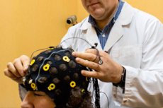
Quantitative (digital, computer, paperless) electroencephalography arose in connection with the rapid development of electronic computing technology as a further development of the EEG method.
The beginning of this new method in the late 1950s was laid by the works of Gray Walter, M.N. Livanov and V.M. Ananyev, who created the encephaloscope - a device that displayed a map of the distribution of EEG amplitudes on the scalp on a light board (in later versions on a cathode-ray tube screen) in the form of dots glowing with different brightness. Later, the method was improved by Japanese scientists, who implemented it on the basis of the first laboratory and personal electronic computers. Quantitative EEG became widely known after the description of the method of mapping the electrical activity of the brain.
Modern hardware and software systems for quantitative analysis and topographic mapping of EEG include an EEG amplifier with digital filters (usually controlled by software), an analog-to-digital converter for recording EEG signals on magnetic or other storage media in digital form, a central processor (usually a serial personal computer) that performs special types of EEG analysis (spectral-coherent, periodometric, nonlinear), and information display means (video monitor, printer, etc.).
The software usually supports a database, provides statistical processing, and also contains text and graphic editors for preparing conclusions and illustrations, which are displayed in the form of visual EEG maps of the brain.
 [
[