Medical expert of the article

New publications
Aneurysm of the hamstring artery
Last reviewed: 12.07.2025

All iLive content is medically reviewed or fact checked to ensure as much factual accuracy as possible.
We have strict sourcing guidelines and only link to reputable media sites, academic research institutions and, whenever possible, medically peer reviewed studies. Note that the numbers in parentheses ([1], [2], etc.) are clickable links to these studies.
If you feel that any of our content is inaccurate, out-of-date, or otherwise questionable, please select it and press Ctrl + Enter.
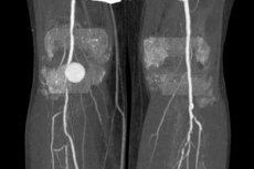
The diagnosis of aneurysm of the popliteal artery means focal dilation of this vessel - abnormal expansion of its wall (in the form of a protrusion), leading to an increase in the lumen relative to the normal diameter of at least 150%.
This is a disease of the circulatory system, of which the arteries are a part, and according to ICD-10 its code is I72.4 (Aneurysm and dissection of the artery of the lower extremities).
Epidemiology
Popliteal artery aneurysm is considered a rare disease, and its incidence in the population is estimated at 0.1–1%. However, among peripheral arterial aneurysms, it is the most common: it accounts for 70–85% of lower extremity aneurysms. [ 1 ]
As clinical statistics show, the prevalence of this pathology increases with age, reaching a maximum of cases after 60-70 years. The main patients (95-97%) are men (most likely due to their predisposition to atherosclerosis). [ 2 ]
The presence of an aneurysm of the popliteal artery in 7-20% of cases (according to other data, in 40-50%) is associated with an aneurysm in other vessels. In particular, in individuals with an aneurysm of the abdominal aorta, the incidence of aneurysms of the popliteal artery is 28% higher than in the general population.
In addition, 42% of patients (according to other data, 50–70%) have contralateral (bilateral) popliteal aneurysms. [ 3 ]
Causes aneurysms of the hamstring artery
The popliteal artery (Arteria poplitea) is a direct continuation of the superficial femoral artery (Arteria femoralis) - it passes between the medial and lateral heads of the gastrocnemius muscle (behind the popliteal muscle) and supplies blood to the tissues of the distal lower limb. Passing through the popliteal fossa, smaller vessels branch off from the artery to the knee joint area, forming anastomoses that supply blood to this joint. Further, below the knee joint, the popliteal artery bifurcates with division into the anterior tibial artery (Arteria tibialis anterior) and the tibioperoneal or tibiofibular trunk (Truncus tibiofibularis).
To date, the exact causes of aneurysms, including popliteal artery aneurysms, are unknown. Researchers suggest that the cause may be genetic or acquired defects of the media (Tunica media) - the middle layer of arterial vessels, as well as inflammatory processes, in particular, inflammatory arteritis. Perhaps the tendency of this artery to focal dilation is associated with the tension of the vessel walls during flexion and extension of the knee joint.
But most experts believe that the cause of popliteal aneurysm in 90% of cases is atherosclerosis. [ 4 ], [ 5 ], [ 6 ]
Risk factors
Modifiable risk factors include dyslipidemia (high blood cholesterol and triglyceride levels), which is associated with atherosclerosis, as well as hypertension, connective tissue disorders (such as Marfan syndrome and Ehler-Danlos syndrome), smoking, diabetes, and injury. [ 7 ]
Non-modifiable risk factors include older age, male gender, Caucasian race, and family history of aneurysmal disease.
It is also important to consider the presence of aneurysm in the family history, which may be indirect evidence of a mutation in the elastin gene or associated proteins necessary for the formation and maintenance of elastic fibers that affect the mechanical properties of arterial walls.
The formation of a false aneurysm [ 8 ], [ 9 ] is caused by repeated trauma to the arterial wall by the osteochondroma spike during flexion and extension of the knee. This repeated trauma leads to chronic abrasion of the popliteal artery and the development of an adventitial defect with subsequent pseudoaneurysm. [ 10 ], [ 11 ]
Treatment of false aneurysm of the popliteal joint consists of surgical removal of the exostosis [ 12 ] and restoration of the vascular axis. Some authors suggest prophylactic removal of exostoses located on the vascular axis to prevent the onset of such accidents, while others suggest that surgical removal is indicated in case of malignant change or when the vascular axis is disrupted. [ 13 ]
Pathogenesis
The popliteal artery is an extraorgan distributing artery of the muscular type; normally, its diameter varies from 0.7 to 1.5 cm, but it varies along the entire length of the vessel. And the average diameter of the dilated section in most cases reaches 3-4 cm, although more significant dilations are not excluded - up to giant aneurysms. [ 14 ]
The true pathogenesis of popliteal artery aneurysm formation is unknown and is associated with several factors.
More and more studies confirm the connection between the pathogenesis of aneurysms and changes in the structure of the vascular wall and its biomechanical properties. The latter directly depend on the components of the extracellular matrix of the arterial wall, in particular, elastin and collagen fibers, which (together with smooth muscle tissue) form the middle layer of the artery (the middle layer of its wall) - the media (Tunica media).
The dominant protein of the extracellular matrix of the media is mature elastin, a hydrophobic connective tissue protein, structurally organized in the form of plates, which also have smooth muscle cells (arranged in concentric rings) and collagen fibers. Thanks to elastin, the walls of the vessels can be reversibly stretched, and the strength of the vascular wall is provided by collagen fibers.
The process of vessel wall formation, including elastogenesis – the transformation of the soluble monomeric protein tropoelastin (produced by fibro and chondroblasts, smooth muscle cells and endothelium), occurs during embryonic development, and their structure is constant throughout life.
However, with age or due to pathological effects, the structure of elastic fibers can change (due to destruction and fragmentation). In addition, inflammatory processes induce the synthesis of tropoelastin, which in adults is not capable of transforming into elastin. All this affects the biomechanics of arteries in the direction of reducing the elasticity and resilience of their walls.
As for arterial hypertension and atherosclerosis, increased pressure causes stretching of the walls of the artery passing through the popliteal fossa. And cholesterol deposits on the intima of the vascular wall create areas of narrowing of the artery, leading to local turbulence of blood flow, which increases the pressure on the nearest section of the vessel and entails a decrease in the thickness of its wall and a change in the structure of the medial layer.
Symptoms aneurysms of the hamstring artery
The first signs of a popliteal aneurysm, which is asymptomatic in almost half of patients at the initial stage, are the presence of a palpable pulsating mass in the popliteal fossa.
Clinical manifestations of aneurysms include: ruptures (5.3%); deep vein thrombosis (5.3%); sciatic nerve compression (1.3%); leg ischemia (68.4%) and asymptomatic pulsatile lesions 15 (19.7%).[ 15 ]
According to a 2003 study, small popliteal artery aneurysms were associated with a higher incidence of thrombosis, clinical symptoms, and distal occlusion.[ 16 ]
As the pathological process progresses, paresthesia in the leg and pain under the knee are noted, which are a consequence of compression of the peroneal and tibial nerves. Pain may also occur in the skin of the medial side of the shin, ankle or foot.
Due to compression of the popliteal vein, the soft tissues of the lower leg swell. And with progressive narrowing of the lumen of Arteria poplitea, associated with the formation of a thrombus, a symptom such as intermittent claudication appears.
In cases of acute aneurysm thrombosis, the pain intensifies and becomes more severe, the skin on the leg turns pale (due to ischemia), the toes become cold and bluish (their cyanosis develops).
Forms
An aneurysm of the artery below the knee may affect one or both limbs and will be diagnosed as unilateral or bilateral, respectively.
According to shape, there are such types of popliteal artery aneurysms as fusiform and saccular (in the form of a sac). Most popliteal artery aneurysms are fusiform, and up to a third of cases are bilateral.
Complications and consequences
Popliteal artery aneurysms cause thrombosis (formation of a blood clot) and embolization (movement of clot fragments into smaller vessels) – with a high risk of limb loss. And these are their main consequences and complications.
According to some data, thrombosis of the aneurysmal sac occurs in 25-50% of cases, which causes limb tissue ischemia with a limb loss rate of 20% to 60% and mortality of up to 12%. [ 17 ] And distal embolism, leading to vascular occlusion, is detected in 6-25% of patients with popliteal artery aneurysm. [ 18 ]
In every fourth case of thromboembolism, there is a need for amputation of the affected limb.
Rupture of a popliteal artery aneurysm occurs in 3-5% of cases on average. Popliteal aneurysms usually rupture into the popliteal space, which is bounded by muscles and tendons. The main symptoms are pain and swelling. [ 19 ]
Diagnostics aneurysms of the hamstring artery
Imaging is critical in diagnosing popliteal artery aneurysm.
Instrumental diagnostics uses:
- standard angiography with contrast;
- two-dimensional ultrasonography or duplex scanning of the arteries of the lower extremities;
Ultrasound methods are very effective in screening painful lesions of the popliteal space. These methods easily differentiate popliteal cysts from thrombophlebitis and, in addition, allow for consistent evaluation without discomfort to the patient. [ 20 ]
- CT or MR angiography.
Peripheral arterial blood flow is examined using ultrasound Dopplerography of the vessels of the lower extremities.
Differential diagnosis
Differential diagnosis takes into account the possibility that patients may have similar symptoms:
- cystic adventitial disease - a cyst of the outer lining of the popliteal artery wall (or Baker's cyst);
- inflammation of the popliteal lymph node;
- varicose veins of the popliteal vein;
- adventitial cyst (outer lining of the wall) of the popliteal artery,
- dystopic popliteal artery entrapment syndrome (trapped artery syndrome).
Who to contact?
Treatment aneurysms of the hamstring artery
Asymptomatic aneurysms (up to 2 cm in size) are monitored under duplex ultrasound control, and conservative treatment is carried out for those diseases that are involved in the development of the aneurysm.
Read more:
Recently, if the operation does not expose the patient to high risk, vascular surgeons recommend eliminating even asymptomatic aneurysms due to frequent complications that arise even with small aneurysms.
Many clinicians use a diameter of 2 cm, with or without evidence of thrombosis, as an indication for prophylactic surgery, which is supported by the 2005 American College of Cardiology/American Heart Association guidelines on peripheral arterial disease.[ 21 ] Asymptomatic aneurysms larger than 4–5 cm require surgical intervention because they may cause acute limb ischemia secondary to vessel kinking.
If symptoms are present, surgical treatment is required, either by open surgery or by endovascular stent grafting.
- Open surgical approach
In an open operation, the popliteal artery is ligated above the knee and below the aneurysm, excluding this area from the blood flow, and then reconstructing it (revascularizing) using an autologous graft from the patient’s subcutaneous vein or an artificial vascular prosthesis. [ 22 ]
Surgical bypass is considered the gold standard for the treatment of popliteal artery aneurysm (PAA), especially in young patients. [ 23 ] The great saphenous vein (GSV) is an ideal material, and prosthetic grafts are a reliable alternative to GSV for surgical bypass.
- Endovascular approach
Recently, endovascular techniques have gained popularity in popliteal artery reconstruction as an alternative to the open surgical approach. This is achieved by excising the aneurysm sac with implantation of a stent graft. Recent studies suggest that popliteal artery stenting is a safe alternative treatment for popliteal aneurysm, especially in high-risk patients. Advantages of the endovascular technique include a shorter hospital stay and reduced operative time compared to open surgery. Disadvantages include higher 30-day graft thrombosis rates (9% in the endovascular group vs. 2% in the open surgical group) and higher 30-day reintervention rates (9% in the endovascular group vs. 4% in the open surgical group). [ 24 ]
Acute thrombosis is treated with heparin (administered intravenously and by continuous infusion). And in case of threatening ischemia, thrombectomy is used followed by bypass of the popliteal artery.
According to a 2007 Swedish Nationwide Study, the rate of limb loss within 1 year after surgery was about 8.8%; 12.0% for symptomatic and 1.8% for asymptomatic aneurysms (P < 0.001). Risk factors for amputation were: presence of symptoms, previous thrombosis or embolism, urgent treatment, age over 70 years, graft replacement, and no preoperative thrombolysis for acute ischemia. The amputation rate decreased with time (P = 0.003). Primary patency at 1, 5, and 10 years was 84%, 60%, and 51%, respectively. Overall survival was 91.4% at 1 year and 70.0% at 5 years.[ 25 ]
Prevention
Specific measures to prevent the development of aneurysms have not been developed, but it is important for vascular health to quit smoking, lose excess weight, control high blood pressure, cholesterol and blood sugar levels, and also eat right and exercise more.
Early diagnosis of popliteal artery aneurysm and surgical treatment before embolism, thrombosis and rupture are necessary to prevent serious complications. [ 26 ]
Forecast
Early detection of popliteal artery aneurysm and its treatment provide a favorable prognosis. Lack of treatment increases the risk of complications by 30-50% over 3-5 years.
The worst outcome is amputation of the limb if the aneurysm ruptures.