Medical expert of the article
New publications
Pneumococci
Last reviewed: 04.07.2025

All iLive content is medically reviewed or fact checked to ensure as much factual accuracy as possible.
We have strict sourcing guidelines and only link to reputable media sites, academic research institutions and, whenever possible, medically peer reviewed studies. Note that the numbers in parentheses ([1], [2], etc.) are clickable links to these studies.
If you feel that any of our content is inaccurate, out-of-date, or otherwise questionable, please select it and press Ctrl + Enter.

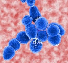
A special position in the genus Streptococcus is occupied by the species S. pneumoniae, which plays a very important role in human pathology. It was discovered by L. Pasteur in 1881. Its role in the etiology of lobar pneumonia was established in 1886 by A. Frenkel and A. Weichselbaum, as a result of which S. pneumoniae is called pneumococcus. Its morphology is unique: the cocci have a shape resembling a candle flame: one end of the cell is pointed, the other is flattened; they are usually located in pairs (the flat ends face each other), sometimes in the form of short chains. They do not have flagella and do not form spores. In the body of humans and animals, as well as on media containing blood or serum, they form a capsule. They are gram-positive, but in young and old cultures they are often gram-negative. Facultative anaerobes. The optimum temperature for growth is 37 °C; they do not grow at temperatures below 28 °C and above 42 °C. The optimum pH for growth is 7.2-7.6. Pneumococci produce hydrogen peroxide, but they do not have catalase, so they require the addition of substrates containing this enzyme (blood, serum) for growth. On blood agar, small round colonies are surrounded by a green zone formed as a result of the action of the hemolysin exotoxin (pneumolysin). Growth on sugar broth is accompanied by turbidity and the formation of a small sediment. In addition to the O-somatic antigen, pneumococci have a capsular polysaccharide antigen, which is distinguished by great diversity: according to the polysaccharide antigen, pneumococci are divided into 83 serovariants, 56 of which are divided into 19 groups, 27 are represented independently. Pneumococci differ from all other streptococci in their morphology, antigen specificity, and also in that they ferment inulin and are highly sensitive to optochin and bile. Under the influence of bile acids, pneumococci activate intracellular amidase. It breaks the bond between alanine and muramic acid of peptide glycan, the cell wall is destroyed, and pneumococci lysis occurs.
The main factor of pneumococcal pathogenicity is the capsule of polysaccharide nature. Uncapsulated pneumococci lose virulence.
Pneumococci are the main causative agents of acute and chronic inflammatory lung diseases, which occupy one of the leading places in morbidity, disability and mortality of the population throughout the world.
Pneumococci, along with meningococci, are the main culprits of meningitis. In addition, they cause creeping ulcer of the cornea, otitis, endocarditis, peritonitis, septicemia and a number of other diseases.
Laboratory diagnostics
Diagnosis of pneumococcal diseases is based on the isolation and identification of S. pneumoniae. The material for the study is sputum and pus. White mice are very sensitive to pneumococci, so a biological sample is often used to isolate pneumococci. In dead mice, pneumococci are found in a smear preparation from the spleen, liver, lymph nodes, and when sowing from these organs and from the blood, a pure culture is isolated. To determine the serotype of pneumococci, an agglutination reaction on glass with typical serums or the phenomenon of "capsule swelling" is used (in the presence of homologous serum, the pneumococcal capsule swells sharply).
Specific prevention
Prevention of pneumococcal diseases is carried out using vaccines prepared from highly purified capsular polysaccharides of those 12-14 serovariants that most often cause diseases (1, 2, 3, 4, 6A, 7, 8, 9, 12, 14, 18C, 19, 25). The pneumococcal vaccine is highly immunogenic.


 [
[