Medical expert of the article
New publications
Pericardial tumors
Last reviewed: 29.06.2025

All iLive content is medically reviewed or fact checked to ensure as much factual accuracy as possible.
We have strict sourcing guidelines and only link to reputable media sites, academic research institutions and, whenever possible, medically peer reviewed studies. Note that the numbers in parentheses ([1], [2], etc.) are clickable links to these studies.
If you feel that any of our content is inaccurate, out-of-date, or otherwise questionable, please select it and press Ctrl + Enter.

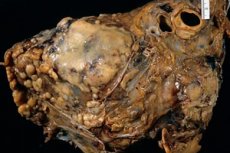
Pericardial tumors are a serious problem. Conventionally, all pericardial tumors can be divided into primary and secondary tumors. However, primary tumors are relatively rare. Secondary tumors are much more frequently observed. According to the histological structure, tumors can be divided into benign and malignant.
Of the benign tumors, the most common are fibroma, or fibromatosis, fibrolipoma, hemangioma, lymphagioma, dermoid cyst, teratoma, and neurofibroma. All of these tumors have some common features. As a rule, these tumors hang directly into the pericardium. Their weight is quite large. There are known cases when the weight of benign pericardial tumors reached 500 grams.
It is also not uncommon to see pseudotumors (thrombotic masses). Such tumors are also called fibrinous polyps.
Tumors, especially small ones, are quite difficult to recognize. For example, they are practically not visualized on ultrasound, are not seen on X-rays. The danger of them is that they can grow, gradually accompanied by symptoms that are similar to disorders of the respiratory system. For example, there is often compression of the airways, esophagus. In this case, respiratory function, digestion, swallowing are disturbed. As a rule, this makes diagnosis even more difficult. Gradually irritation occurs, coughing, dyspnea develops. At the same time, generalized compression occurs, heart failure develops. If aortic compression occurs, symptoms such as a systolic murmur appear. At the same time, it is most often heard above the compressed area. Despite the fact that the vessels are compressed insignificantly, blood circulation is significantly disturbed.
Angiomas and teratomas are quite dangerous. They can be fatal. The cause in most cases is fatal bleeding that cannot be stopped. The complications are often hemorrhagic pericarditis, as well as the risk of malignization.
The main method of treatment is surgery. The question of the expediency of surgery is decided based on the severity of the condition, the severity of clinical symptoms. If the tumor grows quite rapidly, it must be removed.
Malignant tumors, or cancerous tumors, are considered the most dangerous type of tumors.
Pericardial cancer
Malignant tumors, or cancer of the pericardium, are also observed. They are much more common than benign tumors and are more dangerous. The risk of a fatal outcome increases manifold. As primary tumors of malignant character, it is necessary to name sarcoma, angiosarcoma, mesothelioma. Histological variants of such tumors can be many. Malignant tumors are cancerous tumors, the cells of which are characterized by the ability to unlimited growth, rapid multiplication, inability to apoptosis.
Here are some of the characteristics of this disease:
- Rarity: Pericardial cancer accounts for only about 1% of all newly diagnosed cases of heart and pericardial cancer.
- Symptoms: Patients with pericardial cancer may experience a variety of symptoms including chest pain, difficulty breathing, palpitations, fatigue, general malaise, and weight loss.
- Diagnosis: Various examination methods such as echocardiography, computed tomography (CT), magnetic resonance imaging (MRI) and biopsy are used to diagnose pericardial cancer.
- Treatment: Treatment for pericardial cancer may include surgical removal of the tumor, chemotherapy, radiation therapy, or a combination of these. Because it is a rare disease, the optimal treatment approach may vary depending on individual patient characteristics and stage of disease.
- Prognosis: Prognosis depends on many factors, including the stage of the cancer at diagnosis, the size and location of the tumor, and the effectiveness of treatment. In general, the prognosis for pericardial cancer is often unfavorable due to its rarity and tendency to be diagnosed in the later stages of the disease.
- Support and care: Patients with pericardial cancer may need support from medical professionals as well as from family and friends. The support of a psychologist or support group can also be helpful in helping patients cope with the emotional aspects of the disease.
Pericardial mesothelioma
Pericardial mesothelioma tumor is characterized by the fact that it can secrete mucus, which becomes viscous and thick in the pericardial cavity. At the same time, as a rule, the mucus is colorless. Tumors represent a limited polyposis outgrowth, filled with hemorrhagic exudate. Diffuse tumor infiltration and obliteration of the cavity occurs.
On microscopic examination of mesothelioma, it is noteworthy that it is of three types. The simplest and safest are fibrous, or epithelial tumors represented by epithelial tissue. They are characterized by a high degree of enzymatic activity. Epithelial fibrous tumors are not uncommon. The most common, and the most dangerous type of tumors, are metastatic tumors. It is worth noting that 5% of those who died of breast cancer were diagnosed with metastatic tumors to the pericardium. Many of them are diagnosed posthumously. Such tumors are often complicated by long-term hemorrhagic pericarditis.
Clinical symptomatology depends on how fast the tumor grows and how susceptible it is to metastasis. The most dangerous are metastases to the lungs, pleura, liver. Almost all tumors exert pressure on neighboring organs, cavities. Characteristic symptoms in this case are specific ECG changes peculiar to myocardial infarction.
They are treated exclusively with surgery. Radiation therapy is performed. It is often used for inoperable tumors. As a rule, radiation therapy allows only temporarily suspend the tumor process, reduce the rate of progression of the disease. Slowing tumor growth is possible for months, years, until remission is achieved.

