Medical expert of the article

New publications
Osteonecrosis: Causes, Symptoms, Diagnosis, Treatment, and Prognosis
Last updated: 16.03.2026

All iLive content is medically reviewed or fact checked to ensure as much factual accuracy as possible.
We have strict sourcing guidelines and only link to reputable media sites, academic research institutions and, whenever possible, medically peer reviewed studies. Note that the numbers in parentheses ([1], [2], etc.) are clickable links to these studies.
If you feel that any of our content is inaccurate, out-of-date, or otherwise questionable, please select it and press Ctrl + Enter.
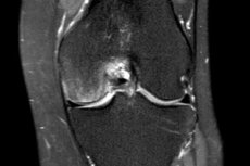
Osteonecrosis is the death of a section of bone tissue due to a disruption of the blood supply. In clinical practice, this condition is also referred to as avascular, aseptic, or ischemic bone necrosis. The disease can affect various bones, but most commonly affects the femoral head, and less commonly the humerus, knee, talus, and bones of the hand and foot. If the process develops in the joint area, over time it leads not only to destruction of the bone itself but also to secondary osteoarthritis. [1]
The key problem with osteonecrosis is that its early stages often proceed latently or are virtually asymptomatic. Meanwhile, irreversible changes are already occurring within the bone: some bone cells die, the trabecular structure weakens, and a zone of increased risk of microfracture and collapse develops beneath the articular cartilage. This is why modern orthopedics emphasizes early imaging, primarily using magnetic resonance imaging, and the earliest possible selection of treatment strategies before articular surface deformation occurs. [2]
It's important to understand one more thing: the most studied form of the disease is osteonecrosis of the femoral head. Therefore, the general sections of this article address osteonecrosis in general, while the details of staging, prognosis, and most modern surgical approaches rely primarily on data for the hip joint, as the most studied location. [3]
Code according to ICD-10 and ICD-11
In the International Classification of Diseases, 10th revision, the basic category for osteonecrosis is group M87 "Osteonecrosis." Within this group, it distinguishes idiopathic aseptic bone necrosis, drug-induced osteonecrosis, posttraumatic osteonecrosis, and other secondary forms. The location of the lesion is also taken into account when coding. The official version of the International Classification of Diseases, 10th revision, specifically specifies that avascular necrosis of bone is included in this group, while drug- or radiation-induced osteonecrosis of the jaw is coded separately. [4]
The International Classification of Diseases, 11th revision, uses the FB81 Osteonecrosis trunk with subcategories for the idiopathic form, dialysis-associated variant, drug-induced osteonecrosis, post-traumatic form, variants associated with hemoglobinopathies, after ionizing radiation, alcohol-induced form, and other specified and unspecified forms. The International Classification of Diseases, 11th revision, also provides for post-coordination, that is, the addition of codes specifying the affected side and anatomical location. [5]
| System | Main code | What does it include? |
|---|---|---|
| ICD 10 | M87 | General section "Osteonecrosis" |
| ICD 10 | M87.0 | Idiopathic aseptic bone necrosis |
| ICD 10 | M87.1 | Drug-induced osteonecrosis |
| ICD 10 | M87.2 | Osteonecrosis after trauma |
| ICD 10 | M87.3 | Other secondary osteonecrosis |
| ICD 11 | FB81 | General section "Osteonecrosis" |
| ICD 11 | FB81.0 | Idiopathic aseptic osteonecrosis |
| ICD 11 | FB81.2 | Drug-induced osteonecrosis |
| ICD 11 | FB81.3 | Osteonecrosis due to trauma |
| ICD 11 | FB81.6 | Alcohol-induced osteonecrosis |
Sources for the table: [6]
Epidemiology
The exact global prevalence of osteonecrosis remains difficult to estimate because the disease encompasses various causes, locations, and stages, and asymptomatic early forms are often not included in statistics. However, modern reviews agree that this is not an exceptionally rare condition: osteonecrosis of the femoral head alone is diagnosed in approximately 10,000-20,000 patients annually in the United States, with some reviews providing even higher estimates. Furthermore, this form accounts for a significant proportion of the reasons for total hip arthroplasty. [7]
According to the American Academy of Orthopaedic Surgeons, osteonecrosis of the hip is most common between the ages of 40 and 65 and is more common in men than in women. The disease is often bilateral, especially in non-traumatic cases associated with systemic risk factors such as long-term glucocorticosteroid use or alcohol abuse. In practice, this means that when a lesion is detected in one femoral head, the physician typically evaluates the opposite joint as well, even if there are no complaints on that side yet. [8]
Certain epidemiological trends in the disease are particularly important after trauma. For example, a 2024 meta-analysis of adolescents after surgery for femoral neck fractures showed a high incidence of subsequent osteonecrosis of the femoral head—approximately 24.02%. This emphasizes that osteonecrosis is not only a "slow" non-traumatic disease of adults, but also a severe late complication of fractures and dislocations. [9]
| Epidemiological landmark | What is known |
|---|---|
| The most common localization | Head of the femur |
| Estimating new cases in the US | About 10-20 thousand per year |
| Typical age at onset of hip joint damage | Most often 40-65 years old |
| Gender differences | Men get sick more often |
| Bilateral defeat | It occurs frequently, especially in the non-traumatic form. |
| Contribution to endoprosthetics | A significant proportion of hip surgeries |
Sources for the table: [10]
Reasons
The causes of osteonecrosis are conventionally divided into two broad groups: traumatic and non-traumatic. The traumatic variety develops after fractures, dislocations, and other injuries that damage the blood vessels supplying the bone. A classic example is a femoral neck fracture or hip dislocation, which dramatically impairs the blood supply to the femoral head. In such cases, the mechanism is relatively straightforward: a blood vessel is damaged, blood flow is reduced, and bone tissue dies. [11]
Non-traumatic osteonecrosis develops more complexly. The cause is often multifactorial: intravascular thrombi, fat embolization, increased intraosseous pressure, endothelial dysfunction, impaired venous outflow, and impaired bone regeneration. Therefore, in some patients, a single trigger cannot be identified, and the disease is considered the result of a combination of vascular, metabolic, and mechanical disorders. [12]
Among the most well-documented causes and associations, modern literature cites long-term use of glucocorticosteroids, chronic alcohol abuse, hemoglobinopathies, systemic lupus erythematosus, antiphospholipid syndrome, Gaucher disease, vasculitis, thrombosis, decompression sickness, conditions after organ transplantation, radiation therapy, and some other severe chronic conditions. In some patients, an idiopathic form develops, when a clear cause cannot be established. [13]
Risk factors
The most well-known and practically significant risk factors are considered to be the use of high doses of glucocorticosteroids, alcohol abuse, and a history of joint or bone trauma. These factors are constantly repeated in guidelines, reviews, and clinical materials, as they have accumulated the most observations. Situations where a person has multiple risk factors simultaneously are considered particularly concerning: for example, a systemic disease, treatment with glucocorticosteroids, and smoking. [14]
In addition, blood clotting disorders, hemoglobinopathies, systemic autoimmune diseases, renal failure with dialysis, organ transplantation, obesity, smoking, and repeated joint overload are also significant. According to a 2025 review of femoral head collapse risk, an unfavorable prognosis was associated, in particular, with alcohol exposure and a large lesion size. This is important because risk factors are responsible not only for the onset of the disease but also for the rate of its progression. [15]
| Risk factor | How does it increase the risk? |
|---|---|
| Glucocorticosteroids | Associated with impaired lipid metabolism, vascular and bone marrow changes |
| Alcohol | Promotes fatty deposits in blood vessels and disrupts blood flow |
| Fracture, dislocation, severe injury | May directly damage feeding vessels |
| Hemoglobinopathies | Increase the risk of vascular occlusion |
| Systemic lupus erythematosus and autoimmune diseases | Associated with both the disease itself and glucocorticosteroid therapy |
| Dialysis, transplantation | Reflects severe systemic background and vascular-metabolic disorders |
| Smoking, obesity | Increase the vascular and metabolic risk components |
| Large lesion | Increases the risk of collapse and adverse outcome |
Sources for the table: [16]
Pathogenesis
Osteonecrosis is caused by a mismatch between bone tissue's need for oxygen and nutrients and the actual blood supply. When arterial inflow is reduced or venous outflow is disrupted, ischemia occurs within the bone. This causes osteocytes and bone marrow cells to die, and the architecture of the spongy bone loses its strength. This sets off a chain of events that may remain undetectable on X-rays for a long time, but which creates the conditions for subsequent fracture of the subchondral zone. [17]
The body then attempts to repair the damage: a repair zone forms, new vessels grow, and resorption and remodeling processes are activated. However, it is precisely this stage that often weakens the bone even further, because the rate of destruction of non-viable trabeculae can temporarily exceed the rate of full recovery. As a result, a microfracture appears under the articular cartilage, which in orthopedics is often called a subchondral fracture or a precursor to collapse. After this, the articular surface flattens, the cartilage is destroyed, and the disease progresses to the stage of secondary arthrosis. [18]
Symptoms
In the early stages, osteonecrosis may not cause any sensations. As it progresses, pain develops, initially only with weight-bearing activity, later also at rest. Typical hip pain is in the groin, buttock, or thigh; shoulder pain is with arm movement; and knee pain is a deep, aching pain in the condyles and limited support. The pain can be dull, aching, or throbbing. [19]
Gradually, it becomes difficult for a person to stand for long periods, walk, climb stairs, turn, and abduct the limb. In the early stages, range of motion may be relatively well maintained, sometimes creating the false impression that the problem is not very serious. However, after the articular surface collapses, stiffness, lameness, limited motion, and signs of secondary arthrosis rapidly increase. This sequence—first pain, then functional limitations, then joint deformity—is considered typical of progressive osteonecrosis. [20]
Classification, forms and stages
Osteonecrosis is primarily divided into traumatic and non-traumatic. From a practical standpoint, it is also classified as idiopathic, drug-induced, alcohol-induced, radiation-induced, dialysis-associated, and hemoglobinopathies-related. This etiologic classification is important because it influences both relapse prevention and prognosis: for example, with continued high-dose glucocorticosteroid use or persistent alcohol consumption, the risk of progression is higher. [21]
Several staging systems are used for the femoral head, but the International Association for the Study of Bone Circulation classification is particularly frequently cited in modern reviews because it clearly distinguishes between precollapse and postcollapse stages. In the precollapse stages, the articular surface is not yet destroyed, and this is where the chance for joint-preserving treatment remains. After a subchondral fracture and collapse, the chances of preserving the natural joint are greatly reduced. [22]
The size and location of the lesion are no less important than the stage itself. The greater the volume of necrosis and the more heavily loaded the lateral aspect of the femoral head is involved, the higher the risk of collapse. Therefore, modern assessment always considers not only the stage number but also the morphology of the lesion based on magnetic resonance imaging and computed tomography data. [23]
| Stage | What's happening |
|---|---|
| Stage 0 | Research may still be without obvious changes |
| Stage I | The X-ray is normal, but the magnetic resonance imaging already shows a lesion |
| Stage II | Sclerosis, cysts, and bone remodeling without collapse appear. |
| Stage III | A subchondral fracture occurs, the “half-moon sign” appears, and collapse begins |
| Stage IV | Secondary arthrosis develops with joint deformation and loss of function. |
Sources for the table: [24]
Complications and consequences
The main complication of osteonecrosis is the collapse of the subchondral zone and deformation of the articular surface. After this, the joint no longer distributes the load normally, the cartilage wears quickly, and secondary deforming arthrosis develops. For the patient, this means increased pain, lameness, decreased walking distance, loss of independence, and the subsequent need for endoprosthetics. [25]
Additional consequences include chronic pain, muscle atrophy, limited mobility, and a reduced quality of life. The problem is particularly severe in young patients, as the disease develops during working age and often requires major surgery decades earlier than typical osteoarthritis. In cases of bilateral involvement, functional loss can mount very rapidly. [26]
When to see a doctor
A doctor should be consulted for any persistent joint pain, especially if the pain intensifies with activity, develops after an injury, or is accompanied by lameness or limited range of motion. Immediate evaluation is especially important for people receiving glucocorticosteroids, abusing alcohol, having suffered a femoral neck fracture, hip dislocation, or having systemic diseases that increase the risk of vascular disorders. [27]
If you experience sharp pain after an injury, an inability to bear weight on your leg, a sudden limitation of movement, or a suspected fracture or dislocation, you need immediate medical attention. Early diagnosis does change the prognosis: in the pre-collapse stages, methods aimed at preserving your own joint can still be considered, whereas late treatment often results in endoprosthetics. [28]
Diagnostics
Diagnosis begins not with equipment, but with a correct clinical suspicion. The doctor determines the location of the pain, its relationship to stress, the presence of injury, glucocorticosteroid use, alcohol abuse, autoimmune diseases, hemoglobinopathies, episodes of decompression sickness, dialysis, and transplant surgery. At this stage, it is already possible to determine the likelihood of osteonecrosis and the risk of bilateral involvement. [29]
The next step is an examination. The doctor evaluates gait, range of motion, pain with rotation and weight bearing, the presence of contractures, limb shortening, and muscle weakness. However, a clinical examination alone does not confirm the diagnosis, as early symptoms can be subtle and resemble many other joint diseases. [30]
Radiography is typically the first instrumental examination. It is useful as a starting method and helps rule out fracture, severe osteoarthritis, and gross deformity, but its sensitivity is insufficient in the early stages. If radiography is normal and clinical suspicion remains high, magnetic resonance imaging is the current standard. According to a 2026 systematic review, it demonstrated the best overall diagnostic accuracy for the early detection of osteonecrosis: sensitivity of approximately 0.91 and specificity of approximately 0.96. [31]
Computed tomography (CT) is particularly useful for assessing the presence of a subchondral fracture, initial collapse, and the extent of disruption of bone architecture. Current data show that CT and MRI are superior to conventional radiography for detecting subchondral fractures. Radioisotope techniques and angiographic studies are used less frequently but can be useful in selected cases, particularly when planning joint-preserving surgeries and searching for multiple lesions. [32]
Laboratory tests don't directly confirm osteonecrosis, but they help identify its cause and rule out other conditions. Depending on the situation, a complete blood count, inflammation markers, coagulation profile, lipid profile, liver and kidney function tests, autoimmune markers, and, if there's a relevant history, a hemoglobinopathies screening are prescribed. In other words, tests are needed not to "see necrosis," but to understand why it occurred and how to reduce the risk of further progression. [33]
| Stage | What does a doctor do? | Why is this necessary? |
|---|---|---|
| 1 | Collection of anamnesis | Identify trauma, medications, alcohol, systemic diseases |
| 2 | Inspection and evaluation of function | Understand the severity of pain and limitation of movement |
| 3 | X-ray | Rule out fracture, arthrosis, and look for late signs |
| 4 | Magnetic resonance imaging | Find an early lesion, assess the volume and location of the lesion |
| 5 | Computed tomography | Specify the subchondral fracture and the degree of collapse |
| 6 | Blood tests and additional examinations | Find the cause and factors of progression |
Sources for the table: [34]
Differential diagnosis
Osteonecrosis must be distinguished primarily from osteoarthritis, especially when joint deformity and pain with weight-bearing activity already exist. However, there is a fundamental difference between these conditions: in osteonecrosis, the primary event is a vascular catastrophe within the bone followed by a subchondral fracture, while in ordinary arthrosis, the leading mechanism is chronic degeneration of the cartilage and articular surfaces. In the early stages, magnetic resonance imaging allows this distinction to be made most reliably. [35]
The second important condition for differential diagnosis is bone marrow edema syndrome and transient osteoporosis. These also cause pain and edematous changes on magnetic resonance imaging, but they usually have a different signal pattern and often regress within a few months without bone collapse. Guidelines emphasize that in this condition, the characteristic band-like boundary between viable and non-viable bone typical of osteonecrosis is absent. [36]
The doctor also rules out subchondral stress fractures, tumors, dysplastic coxarthrosis, ankylosing spondylitis with hip involvement, infection, and the consequences of previous radiation therapy. In practice, differential diagnosis is almost always based on a combination of history, clinical presentation, and magnetic resonance imaging data, rather than on a single isolated symptom. [37]
Treatment
Treatment for osteonecrosis is always tailored to the stage, location, and extent of the lesion, the patient's age, and the underlying cause. The main decision is simple: whether the articular surface has already collapsed. Until collapse occurs, the goal is to preserve the natural bone and joint as much as possible. After collapse, the focus increasingly shifts to reconstructive surgery and endoprosthetics. [38]
Non-pharmacological conservative management includes limiting impact loads, using a cane or crutches if supporting bones are affected, adjusting mobility, controlling body weight, and avoiding factors that contribute to the disease. This approach can reduce pain and decrease mechanical pressure on the affected area, but rarely stops the progression of an already established lesion. This is especially true for osteonecrosis of the femoral head, where the load on the subchondral bone is very high. [39]
Drug treatment plays a primarily symptomatic and supportive role. Anti-inflammatory medications reduce pain, physical therapy helps maintain mobility and muscle balance, and treatment of the underlying condition reduces the risk of further bone damage. However, current government and professional sources emphasize that no conservative treatment has proven universally effective in "curing" osteonecrosis as an independent structural problem. [40]
If the disease is detected early, decompression of the lesion remains the most studied joint-preserving surgery. This method involves the surgeon creating one or more canals in the affected bone, reducing intraosseous pressure and creating conditions for improved blood supply and repair. For early stages, this approach is considered the most common surgical option. In some patients, it can delay collapse and reduce pain, especially in small or moderate lesions without deformity of the articular surface. [41]
Decompression is increasingly being combined with bone grafting or biological reinforcement. A 2026 systematic review found that adding bone marrow concentrate to decompression was associated with a lower risk of collapse compared to decompression alone, although the authors rated the quality of the evidence as low. This indicates that this approach is promising, but it does not yet guarantee the same good outcome for all patients. In practice, such methods are more often considered in young patients at a pre-collapse stage who desire to preserve their natural joint as much as possible. [42]
Bone grafting can be either non-vascularized or vascularized. In the former case, a bone graft or synthetic material is inserted into the lesion for support and reconstruction. In the latter case, a bone fragment is transplanted along with its supplying vessels, which appears more biologically convincing. A 2026 review found that vascularized bone grafting had a lower risk of collapse compared to non-vascularized bone grafting, but the authors also emphasized the limitations and heterogeneity of the available studies. [43]
Osteotomies remain an option for carefully selected patients, usually relatively young, when it is necessary to redistribute the load so that the less damaged portion of the bone bears the brunt of the stress. This is not a "simple operation," but a reconstructive intervention that requires a precise understanding of the lesion's geometry and extensive surgical experience. Contemporary reviews acknowledge the benefit of osteotomies in some patients, but emphasize that direct comparative studies remain insufficient, and patient selection is critical. [44]
New techniques are currently being developed primarily in the field of regenerative orthopedics: bone marrow concentrate, mesenchymal cells, growth factors, biomaterials, combinations with decompression and guided bone grafting. A 2025 meta-analysis of 954 hip joints showed that the combination of decompression with regenerative technologies improved pain and function, with the most convincing data obtained for bone marrow concentrate. However, the authors themselves cautioned about the high heterogeneity of the studies and the limited ability to draw definitive conclusions about slowing disease progression. [45]
When the articular surface has already been destroyed or pain and functional limitation have become severe, total hip replacement is usually the best definitive treatment option. Total hip replacement involves removing the destroyed articular surfaces and replacing them with artificial components, which significantly reduces pain and restores support. Current guidelines and clinical literature agree that in advanced stages with collapse, this is the most predictable and effective approach. It is particularly often required for femoral head lesions. [46]
Treatment doesn't end after surgery. Rehabilitation, restoration of range of motion, muscle strengthening, pain control, thrombosis prevention, correction of underlying risk factors, and consistent imaging monitoring are all important. Even after successful joint-preserving treatment, the patient requires long-term monitoring, as progression can be slow and not immediately apparent. And after endoprosthetics, the goal shifts from preserving the bone head to ensuring long-term implant stability and a good functional outcome. [47]
| Approach | When considered | The main goal |
|---|---|---|
| Load limitation, support, rehabilitation | Early stages, symptom control | Reduce pain and mechanical stress |
| Decompression | Pre-collapse stages | Reduce intraosseous pressure, delay collapse |
| Biologically assisted decompression | Early stages in selected patients | Improve repair and support of the lesion |
| Bone grafting | Larger lesions without gross deformation | Support the subchondral bone |
| Vascularized graft | In young and motivated patients | Biological and mechanical support |
| Osteotomy | Strictly selected cases | Redistribute the load |
| Endoprosthetics | Collapse, secondary arthrosis, severe pain | Eliminate pain and restore function |
Sources for the table: [48]
Prevention
Prevention of osteonecrosis primarily involves controlling manageable risk factors. If possible, avoid long-term use of high doses of glucocorticosteroids or use the lowest effective dose under strict supervision. Avoiding alcohol abuse, stopping smoking, treating lipid metabolism and coagulation disorders, and promptly managing systemic diseases, which themselves increase the risk of bone ischemia, are important. [49]
After fractures and dislocations of large joints, prevention means the earliest and highest quality restoration of anatomy, observation by an orthopedist, and control imaging if pain occurs. For high-risk patients, a low threshold for magnetic resonance imaging is justified, as early detection has the greatest impact on prognosis. For those who have already suffered osteonecrosis, recurrence prevention is not a formality but a mandatory part of management: controlling the underlying cause of the disease is often as important as the surgery itself. [50]
Forecast
The prognosis for osteonecrosis depends primarily on the stage, size, and location of the lesion, the presence of a subchondral fracture, the underlying cause, and the speed of elimination of risk factors. Small, pre-collapse lesions have a significantly better prognosis than large, lateral lesions that have already collapsed. Once a subchondral fracture occurs, the likelihood of further deformity increases sharply, and the chances of preserving the joint decrease. [51]
Early treatment usually offers the best chance of delaying joint destruction, but it does not guarantee a complete cure. Even with successful joint-preserving treatment, some patients still eventually undergo joint replacement. On the other hand, modern data show that the prognosis should not be overly simplified: in some patients with mild collapse, the joint may still retain function, while in others, progression occurs more rapidly. Therefore, the prognosis is always individual and should be regularly reviewed based on clinical and imaging data. [52]
FAQ
Can osteonecrosis resolve on its own?
Sometimes small lesions in non-weight-bearing areas may partially stabilize, but in most clinically significant cases, especially in weight-bearing joints, the disease tends to progress. Current sources emphasize that a wait-and-see approach without monitoring is not always acceptable. [53]
Which test is best for detecting early osteonecrosis?
Magnetic resonance imaging (MRI) is considered the best method for early detection. A 2026 systematic review found that it demonstrated the highest sensitivity and specificity among the main imaging modalities. [54]
Is surgery always necessary?
Not always immediately, but with progressive osteonecrosis, many patients require surgery. Conservative measures reduce symptoms but are often unable to completely stop the structural destruction of the bone. [55]
What is lesion decompression?
It's a procedure in which a canal or canals are created in the affected bone to reduce intraosseous pressure and improve healing conditions. It works best in the early stages, before the articular surface collapses. [56]
Do cellular and regenerative technologies help?
They appear promising, especially when combined with decompression, and some meta-analyses show improvements in pain and function. However, the quality of the evidence is still limited, so these methods should be considered an evolving approach rather than a universal standard. [57]
When is an artificial joint needed?
Typically, this is when the articular surface has collapsed, secondary arthrosis has developed, and pain and loss of function have become severe. For later stages, this is the most predictable solution. [58]
Is it possible to play sports?
The exercise regimen is selected individually. Typically, high-impact and overloading activities are avoided, with emphasis placed on gentle rehabilitation, mobility exercises, and muscle support under the supervision of a physician and rehabilitation specialist. [59]
Is bilateral involvement dangerous?
Yes, because it more quickly limits walking and daily activities. If there is only one lesion, especially in the presence of systemic risk factors, the doctor often evaluates the opposite joint as well. [60]
Key points from experts
Dewei Zhao, Professor, Department of Orthopedics, Zhongshan Affiliated Hospital, Dalian University. In a guideline for the diagnosis and treatment of osteonecrosis of the femoral head, his group emphasizes that early staging and the appropriate choice of joint-preserving surgery depend on the extent of necrosis, stage, age, and blood supply. The practical implication of this position is simple: osteonecrosis cannot be treated "with a one-size-fits-all approach." [61]
Edward Y. Cheng, MD, professor of orthopedic surgery, University of Minnesota Medical School. In a systematic review by the International Association for the Study of Bone Blood flow task force published in 2026, he and co-authors emphasize that magnetic resonance imaging remains the preferred method for early diagnosis, and many joint-preserving interventions are still based on a limited evidence base. This is a particularly important point for clinical practice: the novelty of a method does not necessarily equate to its proven universal effectiveness. [62]
Zongke Zhou, MD, PhD, professor and director of the orthopedic department at West China Hospital, Sichuan University, said: "In a 2025 meta-analysis, his group demonstrated that combining decompression with regenerative technologies improves pain and function, with the most compelling results obtained for bone marrow concentrate. However, the authors also emphasize the heterogeneity of the studies and the need for higher-quality trials, maintaining a cautious, scientifically sound approach to new technologies. [63]
Conclusion
Osteonecrosis is not simply "poor bone circulation," but a progressive structural disease that can result in joint collapse and early arthroplasty. The most important practical considerations today are timely recognition of risk factors, early magnetic resonance imaging if the disease is suspected, and stage-by-stage treatment. The earlier the diagnosis, the greater the chance of preserving the joint. [64]
According to current data, early stages are more often treated with a joint-preserving strategy, focusing on decompression, sometimes combined with bone grafting and biological reinforcement. Later stages with collapse and secondary arthrosis more often require endoprosthetics. New cellular and regenerative technologies show promise, but still require a stronger evidence base. [65]