Medical expert of the article

New publications
Osteoma of the right and left frontal sinus: signs, removal
Last reviewed: 04.07.2025

All iLive content is medically reviewed or fact checked to ensure as much factual accuracy as possible.
We have strict sourcing guidelines and only link to reputable media sites, academic research institutions and, whenever possible, medically peer reviewed studies. Note that the numbers in parentheses ([1], [2], etc.) are clickable links to these studies.
If you feel that any of our content is inaccurate, out-of-date, or otherwise questionable, please select it and press Ctrl + Enter.
A tumor-like bone formation that occurs in the air cavity (frontal sinus) localized in the spongy substance of the frontal bone of the cranial part of the skull is defined as osteoma of the frontal sinus. Osteoma is benign, the pathology code according to ICD-10 is D16.4.
 [
[ Epidemiology
Domestic clinical statistics of osteoma of the frontal sinuses are unknown. It is noted that asymptomatic osteoma is detected in a maximum of 3% of patients aged 20 to 50 years during CT of the paranasal sinuses - completely by chance. This pathology develops in men 2-2.5 times more often.
Causes osteomas of the frontal sinus.
To date, the exact causes of frontal sinus osteoma have not been established, but doctors associate the etiology of locally limited proliferation of bone tissue cells (osteocytes) with a disruption in the processes of its formation (osteogenesis) and resorption due to increased activity of osteoblasts and osteoclasts - osteogenic bone cells.
The causes of such disorders may include not only a genetic predisposition, but also infections: approximately 30% of patients had a history of chronic rhinosinusitis, although its causal relationship with the formation of osteoma could not be established.
It is assumed that risk factors for this formation may include traumatic brain injuries (including birth injuries), metabolic pathologies (in particular, calcium), and autoimmune diseases (systemic collagenoses).
Very rarely, frontal sinus osteoma is associated with Gardner's syndrome (disease), the development of which is provoked by gene mutations.
Pathogenesis
While studying the pathogenesis of benign bone tumors and bone tissue defects, scientists have identified a number of disorders of its metabolism, the regulation of which is a complex biochemical process. It occurs with the participation of the pituitary somatotropic hormone; thyroxine and calcitonin produced by the thyroid gland; parathyroid hormone (PTH); cortisol produced by the adrenal cortex; osteoprotegerin (a receptor protein regulating the activity of osteogenic cells) and other enzymes and hormones.
For example, for reasons that are still unknown, in adults – especially in cases of non-closure of the sutura metopica (frontal, i.e. metopic suture) – the activity of the bone isoenzyme alkaline phosphatase, which ensures the development of the head skeleton and bone growth in children and adolescents, may be increased.
By the way, the air-bearing frontal bone of the skull is formed in the fetus from mesenchyme cells (connective tissue of the embryo) and consists of two parts. Over time, the mesenchyme is transformed into bone tissue (by ossification from ossification points located in the area of the eye sockets and brow ridges). The frontal bone becomes a single whole only by the age of six or seven due to the fusion of the frontal suture. And the development of the frontal sinuses is activated during puberty and continues until the age of 20.
There is also a connection between the formation of osteomas of the craniofacial spongy bones and abnormalities in the catabolism of collagen proteins of the intercellular matrix, with an imbalance of non-collagenous bone tissue proteins synthesized by osteoblasts (osteocalcin, osteopontin, osteonectin, thrombospondin), as well as a violation of the metabolism of calcitriol and cholecalciferol (vitamin D3).
Symptoms osteomas of the frontal sinus.
Superficial osteoma, the first signs of which are a slowly increasing dense bulge (exostosis) of a rounded shape on the forehead, is painless. According to histological studies, it consists of mature, largely mineralized lamellar bone and is defined as a compact osteoma of the frontal sinus. Usually the formation is unilateral, located near the cranial sutures: osteoma of the left or osteoma of the right frontal sinus.
If the formation consists of a spongy (diploic) bone component with an admixture of fibrous tissue and fat cells, it is a spongy or spongy osteoma of the frontal sinus. It may also be a mixed osteoma.
A formation growing intracranially on the posterior wall of the frontal sinus or on the inner side of the frontal bone on the left side is an osteoma of the basal parts of the left frontal sinus, on the right - respectively, of the right frontal sinus. Most of them are formed by dense immature bone tissue, often with a fibrous core and the presence of active osteoblasts and osteoclasts, due to which their growth is supported.
It is in such cases that the bone tumor, gradually increasing in size, presses on the localized nearby nerves, structures of the brain and facial skull, causing symptoms of osteoma of the frontal sinus:
- persistent headaches (often with nausea and vomiting) due to increased intracranial pressure;
- pain in the face;
- protrusion of the eyeball (exophthalmos or proptosis);
- inability to open the eye normally (due to drooping of the upper eyelid – ptosis);
- unilateral deterioration of vision with possible double vision (with compression of the supraorbital nerve);
- hearing loss, ringing and noise in one ear (if the formation is located closer to the sphenoid-frontal suture).
Complications and consequences
Although osteoma invasion into the cerebral part of the skull is quite rare, the larger its size, the more likely serious consequences and complications associated with pressure on the frontal lobes of the brain with irritation of areas of the motor cortex (primary motor and premotor), the frontal oculomotor field and other structures. This can lead to impaired coordination of movements, convulsions, and psychogenic disorders.
Even less frequently, the consequence of such osteoma is erosion of the dura mater or intracranial infection (meningitis, brain abscess).
Most often, the localization of osteoma closer to the nasal cavity is manifested by deterioration of drainage of one or more paranasal sinuses (leading to chronic sinusitis), as well as difficulty in nasal breathing.
Diagnostics osteomas of the frontal sinus.
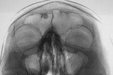
In the diagnosis of frontal sinus osteoma, instrumental diagnostics play a major role: radiography, computed tomography and magnetic resonance imaging.
In this case, an X-ray of the frontal sinus osteoma gives a precisely outlined, smooth-contour shadow of high intensity, adjacent to one of its walls.
Differential diagnosis
Differential diagnosis should exclude the presence of:
- osteomyelitis;
- ossified fibrous dysplasia;
- osteopoikilosis;
- osteogenic sarcoma;
- osteoblastomas;
- osteoblastic metastases.
Treatment osteomas of the frontal sinus.
Methods of drug therapy for this pathology have not been developed, and in the absence of symptoms, treatment of a small frontal sinus osteoma is not carried out.
A significant size of the formation located on the outer side of the frontal bone is considered as an indication for its removal as an aesthetic defect of the facial part of the skull.
If the osteoma spreads into the skull and there are symptoms due to compression of nearby brain structures, surgical intervention is indicated - either by surgical excision of the formation or by endoscopic laser vaporization.
Forecast
With a superficial location of the osteoma, the prognosis is positive, since these formations do not become malignant. Also, specialists consider the outcome of the frontal sinus osteoma to be favorable if, with its growth into the cranium, accompanied by neurological symptoms, high-quality surgical intervention is performed in a timely manner.