Medical expert of the article

New publications
Mouth amoeba
Last reviewed: 06.07.2025

All iLive content is medically reviewed or fact checked to ensure as much factual accuracy as possible.
We have strict sourcing guidelines and only link to reputable media sites, academic research institutions and, whenever possible, medically peer reviewed studies. Note that the numbers in parentheses ([1], [2], etc.) are clickable links to these studies.
If you feel that any of our content is inaccurate, out-of-date, or otherwise questionable, please select it and press Ctrl + Enter.
The oral amoeba (Entamoeba gingivalis) is a type of unicellular organism (protist) of the sarcode type. It belongs to the suborder of amoebozoa and is one of six species of endoparasites of this group that can live inside a person. Unlike the dysenteric amoeba, the oral amoeba is not recognized as a pathogenic protozoan (protozoan) and is considered a non-pathogenic commensal in medical parasitology. Although scientific research into the possible pathogenic action of this type of amoeba has been conducted since its discovery in the mid-19th century.
The oral amoeba's habitat is soft dental plaque and periodontal (gum) pockets at the base of the teeth, and is also found in carious teeth and lacunae of the palatine tonsils. It is believed that these protists live in the mouth of almost every adult.
 [
[ Structure mouth amoeba
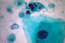
In its structure, the oral amoeba is a trophozoite, that is, it has a vegetative form of a unicellular body.
This amoeba does not form cysts, and its entire life cycle occurs only in the trophozoite stage, ranging in size from 5 to 50 µm in diameter, but usually does not exceed 10-20 µm.
The structure of the oral amoeba is distinguished by the fact that its cell does not have a constant configuration and is limited by a compacted layer of transparent and viscous ectoplasm - the plasma membrane. Under this layer is a more liquid granular endoplasm, and both layers are distinguished at high magnification only when the amoeba is in motion.
The endoplasm contains one small and inconspicuous spherical nucleus covered by a membrane, and inside it are unevenly distributed small chromatin clusters (karyosomes) consisting of proteins and RNA.
The organelles of E. gingivalis movement are pseudopodia (false legs) in the form of cytoplasmic outgrowths that appear when the amoeba needs to move. With these same outgrowths it captures food - polymorphonuclear leukocytes (neutrophils), the remains of dead mucosal cells (cellular detritus) and bacteria that form dental plaque.
The food ends up inside the amoeba's body (in the cytoplasm) and is digested in phagosomes - digestive vacuoles. This process is called phagocytosis. And the undigested remains are excreted through any part of the protist's body.
E. gingivalis reproduces by binary fission, producing two smaller daughter cells.
Symptoms
In fact, there are no symptoms of oral amoeba, that is, signs of its presence in the oral cavity.
The final verdict of parasitologists regarding the actual pathogenicity of the oral amoeba has not yet been made. The issue continues to be debated, and the starting point for the negative attitude towards the oral amoeba is its detection in people with such gum pathology as periodontosis (alveolar pyorrhea). As reported in the journal Dental Research, the oral amoeba is present in 95% of patients with this disease, but E. gingivalis has also been detected in half of patients with healthy gums…
To date, there is no convincing evidence that the oral amoeba is involved in the development of periodontal disease and can cause the release of pus.
Oral or mouth amoeba is a synanthropic organism, that is, it coexists with humans, and, as researchers note, the host in whose mouth E. gingivalis lives provides it with “home and food.” And the trophozoites of this amoeba do not cause direct harm to the host’s health. There is even a theory that this protozoan helps reduce or prevent the increase of other, potentially harmful microorganisms, since bacteria are part of its “diet.” Looking at the situation from this point of view, we can assume that the oral amoeba brings certain benefits to the human host.
Diagnostics
E. gingivalis can be found in the human oral cavity only through laboratory testing of smears from periodontal pockets and scrapings of dental plaque. There are also cases of detection of oral amoeba in sputum.
In this case, according to experts, the oral amoeba can be confused with the dysenteric amoeba (Entamoeba histolytica) in a lung abscess. But the distinguishing feature of Entamoeba gingivalis is that its trophozoites often contain engulfed leukocytes.