Medical expert of the article
New publications
Phlegmona dissection
Last reviewed: 06.07.2025

All iLive content is medically reviewed or fact checked to ensure as much factual accuracy as possible.
We have strict sourcing guidelines and only link to reputable media sites, academic research institutions and, whenever possible, medically peer reviewed studies. Note that the numbers in parentheses ([1], [2], etc.) are clickable links to these studies.
If you feel that any of our content is inaccurate, out-of-date, or otherwise questionable, please select it and press Ctrl + Enter.

Before we understand how phlegmon is opened, we should first explain what this pathology is.
So, phlegmon is an acute limited purulent inflammatory reaction in tissues, accompanied by their melting, with further formation of a cavity. In fact, this is the same abscess, but without clearly defined contours, which is explained by the same melting of tissues. Pus in phlegmon often spreads, affecting nearby organs and tissues.
To treat phlegmon, surgeons use the so-called opening procedure, which is performed under general or local anesthesia. The pathological cavity is opened, the purulent contents are pumped out, sanitization is performed, and the phlegmonous capsule is removed. [ 1 ]
Indications for the procedure
Phlegmon is a bacterial infection affecting subcutaneous tissue. Most often, the inflammatory process develops under the influence of streptococci or staphylococci. The main clinical signs of phlegmon are clearly defined pain, attacks of heat, rapidly spreading redness and swelling. Fever often occurs against the background of progression, and in serious cases, an increase and compaction of nearby lymph nodes can be noticed.
Opening of phlegmon is always prescribed when the inflammatory process progresses, occurring against the background of elevated temperature and softening of the infiltrate. Conservative treatment for phlegmon is prescribed only in isolated cases - for example, if the painful reaction is at the very initial stage of serous inflammation, and the local clinical picture is not yet sufficiently expressed: the patient's condition is satisfactory, the temperature is maintained within subfebrile limits, and there are any contraindications to the operation of opening.
In all other cases of phlegmon and other purulent processes in the skin, surgical intervention is indicated, and on an emergency basis.
Preparation
Opening of the phlegmon is performed after examination and consultation with a medical specialist - usually a surgeon, who examines and diagnoses the pathological formation. Standard stages of preparation for opening the abscess include:
- thorough examination by a surgeon;
- conducting an ultrasound examination;
- if necessary, performing a diagnostic puncture to collect the contents of the phlegmonous cavity with subsequent examination (determining the pathogen and its sensitivity to antibiotic therapy);
- laboratory tests (usually allow us to assess the severity of the inflammatory reaction).
In addition, the doctor must clarify with the patient information about the presence of allergies to anesthetics and other medications.
Instruments for opening phlegmon
Opening of the phlegmon is performed using a strictly defined set of instruments. This set includes:
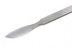
- one scalpel each - pointed and bellied;
- two pairs of scissors - pointed and Cooper;
- four Kocher clamps and the same number of Bilroth clamps;
- two Mosquito clamps;
- two anatomical and surgical tweezers;
- four clothes pins;
- a pair of forceps;
- two hooks each - toothed and plate Farabeuf;
- one probe each - grooved and button-shaped.
All sterile instruments are laid out on a large tray and given to the surgeon by the nurse during the operation to open the phlegmon.
Technique phlegmons
Opening of phlegmon, as well as other superficial purulent formations, can be performed both under local and intravenous anesthesia. The type of anesthesia is chosen by the doctor: the anesthesia should be sufficient to conduct a thorough revision of the phlegmonous focus. Sometimes local anesthesia may be contraindicated due to the high probability of infection spread.
The nuances of surgical access depend on the anatomical and topographic features of the affected area. If possible, the surgeon performs an incision along the lower pole of the phlegmon to ensure optimal conditions for the release of purulent contents. Most often, layer-by-layer tissue dissection is performed, the phlegmon is opened, necrotic tissue and secretions are removed using tampons or a special suction device. After this, a high-quality revision of the lesion is performed, the present layers are isolated, and tissue sequesters are excised. The cavity is washed with an antiseptic solution, drainage is installed using a basic incision or counter-opening.
The surgeon performs the opening and drainage of the phlegmon. The drains are removed the next day, if there are no pathological discharges. The stitches are removed on the 5th-6th day.
- The incision for opening the phlegmon of the hand is carried out using different methods, depending on the localization of the problem:
- in case of commissural phlegmon, an incision is made over the site of inflammation from the interdigital fold to the border of the base of the heads of the metacarpal bones; if purulent discharge is present between the metacarpal bones to the back of the wrist, then an incision is made symmetrically with drainage;
- in case of deep mid-palmar phlegmon, a longitudinal-midline incision is made on the border of the inner edge of the thenar; using a grooved probe, the palmar aponeurosis is dissected, and the purulent contents are removed; if the pus has spread to the hypothenar, the next incision with drainage is made;
- In case of deep phlegmon of the carpal dorsum, a longitudinal midline incision is made on the dorsal side.
- Opening of the phlegmon of the foot from the dorsal side is performed by making two or three longitudinal incisions parallel to the extensor tendons. The skin and subcutaneous tissue, superficial and deep dorsal fascia are dissected. If the phlegmon is localized in the sole area, the opening is performed by means of two typical Delorme incisions. The external and internal incisions run along the sides of the densest section of the plantar aponeurosis. The lines are marked as follows: one of them runs three fingers from the posterior heel edge. Its middle connects with the third interdigital space (second line). The third line is the connection of the midpoint from the medial half of the transverse heel line with the first interdigital space. This type of opening of subaponeurotic phlegmon of the sole is called Voino-Yasenetsky: Incisions in soft tissues in this way do not lead to damage to the plantar aponeurosis and the short digital flexor. [ 2 ]
- Opening of phlegmon of the neck depends on the localization of the process. In case of deep paraesophageal phlegmon, an incision is made along the medial border of the sternocleidomastoid muscle. With orientation to the lateral tracheal wall, a deeper revision is carried out, with the displacement of the vascular cluster outward. Opening of vaginal phlegmon is also carried out, with separation of the adhesion and fascia outward from the esophageal tube below the sternocleidomastoid muscle. When opening phlegmon of the lateral cervical triangle, an incision is made along a line two centimeters above the contour of the clavicle. The platysma is dissected, the buccal cellular space is exposed. If necessary, a deeper revision is carried out, with separation of the third fascia of the neck. [ 3 ]
- The submandibular phlegmon is opened by incision of the skin and platysma along a line parallel to the horizontal mandibular branch. After exposure of the submandibular gland, a deeper revision is performed, if necessary, to the mandibular edge. [ 4 ]
- Opening of the phlegmon of the thigh of the medial bed is performed by longitudinal incisions in the area of the anteromedial femoral surface. The superficial tissues are cut layer by layer two or three centimeters medial to the location of the femoral artery. After opening the broad fascia, the median border of the long adductor muscle is isolated, and access to the phlegmon is opened through the intermuscular spaces. Opening of the phlegmon of the posterior bed is performed by longitudinal incision along the lateral border of the biceps muscle, or along the semitendinosus muscle. The broad fascia of the thigh is opened, access to the purulent focus is opened. [ 5 ]
- Opening of perineal phlegmon involves making an incision in the perineal skin to the deep fascial muscle sheaths. The surgeon determines the degree of adhesion of the fascial structures to each other. In the absence of a necrotizing process, the fascial sheets are peeled off from adjacent tissues using digital revision, exposing access to the phlegmon. Opening of phlegmon of the penis and pubic area is performed similarly. [ 6 ]
- The opening of the forearm phlegmon in the flexor bed is performed using a longitudinal incision, with orientation toward the projection of the radial and ulnar vessels. The skin, PC, and proper fascia of the forearm are dissected, and the superficial digital flexor is dissected. If the phlegmon is located deeper, the deep leaf of the forearm fascia is also dissected, the elements of the deep digital flexor are moved apart, and Pirogov's cellular space is exposed. According to Voyno-Yasenetsky, radial and ulnar incisions are used to access Pirogov's space.
- The Pirogov method of incision of axillary phlegmon is performed with the arm abducted upward and laterally. The limb is placed on a separate surface. The apical phlegmon is incised by cutting parallel to and below the clavicular line. The skin, PC and proper fascia are dissected, the bundles of the pectoralis major muscle are separated, and the deep fascia is opened. The tissue is separated in the same way and the phlegmon is opened. Sometimes it is necessary to transect or undercut the pectoralis major and minor muscles. If pus is detected in the axillary fossa, additional incisions are made. [ 7 ]
Consequences after the procedure
If the phlegmon is opened in time, there are no negative consequences: complete healing is observed within a couple of weeks. In rare cases, lymphangitis, regional lymphadenitis, thrombophlebitis, sepsis, meningitis and encephalitis occur after opening if the lesion is localized in the facial area. These problems are usually associated with the initial advanced state of the phlegmon. However, in such cases, the patient is required to take a course of antibiotics, antihistamines and vitamins, as well as detoxification treatment.
- Why does the temperature rise after opening the phlegmon? During the first three days after the intervention, the patient may have a slight subfebrile temperature. This condition is considered normal and should not cause concern. But cases when the temperature persists for more than three days, or suddenly "jumps" to high values (above 38 ° C), this indicates a recurrence of inflammation and requires emergency surgical assistance.
- If after opening the phlegmon the platelets in the blood are elevated, then there is no need to panic: this happens during inflammatory processes caused by infection, as well as during injuries and surgeries. Against the background of the disappearance of inflammation symptoms, simultaneously with the improvement of other clinical and laboratory indicators, the platelet level always decreases.
Complications after the procedure
First of all, I would like to point out the possible complications if the patient does not want to undergo an autopsy of the phlegmon, or does not seek medical help at all.
- Failure to seek timely treatment for opening the phlegmon may lead to further spread of the disease process, including to large vessels, which may cause damage and bleeding.
- If the autopsy is delayed, the process may affect the nerve trunks (neuritis) and the bone apparatus (osteomyelitis).
- Phlegmon can easily spread to adjacent tissues, and the purulent process can spread throughout the body. This is a very dangerous complication that requires emergency medical intervention.
To avoid such troubles, it is important to consult a doctor at the first signs of phlegmon development. By the way, at the early stages of development - namely, at the stage of serous phlegmon - the inflammatory process can be cured without opening, using conservative therapy.
The operation to open the phlegmon itself rarely results in complications, but they still occur in approximately 3-4% of patients:
- relapse of the inflammatory process;
- hemorrhage or hematoma;
- compaction in the area of phlegmon opening, formation of a rough scar.
Such complications are not critical and are resolved with the help of additional treatment measures. Thus, if the inflammatory process develops again, an autopsy is performed again, the tissues are additionally cleaned and processed, and antibiotic therapy is prescribed. Hematomas often resolve on their own, sometimes it is possible to connect physiotherapy procedures and methods of external therapy. If the operated area is compacted, drugs are prescribed that improve microcirculation.
Care after the procedure
Depending on the size and location of the phlegmon, the recovery period can last from several days to two weeks. As a rule, after the phlegmon has been opened, the attending physician prescribes a course of drug treatment to the patient to speed up healing and prevent complications. Such treatment usually includes:
- analgesics, antipyretics;
- antibiotics;
- immunostimulants.
Care for the site of phlegmon opening consists of the following stages:
- maintaining hygiene of the body and the operated area;
- regular dressings;
- the patient's compliance with all medical prescriptions and monitoring of healing by the doctor.
How phlegmon heals after opening depends on several factors at once:
- from the size of the pathological focus, its depth and degree of neglect;
- from the localization of phlegmon (the wound heals faster in areas with better blood supply and thinner skin);
- from the general health condition and age of the patient (in young people who do not suffer from chronic diseases and diabetes, healing occurs faster).
On average, complete healing of the operated tissues after opening the phlegmon occurs within 2-3 weeks.

