Medical expert of the article

New publications
Open oval window in the heart: what is dangerous, signs, diagnosis, treatment
Last reviewed: 04.07.2025

All iLive content is medically reviewed or fact checked to ensure as much factual accuracy as possible.
We have strict sourcing guidelines and only link to reputable media sites, academic research institutions and, whenever possible, medically peer reviewed studies. Note that the numbers in parentheses ([1], [2], etc.) are clickable links to these studies.
If you feel that any of our content is inaccurate, out-of-date, or otherwise questionable, please select it and press Ctrl + Enter.
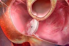
The gap in the wall between the right and left atrium is the open oval window of the heart. Let's consider the causes and pathogenesis of this phenomenon, methods of treatment and prevention.
According to the international classification of diseases ICD-10, congenital communication between the right and left atria is included in class XVII: Q00-Q99 Congenital anomalies (malformations), deformations and chromosomal abnormalities.
Q20-Q28 Congenital anomalies of the circulatory system.
Q21 Congenital anomalies (malformations) of the cardiac septum.
- Q21.1 Atrial septal defect:
- Coronary sinus defect.
- Unclosed or preserved: foramen ovale, secondary foramen.
- Venous sinus defect.
The heart has a complex structure and performs many important functions. The organ contracts rhythmically, providing blood flow through the vessels. It is located behind the sternum in the middle section of the chest cavity and is surrounded by the lungs. Normally, it can shift to the side, as it hangs on the blood vessels and has an asymmetrical localization. Its base is turned toward the spine, and the apex faces the fifth intercostal space.
Anatomical features of the heart muscle:
- The heart of an adult consists of 4 chambers: 2 atria and 2 ventricles, which are separated by partitions. The walls of the ventricles are thickened, and the walls of the atria are thin.
- The pulmonary veins enter the left atrium, and the cava veins enter the right atrium. The pulmonary artery exits the right ventricle, and the ascending aorta exits the left ventricle.
- The left ventricle and left atrium are the left section, which contains arterial blood. The right ventricle and atrium are the venous heart, that is, the right section. The right and left parts are separated by a solid partition.
- The left and right chambers are separated by the interventricular and interatrial septums. Thanks to them, blood from different parts of the heart does not mix with each other.
Incomplete closure of the septum is a congenital anomaly, i.e. a residual element of embryonic development. In essence, it is a hole between the two atria, through which blood is thrown from one ventricle to the other during contractions.
The interatrial opening with a valve develops in utero and is a necessary condition for the normal functioning of the cardiovascular system at this stage of development. It allows part of the placental and oxygenated blood to penetrate from one atrium to another without affecting the undeveloped and non-working lungs. This ensures normal blood supply to the head and neck of the fetus, as well as the development of the spinal cord and brain.
When the newborn first cries, the lungs open and the pressure in the left atrium increases significantly. Due to this, the valve completely closes the embryonic gap. Gradually, the valve tightly fuses with the walls of the interatrial septum. That is, the gap between the right and left atrium closes.
In about 50% of cases, valve fusion occurs in the first year of a child's life, but in some cases by 3-5 years. If the valve is small, the gap does not close and the atria are not isolated. This pathology is classified as MARS syndrome, that is, a minor anomaly in the development of the heart. In adult patients, this problem occurs in 30% of cases.
Epidemiology
Medical statistics indicate that patent foramen ovale (PFO) in the heart is common in two age categories:
- In children under one year of age, this is normal. When conducting an ultrasound, the anomaly is detected in 40% of newborns.
- In adults, this heart defect occurs in 3.6% of the population.
- In patients with multiple heart defects, PFO is diagnosed in 8.9% of cases.
In 70% of cases, incomplete closure of the septum is detected in infancy. In 30% of adults, this disorder manifests itself as a channel or shunt, which provokes various diseases of the cardiovascular system. In healthy and full-term children, the hole closes by 50% in the first year of life.
Causes open oval window
In most cases, the causes of an open oval window are related to genetic predisposition. As a rule, the anomaly is transmitted through the maternal line, but it can also occur due to other reasons:
- Birth of a premature baby.
- Harmful habits of the mother during pregnancy (alcohol, drug addiction, smoking).
- Congenital heart defects.
- Toxic drug poisoning during pregnancy.
- CNS disorders: severe stress and nervous experiences, emotional exhaustion.
- Connective tissue dysplasia.
- Unfavorable ecology.
- Poor nutrition during pregnancy.
Very often, the pathology is detected in other pathologies of heart development: open aortic duct, congenital defects of the mitral and tricuspid valves.
Risk factors
Atrial septal defect occurs for various reasons. Risk factors for the pathological condition are most often associated with genetic disorders in the first line of kinship.
The development of the disorder is facilitated by:
- Increased physical activity (strength sports, diving, weightlifting, etc.).
- Pulmonary embolism in patients with thrombophlebitis of the lower extremities and pelvic organs.
- Bad habits of women during pregnancy.
- Toxic poisoning.
- Premature birth.
- Reduced immune status of a woman.
- Poor ecological environment.
- Deficiency of vitamins and minerals in the female body during pregnancy due to poor nutrition.
In addition to the above factors, the disorder can be caused by increased pressure in the right part of the heart muscle.
Pathogenesis
The mechanism of development of a through hole between the atria is associated with many reasons. The pathogenesis of the anomaly is based on the interaction of internal and external factors. In most cases, these are deviations in the formation, i.e. dysplasia of connective tissue. The disorder leads to the involvement of the heart valves, subvalvular apparatus and cardiac septum in the pathological process.
During the straightening of the newborn's lungs and the increase in pulmonary blood flow, the pressure in the left atrium increases, which contributes to the closure of the gap. But connective tissue dysplasia prevents this process. If primary pulmonary hypertension is diagnosed against this background, the pathology has a favorable prognosis, increasing the patient's life expectancy.
Hemodynamically insignificant patent foramen ovale
The movement of blood through the vessels is associated with the difference in hydrostatic pressure in different parts of the circulatory system. That is, the blood moves from the area of high pressure to low pressure. This phenomenon is called hemodynamics. The open slit in the wall between the right and left atrium is located at the bottom of the oval groove on the inner left wall of the right atrium. The opening has small dimensions from 4.5 mm to 19 mm and, as a rule, is slit-shaped.
A hemodynamically insignificant patent foramen ovale is an anomaly that does not cause blood supply disorders and does not affect the patient's health. This is observed if the defect is small in size and the valve prevents the shunt of blood from left to right. In this case, people with pathologies are unaware of its presence and lead a normal life.
Symptoms open oval window
In most cases, the symptoms of an open oval window do not manifest themselves in any way. A person learns about the presence of the pathology by chance during a routine examination. But the latent course of the disease has a characteristic symptom complex that can remain without due attention for a long time:
- Cyanosis and increased pallor of the nasolabial triangle during physical exertion.
- Tendency to colds and bronchopulmonary pathologies of an inflammatory nature.
- Slow physical development.
- Slow weight gain in a child.
- Poor appetite.
- Respiratory failure.
- Sudden fainting spell.
- Signs of cerebrovascular accident.
- Frequent headaches and migraines.
- Postural hypoxemia syndrome.
The presence of the above symptoms requires careful diagnosis and medical care. If various neurological disorders are observed, this may indicate complications of the disorder due to its long-term course.
First signs
Congenital communication between the right and left atria has no specific manifestations. The first signs in most cases remain unnoticed. Suspicion of the presence of a problem arises in the following cases:
- Severe headaches and dizziness.
- Blue lips when coughing or during any other physical activity.
- Predisposition to inflammatory lesions of the respiratory system.
- Severe respiratory failure during physical exertion.
- Fainting state.
- Varicose veins and thrombophlebitis of the lower extremities at a young age.
OA has minimal radiological symptoms that allow one to suspect an anomaly: an increase in the volume of blood in the pulmonary vascular bed and an increase in the right chambers of the heart.
Patent foramen ovale in adults
The main vital organ of any living being is the heart. In humans, it has a complex structure and is responsible for many functions. The organ includes left/right ventricles and atria, connected by special valves. An open oval window in an adult is a pathology that is most often diagnosed in newborns and premature babies.
In adults, the unclosed opening is a shunt. Its presence can cause changes in the cardiovascular system and lungs due to the difference in blood pressure in the atria. But the presence of this anomaly is not always a cause for concern. Very often, people live a full life and do not suspect a disorder. Only an ultrasound can reveal the problem.
The correct functioning of the heart and the body as a whole depends on the size of the defect. The size of the hole can be from 2 mm to 10 mm.
- If the window opens by 2-3 mm, but is not accompanied by deviations from the cardiovascular system, then this condition does not affect the functioning of the body.
- If the through hole is 5-7 mm, this indicates that the disorder is hemodynamically insignificant. The deviation manifests itself only with increased physical exertion.
- If the dimensions are 7-10 mm, then the patient is diagnosed with a gaping open window. In its symptoms, this type of disease is similar to a congenital heart defect.
The inferiority of the pelvic organs usually does not have specific symptoms. The doctor can only guess about the causes of the painful condition. To identify the disorder, a comprehensive diagnosis is indicated. The presence of seemingly clinically insignificant symptoms is also taken into account:
- Blue discoloration of the nasolabial triangle in inflammatory diseases and after physical exertion.
- Frequent fainting.
- Cerebral circulatory disorder.
- Varicose veins and thrombophlebitis.
- Shortness of breath.
- Predisposition to colds.
- Tachycardia.
- Migraine.
- Intolerance to physical exertion.
- Increased volume of blood in the lungs.
- Frequent numbness in the limbs and impaired body mobility.
This disorder is diagnosed in 30% of people, it persists from birth. But the risk of developing the disease increases significantly in athletes and with increased physical activity. The risk group includes: divers, patients with pulmonary embolism (PE) and thrombophlebitis.
Treatment of the disease depends on its severity. Many adults are prescribed a set of preventive methods. In particularly severe cases, not only drug therapy is indicated, but also surgical intervention.
Stages
An open gap in the wall between the right and left atriums is a cardiovascular defect. The stages of the pathology are distinguished by the degree of organ damage and the nature of the symptoms that arise. In medical practice, there is such a concept as MARS syndrome (minor anomalies in the development of the heart), which includes this disorder. The group of pathologies includes disorders in the development of the structures of the external and internal structure of the heart muscle and the vessels adjacent to it.
Incomplete closure of the septum is included in the general classification of MARS syndrome:
- Location and shape.
- Atria:
- Patent oval window.
- Enlarged Eustachian valve.
- Aneurysm of the interatrial septum.
- Prolapsing valve of the inferior vena cava.
- Trabeculae.
- Prolapsing pectineal muscles in the right atrium.
- Tricuspid valve - displacement of the septal valve into the cavity of the right ventricle, dilation of the right AV orifice, protrusion of the tricuspid valve.
- Pulmonary artery - prolapse of the pulmonary artery valve cusps and dysplasia of its trunk.
- Aorta – borderline wide/narrow aortic root, bicuspid valve, dilatation of sinuses, asymmetry of valve cusps.
- Left ventricle - small aneurysm, trabeculae, chords.
- Mitral valve.
- Causes and conditions of occurrence.
- Connective tissue dysplasia.
- Vegetative dysfunctions.
- Ontogenesis.
- Cardiogenesis disorders.
- Possible complications.
- Heart rhythm disturbances.
- Pulmonary hypertension.
- Infective endocarditis.
- Cardiohemodynamic disorders.
- Fibrosis and calcification of valve leaflets.
- Sudden death.
Any form or stage of MARS syndrome is a variant of visceral connective tissue dysplasia. It is characterized by a high frequency of changes in the central nervous system and neurovegetative disorders.
After the type of anomaly is established, hemodynamic disturbances and regurgitation, their severity are identified. In 95% of cases, hemodynamic disturbances and side symptoms do not occur. As the child grows older, structural deviations disappear.
Forms
Normally, an open oval window is temporary, as it is necessary for the saturation of the fetus with oxygen during embryonic development. That is, the anomaly exists in all children, but by the time of birth it heals, as there is no longer a need for additional oxygen saturation, since the lungs begin to work.
The types of incomplete closure of the septum depend on the size of the opening:
- 2-3 mm is the norm, which does not cause deviations and consequences.
- 5-7 mm – the characteristics of this pathology depend on the presence of concomitant provoking factors.
- >7 mm is a gaping hole that requires surgical treatment. According to studies, the maximum size can exceed 19 mm.
In addition to the oval window, there are other defects of the cardiac septum. Their differences are that the window has a valve responsible for regulating the blood flow. PFO is not a heart defect, but refers to minor anomalies in the development of the cardiovascular system.
Patent foramen ovale with discharge
In most cases, a through hole between the atria does not cause serious concerns. Since the pressure in the left atrium is higher than in the right, the valve between the septa is held closed. This prevents blood from flowing from the right atrium to the left. As a rule, this is observed when the window size is no more than 5-7 mm.
An open oval window with a discharge indicates large dimensions of the pathology. This is observed with a temporary increase in pressure in the right atrium due to straining, physical exertion, crying or prolonged nervous tension. This condition causes a discharge of venous blood through the OA, manifested by temporary cyanosis of the nasolabial triangle and paleness of the skin.
The disorder can lead to such a complication as paradoxical embolism. Thrombi, gas bubbles, emboli, foreign bodies from the right atrium, having entered the left and continuing further movement, can reach the vessels of the brain. This leads to the development of stroke, thrombosis and infarction. To prevent such disorders, it is necessary to conduct a comprehensive diagnosis and timely treatment.
 [ 17 ]
Patent oval window with left-right discharge
A short channel between the right and left atrium, covered by a valve and with abnormal blood circulation, is an open oval window with a left-right discharge. Normally, fluid discharge occurs in one direction - from right to left. PFO is a physiological feature of the body, which is necessary during the period of embryonic development. But after birth, the need for it disappears and the gap heals, as the lungs begin to work.
The following types of functioning of the oval window are distinguished:
- Without hemodynamic relief.
- With right-left reset.
- With left-right reset.
- With biderectoral bypass.
Left-to-right shunt atrial fibrillation indicates that the pressure in the right atrium is lower than in the left. The main causes of this form of disorder include:
- Perforation of the oval window flap.
- Valve deficiency with left atrial dilation
- Valve failure.
Right-to-left shunt, when the pressure in the right atrium is greater than in the left, occurs due to the following reasons: prematurity and low body weight, increased physical activity and psychoemotional disorders, neonatal pulmonary hypertension, respiratory distress syndrome.
Patent foramen ovale without signs of embolic activity
The patent foramen ovale is a valve communication between the atria. In the embryonic period, it is responsible for the passage of arterial blood into the left atrium from the right, without affecting the undeveloped vessels of the lungs. In most people, the PFO closes after birth, but in 30% it remains open, causing various pathological symptoms.
With this minor cardiac anomaly, there is a high risk of developing paradoxical embolism. The pathology leads to the fact that small gas bubbles and thrombi enter the left atrium and through the left ventricle with the blood flow to the brain. The blockage of the brain vessels provokes a stroke.
An open oval window without signs of embolic activity and other pathologies can be considered as a variant of normal heart structure. But in the presence of provoking factors (physical activity, straining, coughing), the pressure in the right atrium increases and a right-left shunt occurs, causing paradoxical embolism.
Complications and consequences
The lack of timely diagnosis and treatment of the through hole of the atrium is the main reason for the development of various consequences and complications. Patients may face the following problems:
- Heart rhythm disturbances.
- Cerebral circulatory disorder.
- Pulmonary hypertension.
- Paradoxical embolism.
- Fibrosis and calcification of the valve cusps of the heart muscle.
- Cardiohemodynamic disorders.
- Heart attack.
- Stroke.
- Sudden death.
According to medical statistics, the above complications are extremely rare.
Is a patent foramen ovale dangerous?
Many experts consider the congenital communication between the right and left atria to be normal. Whether an open oval window is dangerous depends entirely on the patient's general health and the presence of concomitant pathologies.
If the window is small, then, as a rule, it is not a cause for concern. The patient is prescribed regular examinations by a cardiologist, annual planned ultrasound examinations of the heart and a set of preventive measures. In the presence of concomitant diseases, PFO can cause serious changes in the cardiovascular system. This is due to the transfer of blood from the right atrium to the left, bypassing the lungs. In this case, any physical activity can cause various complications.
This congenital anomaly is dangerous due to the development of embolism. This is a condition when blood clots, gas bubbles and bacterial microorganisms enter the arterial blood from the venous blood and through the left sections of the heart into the arteries of the internal organs. In this case, the coronary arteries, kidneys, spleen and limbs can be affected. Heart rhythm disturbances are dangerous due to strokes and heart attacks.
[ 18 ]
Diagnostics open oval window
Minor cardiac anomalies are characterized by a latent, that is, hidden course. Pathology can be suspected in the presence of characteristic symptoms or during a routine examination of the body. Diagnosis of an open oval window is made by the following methods:
- Collection of anamnesis - the doctor asks about the presence of genetic abnormalities among relatives, about the course of pregnancy, the woman's bad habits and medications administered during pregnancy, and the patient's level of physical activity.
- External examination – this method is ineffective, since PFO does not always make itself known with clearly expressed symptoms. But the blueness of the nasolabial triangle during crying and straining, paleness of the skin, poor appetite and delay in physical development allow us to suspect the disorder.
- Laboratory tests – currently there are no genetic tests that can detect MARS syndrome in newborns. Patients are prescribed the following tests:
- General and clinical blood analysis.
- Prothrombin gene mutation.
- Prothrombin time.
- Factor V (Leiden).
- Determination of homocysteine and antithrombin levels.
- Determination of protein C and protein S levels.
- Instrumental studies – for diagnostics, auscultation is performed, that is, listening to the chest for systolic murmurs. The patient is prescribed an ultrasound of the heart, echocardiography, angiography, MRI and a range of other procedures.
During the diagnosis, the doctor evaluates nutrition, identifies eating disorders and symptoms of anomalies associated with an imbalance of consumed nutrients. The environmental characteristics of the patient's living environment are also taken into account.
Patent foramen ovale noise
One of the methods for diagnosing a through hole between the atria is listening to the chest with a phonendoscope. When the cardiovascular system is working, peculiar tones arise. The heart pumps blood, and the valves regulate its direction.
- Before the heart contracts, the valves between the atria and ventricles close.
- Blood from the left ventricle enters the aorta, and from the right ventricle into the pulmonary artery. When this happens, a tone is formed.
- The tone occurs when the valves close, if some kind of obstacle forms in the heart, and due to many other factors.
Noise with an open oval window of the heart cannot always be detected with a phonendoscope. This is due to the fact that the pressure difference between the atria is small, so the vortex flow characteristic of the anomaly may not form.
Heart murmurs can be: soft, rough, blowing. All murmurs are divided into the following groups:
- Pathological – often act as the first, and sometimes the only sign of anomalies of the cardiovascular system.
- Healthy – associated with the growth characteristics of the heart chambers and vessels, and the structural features of the organ.
To determine the nature of the noise and the reasons for its appearance, the doctor conducts echocardiography and ultrasound examination. These methods allow you to evaluate the structure of the heart and the surrounding vessels and tissues.
Instrumental diagnostics
Examination of the body using special equipment is instrumental diagnostics. If there is a suspicion of incomplete closure of the septum of the heart, the following studies are indicated:
- Radiography – determines possible disturbances in the functioning of the heart caused by increased blood pressure in the right ventricle due to an atrial septal defect.
- Ultrasound of the heart – is performed to determine the boundaries of the PFO and its size. It is prescribed for newborns and older patients.
- Echocardiography – is performed when various cardiac abnormalities are suspected. Allows to detect pathology even when it is latent. It is performed in two conditions: after physical exertion and at rest.
- Transthoracic two-dimensional echocardiography – allows to detect the inadequacy of the oval window valve in newborns. Visualizes the movement of the valve flaps, determines the speed and volume of blood flow from one atrium to another.
- Transesophageal echocardiography – is prescribed if an abnormality is suspected in older children and adolescents. During the examination, an endoscope is inserted into the esophagus, bringing it as close as possible to the heart muscle. To obtain more reliable results, bubble contrast may be prescribed.
- Cardiac probing is one of the most accurate, but aggressive diagnostic methods. It is most often used before surgical interventions. The procedure involves moving a probe through the arterial bloodstream to the heart for its detailed visualization.
Based on the results of instrumental diagnostics, a final diagnosis can be made or additional studies can be prescribed.
Patent foramen ovale on ultrasound
Ultrasound examination of the cardiovascular system is one of the instrumental methods for identifying both congenital and acquired anomalies among newborns and older patients.
An open oval window on ultrasound is characterized by the following symptoms:
- Enlargement of the right cardiac chamber.
- Small hole sizes – from 2 to 5 mm.
- Displacement of the main septum between the atria towards the right atrium.
- Thinning of the walls of the interatrial septum.
Using ultrasound, it is possible to visualize the valves in the left atrium, assess the general condition of the organ and the volume of blood flow, localization and other features of the pathology.
Echographic signs of patent foramen ovale
Echocardiography is a diagnostic method using ultrasound waves. It is used to study and determine the localization of internal organs and structures.
Sonographic signs of a patent foramen ovale can be detected immediately after birth using the following tests:
- Contrast echocardiography – reveals a PFO or atrial septal defect of the smallest size. For diagnosis, the patient is given an intravenous injection with a saline solution. If there is a gap, tiny air bubbles will penetrate through it from the right atrium to the left.
- Transthoracic two-dimensional echocardiography (EchoCG) – visualizes not only the opening, but also the functioning valve. This method is especially informative in newborns and early childhood patients.
In addition to the above methods, transesophageal echocardiography with bubble enhancement may be prescribed to determine the echographic signs of the disorder.
Dimensions of the open oval window
Minor cardiac anomalies can be suspected by their characteristic symptoms, which very often occur in a latent form. The size of the open oval window and the presence of concomitant diseases affect the severity of the pathological signs of the disease.
The open gap in the wall between the right and left atrium can have the following dimensions:
- 2-3 mm – is considered normal and does not cause any symptoms or complications.
- 5-7 mm is a small size of the anomaly. Under the influence of certain factors, it causes a number of unpleasant symptoms that can progress without medical diagnosis and treatment.
- 7 mm and more is a large or gaping window that requires surgical treatment. In rare cases, it can reach maximum dimensions - more than 19 mm.
According to studies, in about 40% of adults, the opening between the atria is not tightly closed. The size of the gap is on average 4.5 mm. If the window remains completely open, then an atrial septal defect is diagnosed, which, unlike PFO, is characterized by the absence of a functioning valve.
[ 23 ]
Open oval window 2, 3, 4, 5 mm
Congenital communication between the right and left atria is very often diagnosed in premature infants and a little less often in healthy children. An open oval window of 2, 3, 4, 5 mm is considered normal, but under the influence of certain factors it can cause pathological symptoms.
Holes larger than 5 mm have characteristic signs that allow one to suspect a violation:
- Blueness of the nasolabial triangle during physical activity, crying, screaming.
- Slowing of mental and physical development.
- Loss of consciousness and dizziness.
- Rapid fatigue.
- Presence of heart murmurs.
- Various disorders of the respiratory system.
- Frequent colds.
The appearance of the above symptoms is a reason to immediately contact a cardiologist. After a set of various diagnostic measures, the doctor will prescribe treatment and give recommendations for correcting the disease.
Differential diagnosis
An abnormal valvular communication between the atria requires a comprehensive examination and, if necessary, treatment. Differential diagnostics of an open oval window is carried out with pathologies with similar symptoms.
First of all, differentiation is necessary with other interatrial communications:
- Atrial septal defect.
- Aneurysm of the interatrial septum.
- Hemodynamic discharge disorders.
Let us consider in more detail the differences between congenital communication between the right and left atria and atrial septal defect:
OOO |
ASD |
|
EchoCG |
Imposition of the primary and secondary septa. Formation of a shunt of interatrial communication. |
Valve tissue deficiency. |
Anatomical features |
Incompetence of the foramen ovale valve or patent atrial valve. |
Absence of a greater or lesser part of the secondary septum. |
Blood dumping |
In 95% of cases, left-to-right shunting; in case of decompensation, right-to-left shunting. The hemodynamic significance of blood shunting is not decisive. |
|
Dimensions |
They are not of fundamental importance. |
|
Based on the results of the studies, the doctor makes a final diagnosis or prescribes additional examinations/tests.
Who to contact?
Treatment open oval window
Such a small anomaly of the heart as a through hole between the atria requires special attention. Treatment of an open oval window depends on many factors:
- Dimensions and clinical significance of the gap.
- Fluctuations in shunt size during physical exertion.
- Features of the septum (increased extensibility, loss of contractility).
- The degree of increase in pressure in the pulmonary artery.
- Enlargement of the right chambers of the heart.
- Risk of embolic/cerebral complications.
- Presence of concomitant diseases.
- General condition of the body.
Treatment tactics are based entirely on the presence or absence of symptoms of PFO:
- In the absence of symptoms, therapy is not required. The patient is recommended to be monitored by a therapist/pediatrician and cardiologist, periodically assess the dynamics of the anomaly using ultrasound. If there is a risk of complications (stroke, heart attack, ischemia, lesions of the veins of the lower extremities), then patients are prescribed medications to thin the blood (Warfarin, Aspirin, etc.).
- In the presence of painful symptoms, not only medicinal but also surgical treatment is indicated. In case of pronounced right-to-left blood flow and risk of embolism, the defect is closed using an occluding device or a special absorbable patch.
Elkar with an open oval window
One of the methods of treating MARS syndrome is drug therapy. Elkar is prescribed for an open oval window of the heart from the first days of the disease. Let's consider in more detail the instructions for this drug and the features of its use.
Elkar is a medicine used to correct metabolic processes in the body. The drug contains L-carnitine, an amino acid that is similar in structure to B vitamins. It participates in lipid metabolism processes, stimulates enzymatic activity and secretion of gastric juice, and increases resistance to physical exertion.
The active component regulates the consumption of glycogen and increases its reserves in the liver and muscle tissue. It has pronounced lipolytic and anabolic properties.
- Indications for use: improving the condition of premature babies and newborns after birth injuries, asphyxia. Prescribed for a weak sucking reflex, low muscle tone, poor development of mental and motor functions, and insufficient body weight. The drug is used in the complex therapy of chronic gastritis and pancreatitis, and dermatological diseases. Accelerates the recovery of the body during intense physical and psycho-emotional stress, with reduced performance and increased fatigue.
- Method of administration: the medicine is taken orally 30 minutes before meals. The dosage and course of treatment are individual for each patient, therefore, they are determined by the attending physician.
- Side effects: isolated cases of dyspeptic disorders, myasthenia, gastralgia, systemic allergic reactions have been recorded.
- Contraindications: hypersensitivity to the components of the drug. If the drug is prescribed to patients under 3 years of age, careful medical supervision is required. Not used to treat pregnant women and during lactation.
- Overdose: myasthenia, dyspeptic disorders. There is no specific antidote, therefore symptomatic therapy is indicated.
Elkar is available in the form of a solution for oral administration in 25, 50 and 100 ml bottles with a dosing device.
[ 24 ]
Is it necessary to operate on a patent foramen ovale?
Faced with such a diagnosis as a through hole between the atria, many patients ask themselves: is it necessary to operate on an open oval window? The need for surgical intervention is determined by the size of the gap, the presence of concomitant diseases, painful symptoms and other characteristics of the body.
Medicine claims that up to two years of age, PFO is the norm. The patient must be observed by a cardiologist and undergo echocardiography and ultrasound of the heart every year. If the window has not closed upon reaching a more mature age, the patient is put on strict monitoring by a cardiologist, who decides on the method of treating the defect. The doctor takes into account the fact of complications: thrombus formation, pulmonary insufficiency, paradoxical embolism, ischemic and cardioembolic stroke.
If the oval window is large, there is no valve (atrial septal defect), or a stroke has been suffered, then surgical intervention is a direct indication.
[ 25 ]
Surgical treatment
One of the most effective methods of eliminating PFO is surgical treatment. It is performed at any age, but only if the following indications are present:
- Gross hemodynamic disturbances.
- High risk of complications.
- Severe pain symptoms.
- The defect diameter is more than 9 mm.
- Blood flow into the left atrium.
- Limited physical activity caused by pathology.
- Contraindications to taking medications.
- Complications of the cardiovascular and respiratory systems.
The main goal of the surgical intervention is to close the defect with a patch. The procedure is performed through the femoral or radial artery using a special endoscope and with the introduction of contrast.
Surgical treatment is contraindicated in case of pathological changes in lung tissue and left ventricular failure. As a rule, the operation is performed after reaching 2-5 years, when the window should physiologically close, but this does not happen. Each case is individual and requires a comprehensive comprehensive diagnosis to assess all possible risks of the operation.
Patent foramen ovale surgery
The only and most effective method of treating a residual fetal heart element in adult patients is surgery. In the case of an open oval window, the following surgical interventions may be prescribed:
- Open heart surgery.
Through an incision in the chest, the surgeon disconnects the heart from the vessels. The functions of the heart are taken over by a special device that pumps blood around the body and enriches it with oxygen. Using coronary suction, the doctor cleans the organ of blood and makes an incision in the right atrium to eliminate the defect. The method has the following indications:
- Hole with a diameter greater than 10 mm.
- Severe circulatory disorder.
- Intolerance to physical exertion.
- Frequent colds and inflammatory diseases.
- Pulmonary hypertension.
The following methods are most often used to close the gap:
- Suturing - the hole in the interatrial septum is sutured. The same manipulations are performed for secondary defects located in the upper part of the septum.
- Applying a patch of synthetic fabric, pericardium (a flap of the outer membrane of the heart) or a special plaster. This method is used for primary heart defects located closer to the ventricles, in the lower part of the septum.
After the operation, the doctor stitches up the incision and connects the heart to its blood vessels. The incision on the chest is closed with a suture.
The advantages of such an operation are high precision of execution and rapid restoration of impaired blood circulation in the lung and the entire body, as well as the ability to eliminate defects of any size and location. The disadvantages of the method include: the need to connect a machine for artificial blood circulation, trauma due to a large incision in the chest, a long recovery period - about 2 months and rehabilitation up to 6 months.
- Endovascular surgery (closure of the defect using a catheter).
These are less traumatic operations that do not require opening the chest. Indications for performing:
- A window less than 4 mm in the central part of the interatrial septum.
- Blood flow from the left atrium to the right.
- Increased fatigue.
- Shortness of breath during physical activity.
During the operation, the doctor inserts a catheter into the openings in the large vessels of the groin or neck area. The endoscope is advanced into the right atrium. A special device for closing the window is attached to the end of the device:
- Button devices - discs are installed on both sides of the interatrial septum and are connected to each other using a nylon loop.
- An occluder is a special device that resembles an umbrella. It is inserted and opened in the left atrium, blocking the flow of blood out of it.
The advantages of such minimally invasive treatment are considered to be: low risk of complications, the possibility of performing under local anesthesia, significant improvement in the condition immediately after the operation, a short recovery period - about a month. The main disadvantage of endovascular surgery is that it is not effective in case of large defects and narrowing of blood vessels. The operation is not performed with a window in the lower part of the septum or at the mouths of the vena cava/pulmonary veins.
Regardless of the surgical intervention chosen, most patients recover completely after the operation. An increase in life expectancy of 20-30 years is also observed.
Indications for occluder
If drug therapy is unable to eliminate the pathological symptoms or complications of MARS syndrome, then surgical intervention is indicated. Many patients are prescribed endovascular surgeries, that is, the introduction of a special device, most often an occluder, into the heart through a vein or large artery.
Main indications for the occluder:
- Small size LLC.
- Localization of the defect in the central part of the interatrial septum.
- Increased fatigue and other symptoms of pathology.
In case of a minor cardiac anomaly, blood from the left atrium enters the right, and then the right ventricle and pulmonary artery. This leads to stretching and overload of these parts of the heart. Normally, the left and right parts of the organ are separated by a thin wall, which prevents blood from flowing back. That is, the main indication for the use of an occluder is precisely the enlargement and overload of the right parts of the heart.
The occluder is an umbrella or miniature mesh. It is inserted into the femoral vein using a catheter and installed at the entrance to the left atrium. Implantation is performed using an X-ray system that visualizes the entire operation process.
The occluder is made of a biologically inert material that does not cause rejection reactions and is well accepted by the body. Six months after the operation, the device is endothelialized, that is, covered with heart cells. In rare cases, after the treatment, patients experience complications such as shortness of breath and chest pain.
Prevention
There are no special methods that would prevent incomplete closure of the cardiac septum. Prevention of patent foramen ovale is based on a healthy lifestyle and following these recommendations:
- Give up bad habits (smoking, alcoholism, drug addiction).
- Stick to a rational and balanced diet that will provide the body with a complex of necessary vitamins and minerals.
- Timely treatment of any diseases.
Women planning to have a child and those who are already pregnant should pay special attention to the prevention of anomalies:
- Avoid infectious diseases. Rubella is especially dangerous, as it provokes PFO and other congenital defects.
- Avoid contact with ionizing radiation, such as X-ray machines and fluorographs.
- Avoid contact with chemicals and their vapors (paints, varnishes).
- Take any medications only as prescribed by your doctor.
There are also preventive recommendations for patients who have already been diagnosed with the anomaly: a balanced diet, adequate sleep and rest, limited physical activity and regular preventive examinations by a cardiologist.
[ 26 ]
Forecast
With timely treatment, following all medical recommendations and being monitored by a cardiologist, the prognosis for an open oval window is quite favorable. The outcome of the anomaly depends on what therapy was prescribed and how effective it is.
Another important prognostic factor is the functional state of the heart muscle. If there was an operation and it was successful, then there is a high chance of avoiding consequences and complications. This improves the prognosis of the defect. For example, endovascular occlusion of the PFO allows you to return to normal life within a short period of time, without any restrictions.
Without timely diagnosis, drug or surgical treatment, the prognosis of a minor cardiac anomaly is negative. The risk of serious complications exists with large window sizes, the development of paradoxical embolism and the presence of concomitant diseases.
[ 27 ]
Outpatient observation of children
An abnormal valvular communication between the atria requires not only timely treatment, but also medical supervision. Outpatient observation of children with an open oval window involves systematic medical examinations and research (ultrasound, echocardiography). This allows us to assess the dynamics of the disorder and the risk of its complications.
Parents also receive special recommendations. The newborn is shown a protective regime with long walks in the fresh air and proper nutrition. This is necessary to harden the body and increase immune protection. Physiotherapy and therapeutic gymnastics are also recommended.
[ 28 ], [ 29 ], [ 30 ], [ 31 ], [ 32 ]
What professions are contraindicated with an open oval window?
Such a physiological feature as incomplete closure of the cardiac septum leaves its mark not only on the lifestyle, but also on the sphere of activity.
Let's consider what professions are contraindicated with an open oval window: pilot, diver, sea diver, driver, machinist, astronaut, caisson worker, army officer or submarine crew member. The above-mentioned professions can be dangerous for patients.
For example, when ascending or descending, blood clots can form, blocking blood vessels and causing death. And caisson work is dangerous because the patient has to breathe compressed air, which also has a negative effect on the cardiovascular system.
Patent foramen ovale and sports
Patients with a congenital through-and-through hole between the atria have many restrictions that are aimed at minimizing the risk of developing complications of the disease.
An open oval window and sports are acceptable if the defect does not cause abnormal blood flow, cyanosis of the nasolabial triangle due to physical exertion, embolism and other complications. When choosing a sporting hobby, the size of the window, as well as the results of the treatment, are taken into account.
Patent oval window and the army
According to the order of the Ministry of Defense of Ukraine dated 14.08.2008 No. 402, an open oval window and the army are incompatible. Patients with this anomaly are partially or completely exempted from military service.
Conscripts from the risk group undergo a special military medical examination. Upon completion of the examination, a category is established:
- Limited fitness - the disease is accompanied by bleeding, the conscript is unfit for service in peacetime.
- Fit with restrictions - anomaly without blood discharge, but there are pathological signs of the disorder and the risk of complications.
An open oval window of the heart is a serious congenital pathology. But the final decision on the possibility of military service is made by the draft board.