Medical expert of the article

New publications
Current view on the pathogenetic mechanisms of hyperuricemia
Last reviewed: 07.07.2025

All iLive content is medically reviewed or fact checked to ensure as much factual accuracy as possible.
We have strict sourcing guidelines and only link to reputable media sites, academic research institutions and, whenever possible, medically peer reviewed studies. Note that the numbers in parentheses ([1], [2], etc.) are clickable links to these studies.
If you feel that any of our content is inaccurate, out-of-date, or otherwise questionable, please select it and press Ctrl + Enter.
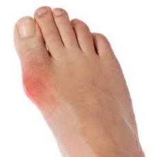
Gout is a systemic tophaceous disease characterized by the deposition of sodium monourate crystals in various organs and tissues and the resulting inflammation in individuals with hyperuricemia caused by environmental and/or genetic factors. The pathogenesis of gout is based on a disorder of uric acid (purine) metabolism and an increase in the content of uric acid (UA) in the blood. The basis of uric acid metabolism is its hyperproduction and decreased excretion by the kidneys. At the same time, only 10% of patients with primary gout have disorders of only endogenous uric acid formation. In other patients, the main factor in the formation of hyperuricemia is a disorder of uric acid excretion by the kidneys.
In addition to damage to the musculoskeletal system, gout is characterized by the presence of visceral manifestations, one of which is urate nephropathy. Urate nephropathy is a variant of chronic tubulointerstitial nephritis, characterized by the accumulation of uric acid crystals in the interstitium with the development of a secondary inflammatory process in it and damage to the epithelium of the tubular apparatus with a violation of its function and reabsorption processes.
Transport of uric acid by the kidneys is a cascade of four processes: glomerular filtration, almost complete reabsorption of filtered uric acid, secretion, and postscretory reabsorption in the proximal tubule. Urate is not protein bound and is therefore freely filtered in the glomeruli. The rate of tubular secretion is much lower than the rate of tubular reabsorption, and therefore the contribution of secreted urate to the total amount of excreted urate is small. Almost 98-100% of filtered uric acid is reabsorbed in the proximal tubule, after which 50% of the filtered urate is resecreted, and then almost 80% of the excreted urate is reabsorbed, and ultimately about 7-10% of the filtered urate is excreted. The reabsorption, secretion, and postscretory reabsorption phases occur in the proximal tubule. The processes of reabsorption and secretion are carried out by specific molecules (transporters) located on the brush border of the epithelium of the proximal tubules.
Most urate transporters belong to the OAT family. Tubular reabsorption of urate is performed by an organic anion transporter (urate anion exchanger) identified as URAT1 (encoded by the SLC22A12 gene). This transporter is present only in humans. Numerous studies, including those in individuals with familial hyporuricemia, indicate a mutation in the SLC22A12 gene encoding the URAT1 transporter. It was found that these patients have virtually no effect of probenecid and pyridinamide (an anti-tuberculosis drug with an antiuricosuric effect) on uric acid excretion.
In addition to URAT1, there are other transporters: URATv1, SLC5A8 encoded sodium-dependent counter-transporter, organic anion transporters of the OAT family (OAT1 and OAT3, OAT2 and OAT4), ABCG2 (urate transporter in collecting tubules), SLC2A3 (sodium/phosphate counter-transporter of proximal tubules). OAT2 and OAT4 are located on the apical membrane of the proximal tubules OAT1 and OAT3 on its basolateral part, their main function is the exchange of organic anions and bicarboxylate, but at the same time there is data on their effect on the transport of urates.
URATv1 (OATv1), which was later named GLUT9, encoded by the SLC2A9 gene, is a voltage-dependent transporter of organic ions, mainly glucose and fructose, as well as a urate transporter, polymorphism of this gene is associated with hypouricemia, which has been confirmed in genetic studies.
Less studied are the mechanisms affecting the secretion of uric acid. Impaired secretion is associated with changes in the ATP-dependent pump, mutations in the MRP4 gene encoding the formation of uromodulin (Tamm-Horsfall protein, ABSG2 gene). The exact mechanism by which uromodulin affects the secretion of urates is still unknown, perhaps it is associated with an increase in the reabsorption of sodium in the proximal tubules and uric acid at the same time.
Impaired renal transporters with increased uric acid reabsorption may lead to hyperuricemia and, ultimately, gout. A number of studies on urate transporter dysfunction have identified genetic mutations, while most of these studies focused on the presence of genetic mutations in urate transporters in patients with hypouricemia, while the issue of the presence of mutations in patients with hyperuricemia remains less studied. Of note are data on the activation of URAT1 and GLUT9 transporters with a purine-rich diet, arterial hypertension, and local ischemia, which in turn causes an increase in uric acid reabsorption. There is evidence that apical tubular reabsorption of urates and sodium via URAT1 is impaired, with subsequent development of hyperuricemia under the influence of diabetic ketoacidosis, ethanolamine intoxication, pyrazinamide treatment, hyperinsulinemia, and metabolic syndrome. Thus, impaired renal uric acid excretion may be a secondary process due to damage to the renal tubular apparatus.
The work of the tubular apparatus in patients with gout can be assessed by daily excretion, clearance, excreted fraction (EF), reabsorption of uric acid, calcium (Ca), phosphorus (P), and ammonia excretion. Moreover, a "standard" examination of the patient does not allow identifying signs of renal dysfunction. The simplest and most accessible method is to assess the clearance of uric acid with subsequent recalculation to the body surface area. Our studies in patients with gout showed a fairly high information content of this test for identifying signs of urate nephropathy, so the value of uric acid clearance less than 7 ml / min / 1.73 m2 has a sensitivity of 90% and a specificity of 66%.
Postgraduate student of the Department of Hospital Therapy Khalfina Tamila Nilovna. Modern view on the pathogenetic mechanisms of hyperuricemia // Practical Medicine. 8 (64) December 2012 / Volume 1
 [
[