Medical expert of the article

New publications
Microphthalmos
Last reviewed: 07.07.2025

All iLive content is medically reviewed or fact checked to ensure as much factual accuracy as possible.
We have strict sourcing guidelines and only link to reputable media sites, academic research institutions and, whenever possible, medically peer reviewed studies. Note that the numbers in parentheses ([1], [2], etc.) are clickable links to these studies.
If you feel that any of our content is inaccurate, out-of-date, or otherwise questionable, please select it and press Ctrl + Enter.
Microphthalmos is diagnosed when the length of the anterior-posterior axis of the eyeball is less than normal and is 21 mm in an adult and 19 mm in a one-year-old child.
Visual acuity in microphthalmos depends on its severity and the presence of concomitant pathology. In some cases, vision may be normal, but in severe pathology it is significantly reduced. The functional prognosis in microphthalmos should be extremely careful to avoid error, and the size of the eyeball is not its only criterion.
 [
[ Isolated microphthalmos
- Idiopathic.
- Hereditary:
- autosomal dominant;
- autosomal recessive;
- X-linked.
Microphthalmos in combination with other pathology of the visual organ
Usually accompanied by the following changes.
- Pathology of the development of two segments of the eyeball - Peters syndrome, Reiger syndrome, etc.
- Cataract.
- Persistent vitreous hyperplasia (PVH).
- Retinal diseases - retinopathy of prematurity (ROP), retinal dysplasia and folding, etc.
- Aniridia.
- Colobomatous microphthalmos. The presence of colobomas is not uncommon both for isolated microphthalmos and for its combination with general diseases.
Microphthalmos in general diseases
- Temple Al-Gazali syndrome: microphthalmos, cutaneous aplasia and sclerocornea in Xp22.2-PTER deletion.
- Chromosomal syndromes. In the vast majority of cases, they are accompanied by colobomas.
- Mentally retarded.
- Macrosomia: cleft palate.
- Head malformations:
- midline defects of the brain;
- oculofacial syndrome in embryonic differentiation disorder;
- frontofacionasal dysplasia.
- Delleman syndrome: papillomas, perforated skin of the auricles, mental retardation, hydrocephalus, orbital cysts, etc.
- Ectodermal dysplasia.
- Intrauterine infections.
- Toxic effect on the fetus.
Microphthalmos in combination with orbital cysts
Microphthalmos (usually very pronounced) in such cases is usually colobomatous in nature and is accompanied by the development of cysts in the orbit. Increasing in volume, the cysts cause swelling and transillumination of the lower eyelid. Since the cysts are connected to the eyeball, their removal is associated with the risk of eye damage.
Cryptophthalmos
- Complete cryptophthalmos. Replacement of the eyelids with skin, while the eyelashes and glands of the eyelids are absent. The conjunctival cavity is not formed.
- Partial cryptophthalmos. Colobomas of the eyelids, adhesions of the eyelids to the cornea. The eye is reduced in size. Fraser syndrome - includes cryptophthalmos, syndactyly, nasal deformity, low hair growth on the lateral surfaces of the forehead and usually mental retardation.
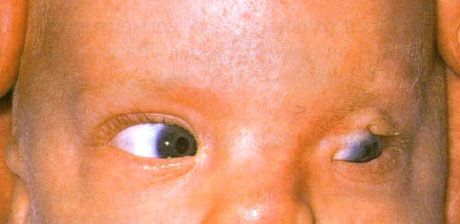
Fraser syndrome. Incomplete cryptophthalmos of the left eye. The eye is reduced in size. Clouding of the cornea. Coloboma and partial fusion of the upper eyelid on the left. Ipsilateral deformity of the nose. Low hair growth on the lateral surfaces of the forehead is characteristic
Nanophthalmos
Nanophthalmos is a special form of microphthalmos, characterized by a decrease in the size of the eyeball, a large thickness of the scleral capsule and high hyperopia. There is a tendency to develop angle-closure glaucoma. When performing abdominal surgery in these patients, the risk of vitreous prolapse is increased.
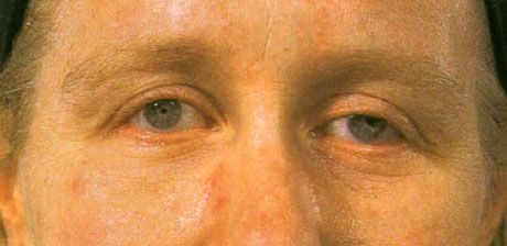
Nanophthalmia. The eyes are reduced in size. The pathological reflex of the fundus is caused by high hyperopia. The anterior chamber is shallow (this is not visible in the illustration). Such eyes are prone to the development of closed-angle glaucoma. The optic disc is often deformed, characterized by the deposition of yellow pigment in the foveolar region, localized between the foveolar zone and the optic disc.
Cyclopia
A condition in which both eyes form as one organ, without developing separately. Vision is usually very poor. The disease is accompanied by severe brain damage.
Diagnosis of microphthalmos
- Visual acuity test.
- Definition of refraction.
- Electrophysiological study to assess visual function.
- General examination to identify associated developmental defects.
- Patients are fitted with cosmetic contact lenses and are recommended to use special devices designed for people with low vision.
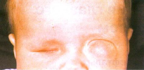
Microphthalmos with associated cyst formation (left eye). Anophthalmos (right eye). The child was diagnosed with anophthalmos on the right and marked colobomatous microphthalmos on the left at birth. Bluish edema was initially mistaken for a vascular anomaly, but the presence of transillumination helped to detect the cyst associated with the microphthalmos.
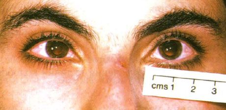
Bilateral microphthalmos without associated colobomas, in adulthood
What do need to examine?
How to examine?