Medical expert of the article

New publications
Methods of electroencephalography
Last reviewed: 04.07.2025

All iLive content is medically reviewed or fact checked to ensure as much factual accuracy as possible.
We have strict sourcing guidelines and only link to reputable media sites, academic research institutions and, whenever possible, medically peer reviewed studies. Note that the numbers in parentheses ([1], [2], etc.) are clickable links to these studies.
If you feel that any of our content is inaccurate, out-of-date, or otherwise questionable, please select it and press Ctrl + Enter.
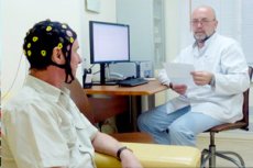
In common practice, EEG is recorded using electrodes placed on intact scalp. Electrical potentials are amplified and recorded. Electroencephalographs have 16-24 or more identical amplification and recording units (channels) that allow simultaneous recording of electrical activity from the corresponding number of pairs of electrodes installed on the patient's head. Modern electroencephalographs are computer-based. Amplified potentials are converted into digital form; continuous EEG recording is displayed on a monitor and simultaneously recorded on a disk. After processing, the EEG can be printed on paper.
Electrodes that conduct potentials are metal plates or rods of various shapes with a contact surface diameter of 0.5-1 cm. Electric potentials are fed to the input box of the electroencephalograph, which has 20-40 or more numbered contact sockets, with the help of which the corresponding number of electrodes can be connected to the device. In modern electroencephalographs, the input box combines an electrode switch, an amplifier and an EEG analog-to-digital converter. From the input box, the converted EEG signal is fed to a computer, with the help of which the device functions are controlled, and the EEG is recorded and processed.
EEG records the potential difference between two points on the head. Accordingly, voltages derived from two electrodes are fed to each channel of the electroencephalograph: one to "input 1" and the other to "input 2" of the amplification channel. A multi-contact EEG lead switch allows you to commutate electrodes for each channel in the desired combination. For example, by setting the correspondence of the occipital electrode to the socket of the input box "1" on any channel, and the temporal electrode to the socket of the box "5", you can thereby record the potential difference between the corresponding electrodes in this channel. Before starting work, the researcher types several lead diagrams using appropriate programs, which are used to analyze the obtained records. To set the bandwidth of the amplifier, analog and digital high- and low-frequency filters are used. The standard bandwidth when recording EEG is 0.5-70 Hz.
Electroencephalogram acquisition and recording
The recording electrodes are positioned so that all the main sections of the brain, designated by the initial letters of their Latin names, are represented in the multichannel recording. In clinical practice, two main EEG lead systems are used: the international 10-20 system and a modified scheme with a reduced number of electrodes. If it is necessary to obtain a more detailed EEG picture, the 10-20 scheme is preferable.
A reference lead is one in which the potential from an electrode located above the brain is fed to "input 1" of the amplifier, and from an electrode located far from the brain to "input 2". The electrode located above the brain is most often called active. The electrode far from the brain tissue is called reference. The left (A 1 ) and right (A 2 ) earlobes are used as reference electrodes. The active electrode is connected to "input 1" of the amplifier, and feeding it a negative potential shift causes the recording pen to deflect upward. The reference electrode is connected to "input 2". In some cases, a lead from two electrodes (AA) shorted together and located on the earlobes is used as a reference electrode. Since the EEG records the potential difference between two electrodes, the position of the point on the curve will be equally but in the opposite direction affected by changes in the potential under each of the pair of electrodes. In the reference lead, an alternating potential of the brain is generated under the active electrode. Under the reference electrode, located far from the brain, there is a constant potential that does not pass into the alternating current amplifier and does not affect the recording pattern. The potential difference reflects without distortion the fluctuations of the electrical potential generated by the brain under the active electrode. However, the area of the head between the active and reference electrodes is part of the "amplifier-object" electrical circuit, and the presence of a sufficiently intense source of potential in this area, located asymmetrically relative to the electrodes, will significantly affect the readings. Consequently, with the reference lead, the judgment about the localization of the potential source is not entirely reliable.
Bipolar is the name given to the lead in which electrodes located above the brain are connected to the amplifier's "input 1" and "input 2". The position of the EEG recording point on the monitor is equally affected by the potentials under each of the pairs of electrodes, and the recorded curve reflects the potential difference of each of the electrodes. Therefore, it is impossible to judge the shape of the oscillation under each of them based on one bipolar lead. At the same time, the analysis of EEG recorded from several pairs of electrodes in various combinations allows us to determine the localization of the potential sources that make up the components of the complex summary curve obtained with bipolar lead.
For example, if there is a local source of slow oscillations in the posterior temporal region, connecting the anterior and posterior temporal electrodes (Ta, Tr) to the amplifier terminals produces a record containing a slow component corresponding to the slow activity in the posterior temporal region (Tr), with faster oscillations generated by the normal brain matter of the anterior temporal region (Ta) superimposed on it. To clarify the question of which electrode records this slow component, pairs of electrodes are switched on two additional channels, in each of which one is represented by an electrode from the original pair, i.e. Ta or Tr, and the second corresponds to some non-temporal lead, for example F and O.
It is clear that in the newly formed pair (Tr-O), including the posterior temporal electrode Tr, located above the pathologically altered brain matter, the slow component will again be present. In the pair, to the inputs of which the activity from two electrodes located above the relatively intact brain (Ta-F) is fed, a normal EEG will be recorded. Thus, in the case of a local pathological cortical focus, connecting the electrode located above this focus in a pair with any other leads to the appearance of a pathological component on the corresponding EEG channels. This allows us to determine the localization of the source of pathological oscillations.
An additional criterion for determining the localization of the source of the potential of interest on the EEG is the phenomenon of oscillation phase distortion. If we connect three electrodes to the inputs of two channels of an electroencephalograph as follows: electrode 1 to "input 1", electrode 3 to "input 2" of amplifier B, and electrode 2 simultaneously to "input 2" of amplifier A and "input 1" of amplifier B; we assume that under electrode 2 there is a positive shift in the electric potential in relation to the potential of the remaining parts of the brain (indicated by the "+" sign), then it is obvious that the electric current caused by this shift in potential will have the opposite direction in the circuits of amplifiers A and B, which will be reflected in oppositely directed shifts in the potential difference - antiphases - on the corresponding EEG records. Thus, the electrical oscillations under electrode 2 in the records on channels A and B will be represented by curves with the same frequencies, amplitudes and shapes, but opposite in phase. When switching electrodes across several channels of an electroencephalograph in the form of a chain, antiphase oscillations of the potential being studied will be recorded along those two channels to whose opposite inputs one common electrode is connected, located above the source of this potential.
 [ 6 ], [ 7 ], [ 8 ], [ 9 ], [ 10 ], [ 11 ]
Rules for recording electroencephalogram and functional tests
During the examination, the patient must be in a light- and sound-proof room in a comfortable chair with their eyes closed. The subject is observed directly or with a video camera. During the recording, significant events and functional tests are marked with markers.
When testing opening and closing the eyes, characteristic electrooculogram artifacts appear on the EEG. The resulting EEG changes allow us to identify the degree of contact of the subject, his level of consciousness, and roughly estimate the reactivity of the EEG.
To detect the brain's response to external influences, single stimuli are used in the form of a short flash of light or a sound signal. In patients in a comatose state, it is permissible to use nociceptive stimuli by pressing a fingernail on the base of the patient's index finger's nail bed.
For photostimulation, short (150 μs) flashes of light close to white in spectrum and of sufficiently high intensity (0.1-0.6 J) are used. Photostimulators allow for the presentation of flash series used to study the rhythm assimilation reaction - the ability of electroencephalographic oscillations to reproduce the rhythm of external stimuli. Normally, the rhythm assimilation reaction is well expressed at a flickering frequency close to the EEG's own rhythms. Rhythmic waves of assimilation have the greatest amplitude in the occipital regions. In photosensitivity epileptic seizures, rhythmic photostimulation reveals a photoparoxysmal response - a generalized discharge of epileptiform activity.
Hyperventilation is performed primarily to induce epileptiform activity. The subject is asked to breathe deeply and rhythmically for 3 minutes. The respiratory rate should be within 16-20 per minute. EEG recording begins at least 1 minute before hyperventilation begins and continues throughout hyperventilation and for at least 3 minutes after it ends.