
Medical expert of the article

New publications
Meibomian glands: structure, functions, diseases and modern treatment methods
Last updated: 27.03.2026

All iLive content is medically reviewed or fact checked to ensure as much factual accuracy as possible.
We have strict sourcing guidelines and only link to reputable media sites, academic research institutions and, whenever possible, medically peer reviewed studies. Note that the numbers in parentheses ([1], [2], etc.) are clickable links to these studies.
If you feel that any of our content is inaccurate, out-of-date, or otherwise questionable, please select it and press Ctrl + Enter.
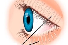
The meibomian glands are modified sebaceous glands located within the cartilaginous plates of the upper and lower eyelids. They open through pores along the edge of the eyelid, behind the lash line, and secrete a fatty substance called meibum. This substance forms the outer lipid layer of the tear film and helps protect the ocular surface from excessive moisture evaporation. [1]
In humans, the meibomian glands are arranged as long tubular-alveolar structures with a central excretory duct and lateral acini, where meibum is synthesized. There are usually more of them in the upper eyelid than in the lower. Various anatomical studies cite approximately 25-40 glands for the upper eyelid, and about 20-30 for the lower eyelid. This explains why the upper eyelid is so crucial for tear film stability and why it is routinely assessed in modern meibography. [2]
These glands do not function in isolation. Their function depends on blinking, the condition of the eyelid margins, the normal anatomy of the mucocutaneous junction, the quality of the tear film itself, and the condition of the ocular surface. With each complete blink, meibum is distributed over the ocular surface in a thin layer. If blinking becomes infrequent or incomplete, the distribution of the secretion is disrupted, and the stability of the tear film begins to deteriorate. [3]
Anatomically, the meibomian glands are important for more than just lubrication. They help reduce eyelid friction, contribute to the smoothing of the optic surface, and prevent tears from leaking over the eyelid margin. Therefore, problems with these glands affect not only comfort but also visual clarity, especially when reading, working at a screen, or during prolonged visual strain. [4]
Modern ophthalmology views the meibomian glands as a central element of ocular surface health. While dry eyes were previously primarily diagnosed with a deficiency in the aqueous portion of tears, it is now well known that the evaporative component is extremely important, and meibomian dysfunction is often the leading cause of complaints. [5]
| Key anatomical fact | What does this mean in practice? |
|---|---|
| The glands are located in the thickness of the eyelids | Their disease often begins at the edge of the eyelid. |
| The outlet openings are located at the edge of the eyelid. | Blockage of the openings quickly disrupts the outflow of secretions. |
| There are usually more glands in the upper eyelid | Examination of the upper eyelid is very important for diagnosis |
| Glands associated with blinking | Incomplete blinking impairs their performance. |
| They form the outer lipid layer of the tear film. | Without them, tears evaporate faster. |
The table is compiled based on modern anatomical and clinical reviews. [6]
How do meibomian glands work and why are they needed by the eye?
The main function of these glands is to produce meibum, a complex mixture of lipids that coats the tear film from the outside. Modern lipidomic studies show that meibum contains cholesterol esters, wax esters, specific acids, and other lipid components. For the patient, the chemical details are less important than the result: this fatty film retains moisture and makes the tear film stable. [7]
When the lipid layer is intact, tears are distributed evenly across the eye and evaporate more slowly. This maintains comfort, reduces irritation, reduces friction during blinking, and helps maintain stable vision between blinks. If the meibum becomes too thick, is deficient, or its composition changes, the tear film breaks down more quickly, and the ocular surface begins to suffer from dryness and inflammation. [8]
This is why the meibomian glands are at the center of the pathogenesis of evaporative dry eye disease. Modern reviews emphasize that meibomian gland dysfunction is the leading cause of the evaporative variant of dry eye disease, and more than 85% of cases of dry eye disease have an evaporative component. This is one reason why a patient complaining of dry eye may not so much have a "lacrimal deficiency" as a poor quality lipid layer. [9]
The glands' function also affects vision. An unstable tear film can cause fluctuations in visual clarity, especially when staring at a computer screen for long periods, reading, or driving. Because of this, patients often report not only dryness but also "blurring," "floating focus," and the need to blink frequently to momentarily restore visual clarity. [10]
It's also important to note that the meibomian glands don't regenerate indefinitely. With prolonged obstruction and chronic inflammation, some of the glands gradually atrophy, which is visible on meibography as gland prolapse. Therefore, modern ophthalmology strives to identify the problem before functional impairment progresses to irreversible morphological tissue loss. [11]
| Function of the meibomian glands | What happens when there is a violation? |
|---|---|
| Form the lipid layer of the tear film | The tear evaporates faster |
| Reduces friction when blinking | A burning sensation and a foreign body sensation appear. |
| Maintains vision stability between blinks | A floating blur occurs |
| Participate in the uniform distribution of tears | The surface of the eye dries unevenly |
| Protect against chronic evaporative stress | Inflammation and damage to the ocular surface increase |
The table reflects the key physiological role of the meibomian glands in maintaining the ocular surface. [12]
Why does the meibomian gland function become impaired?
The most common problem is meibomian gland dysfunction. The classic definition describes it as a chronic, diffuse disorder characterized by obstruction of the terminal ducts and qualitative or quantitative changes in secretion. This leads to changes in the tear film, eye irritation, eyelid margin inflammation, and ocular surface lesions. [13]
The risk increases with age. Modern research shows that aging is associated with glandular atrophy, changes in their structure, and decreased secretion. Even in populations without significant ophthalmologic diseases, age remains one of the most consistent risk factors for glandular loss and deterioration of their morphology. [14]
There are other clinically significant factors. Recent reviews and studies regularly mention contact lens wear, eyelid cosmetics, certain topical medications, refractive surgery, skin conditions, especially ocular rosacea, and demodex mites. In a 2024 study of young healthcare workers, past use of contact lenses and eye cosmetics was associated with greater glandular loss, with eyeliner and eye shadow being particularly prominent. [15]
Incomplete blinking deserves special attention. Recent reviews of screen-related dry eye disease emphasize that prolonged screen time disrupts the frequency and completeness of blinking. Incomplete blinking, in turn, impairs meibum production and increases tear evaporation. This is why some people experience particularly severe symptoms in the evening or after prolonged periods of working in front of a monitor. [16]
Associated conditions also play a significant role. Ocular rosacea is closely linked to posterior blepharitis and meibomian dysfunction, while Demodex is associated with occlusion of the eyelid openings, inflammation of the eyelid margin, and more severe tear film instability. In modern literature, this connection is considered quite convincing, so in chronic blepharitis, physicians are increasingly interested not only in the dryness itself, but also in the condition of the facial skin, eyelid margins, and eyelashes. [17]
| Risk factor | How does it affect the glands? |
|---|---|
| Age | Accelerates atrophy and structural changes |
| Contact lenses | Associated with more pronounced gland loss in some patients |
| Eye cosmetics | May contribute to obstruction and deterioration of morphology |
| Incomplete blinking and screen loading | They disrupt the extrusion and distribution of secretions |
| Ocular rosacea | Maintains chronic inflammation of the eyelid margin |
| Demodex | It increases blepharitis and worsens gland function. |
The table summarizes current data on risk factors and associated conditions. [18]
How does dysfunction manifest itself and why is it dangerous?
The most common complaints associated with meibomian gland dysfunction include burning, tingling, dryness, a gritty sensation, heaviness of the eyelids, intermittent blurred vision, and eye fatigue. These symptoms often worsen toward the end of the day, in air-conditioned rooms, while reading, working at a computer, and wearing contact lenses. The severity of the complaints does not always correspond to the depth of the morphological changes in the glands. [19]
On examination, a doctor may observe redness of the eyelid margin, dilated blood vessels, clogged drainage openings, foamy discharge, changes in the quality of the meibum, inflammation of the posterior eyelid margin, and signs of an unstable tear film. When combined with ocular rosacea, eye redness, photophobia, recurrent chalazia, and even corneal lesions are often present. [20]
The visual component of the disease is particularly distressing for patients. The unstable tear film can quickly disintegrate after blinking, causing vision to blur. This doesn't always reduce the standard line on the vision chart, but it can significantly impair reading quality, working with small text, and visual comfort in real life. [21]
If the problem is left untreated, a vicious cycle develops. A poor lipid layer accelerates evaporation, evaporation increases tear osmolarity, which intensifies inflammation of the ocular surface, and the inflammation further impairs the function of the meibomian glands. Modern reviews highlight this self-perpetuating cycle, which causes the disease to become more persistent over time. [22]
The most important long-term consequence is not just temporary discomfort, but the gradual loss of glandular tissue. Once some glands have atrophied and fallen out, it is difficult to completely restore their structure. Therefore, the modern approach emphasizes early detection, regular care, and treatment before irreversible glandular loss occurs. [23]
| What does the patient feel? | What could be behind this? |
|---|---|
| Burning and dryness | Unstable tear film and evaporation |
| Heaviness of the eyelids and discomfort along the edge of the eyelids | Obstruction and inflammation of the outlet openings |
| Intermittent blurred vision | Rapid breakdown of the tear film |
| Frequent chalazions | Long-term blockage and inflammation of the glands |
| Worsening in the evening | Accumulation of evaporative and screen stress |
The table helps to link typical complaints with the mechanisms of the disease. [24]
How Doctors Evaluate Meibomian Glands
Diagnosis begins with an examination of the eyelid margin and the quality of the secretions. The American Academy of Ophthalmology, in its clinical guidelines, emphasizes that if meibomian gland dysfunction is suspected, examining the eyelid margin for changes and carefully expressing the glands is particularly helpful. This allows one to see how freely the secretions flow and what their appearance has changed. [25]
Next, tear film stability and ocular surface condition are assessed. Depending on the clinic, this may include tear film breakup time, corneal and conjunctival staining, tear meniscus assessment, and other dry eye tests. Recent reviews emphasize that meibomian gland dysfunction cannot be assessed in isolation from the ocular surface, as it is the ocular surface that suffers from evaporative stress. [26]
Meibography has become a very important method. Modern reviews consider it a fundamental tool in the diagnosis of meibomian gland dysfunction and dry eye disease. It can be used to visualize atrophy, shortening, tortuosity, and prolapse of the glands. However, different devices have their advantages and disadvantages, and the results should be interpreted within the context of the clinical picture, not in isolation. [27]
Modern publications increasingly emphasize the importance of examining only the lower eyelid. A 2024 review found that examining the upper eyelid can be clinically important, and in some situations, the upper eyelid better reflects the risk of dryness after surgery, contact lens wear, and systemic diseases. Therefore, a comprehensive assessment today increasingly involves examining both eyelids, not just the lower one. [28]
Finally, the physician should look for underlying causes. This includes rosacea, demodex, hormonal factors, medications, incomplete blinking, screen exposure, and concomitant dry eye disease. In modern ophthalmology, the diagnosis of "meibomian gland dysfunction" is increasingly understood not as a single disease, but as several phenotypes with different mechanisms and, possibly, different responses to treatment. [29]
| Diagnostic step | What does it show? |
|---|---|
| Examination of the eyelid margin | Telangiectasias, blockage, inflammation |
| Expression of glands | How freely does the meibum come out and what is its appearance? |
| Tear film tests | How quickly do tears evaporate and disintegrate? |
| Meibography | Preservation, atrophy and deformation of glands |
| Search for rosacea, demodex, and incomplete blinking | Helps to understand why the disease persists |
The table reflects a modern comprehensive approach to diagnostics, where function, morphology, and the reasons for maintaining the disease are important. [30]
How are meibomian glands treated today?
Basic treatment almost always begins with warmth and eyelid hygiene. The American Academy of Ophthalmology and modern reviews consider warm compresses particularly beneficial for posterior blepharitis and meibomian gland dysfunction. The idea is simple: warming makes the meibum less viscous and facilitates its passage through the ostia. Moreover, the effect depends on regularity, not just one or two treatments. [31]
Home remedies work, but they have limitations. Modern studies emphasize that compresses must be used for a long time, sometimes for weeks or months, and the results depend heavily on how consistently the person adheres to the regimen and how evenly the heat is applied. Therefore, in clinical practice, the problem often stems not from a lack of effect per se, but from poor adherence and inconsistent implementation. [32]
When home care is insufficient, in-office treatments are used. These include mechanical glandular stimulation, thermal pulsation, and intense pulsed light. A 2025 systematic review of thermal devices found improvements in symptoms and tear film stability compared with controls, and a 2025 meta-analysis of intense pulsed light found a likely clinically significant reduction in symptoms compared with placebo, although its role as an adjunct to standard treatment requires further clarification. [33]
Thermal pulsation also plays a prominent role. A 2025 meta-analysis found that this system improves glandular function and reduces ocular surface staining, although the effects on subjective symptoms were less consistent across studies. Another 2025 network meta-analysis found improvements with both thermal pulsation and intense pulsed light, but direct, reliable comparisons between the two are still lacking, so it's too early to draw conclusions that "one is definitely better than the other." [34]
For more pronounced inflammatory components and moderate to severe dysfunction, physicians use medications. A recent review from 2025 indicates that both systemic and topical antibiotics, particularly macrolides and tetracyclines, improve symptoms, meibum quality, and tear film stability in the short term. In a comparison of azithromycin and doxycycline, a short course of azithromycin demonstrated comparable efficacy and better gastrointestinal tolerability. [35]
If Demodex-associated blepharitis is detected concurrently, it also needs to be treated, as without eliminating the underlying parasitic inflammation, glandular dysfunction can persistently recur. If ocular rosacea is pronounced, therapy must address both the skin inflammation and the inflammation of the eyelid margin. This is the modern treatment principle: not simply "squeezing out thick secretions," but breaking the entire pathological cycle from blockage to inflammation and evaporative dryness. [36]
| Therapeutic approach | When it is especially useful |
|---|---|
| Warm compresses | Basic home stage |
| Eyelid margin hygiene | For blepharitis and ciliary margin contamination |
| Expression of glands | When the secretion is thick and difficult to come out |
| Thermal pulsation | In case of persistent obstructive form |
| Intense pulsed light | For evaporative dryness, especially with a vascular inflammatory component |
| Azithromycin or doxycycline | For more severe inflammation and moderate progression |
| Treatment of Demodex and ocular rosacea | When the problem is supported by a concomitant disease |
The table summarizes modern step therapy, in which home care, procedures, and medications are selected according to the disease mechanism. [37]
How to preserve meibomian glands and what to expect in the future
The best prevention is not to wait until dysfunction becomes severe and chronic. If burning, dryness, blurred vision, or recurring chalazia occur regularly, it's better to check the eyelid margins and glandular condition early rather than treat the dryness alone. Recent reviews emphasize that late diagnosis leads to persistent symptoms and chronic deterioration of ocular surface function. [38]
Four simple steps are helpful every day: blinking thoroughly, taking reasonable breaks when working on a screen, carefully applying eyelid makeup, and wearing contact lenses correctly. While this doesn't guarantee perfect protection, it does reduce the number of factors that interfere with normal meibum shedding and accelerate evaporative stress. This is especially important for people with pre-existing dry eye, rosacea, or a tendency toward blepharitis. [39]
The prognosis depends on how early treatment is started and whether glandular prolapse has already occurred. Functional impairments can often be significantly improved early on, especially if the cause is identified and treatment is carried out regularly. However, if glandular tissue has already significantly atrophied, the goal is often not to completely restore the structure, but to maximally preserve the remaining function and protect the ocular surface. [40]
Future developments are already visible. Current reviews point to more accurate disease phenotyping, automated meibography, artificial intelligence for morphological assessment, more targeted drug therapy, and improved patient subtyping. This is important because the single term "meibomian gland dysfunction" currently encompasses several biologically distinct conditions, and treatment is gradually becoming more personalized. [41]
The most practical conclusion is this: the meibomian glands are not a minor detail in the eyelid's structure, but one of the main regulators of comfortable and stable vision. When they function well, a person barely notices their existence. When their function is impaired, they often become the central link in the overall picture of dryness, irritation, and eye fatigue. [42]
| Everyday measure | Why is it needed? |
|---|---|
| Full blink | Helps to squeeze and distribute meibum |
| Breaks when working on a screen | Reduces the frequency of incomplete blinking |
| Careful use of eye makeup | Reduces the risk of blockage and inflammation |
| Control when wearing contact lenses | Helps to notice early deterioration |
| Early examination for dryness and chalazion | Allows treatment up to severe atrophy |
The table reflects practical measures that help reduce the load on the meibomian glands and the ocular surface. [43]
FAQ
Are the meibomian glands and the sebaceous glands of the eyelid the same thing?
No, not exactly. The meibomian glands are modified sebaceous glands, but they are a specialised type located deeper in the eyelid and act on the ocular surface rather than on the skin like regular sebaceous glands. [44]
If your eyes are dry, does that always mean there aren't enough tears?
No. In many cases, the problem isn't that the tears are "low in volume," but that they evaporate too quickly due to a poor lipid layer. This is why a person can have pronounced symptoms of dryness with a relatively normal tear water content. [45]
Is it possible to completely restore prolapsed meibomian glands?
In the early stages, function can often be significantly improved. However, if the gland has already atrophied and prolapsed on meibography, it is difficult to completely restore its structure. Therefore, early treatment is more important than attempting to repair advanced damage. [46]
Do warm compresses really help?
Yes, they do help, especially as a first line treatment at home. But the effect depends on regularity, duration, and technique. Modern reviews show that heat remains the basic method, although in some patients it must be supplemented with in-office procedures or medications. [47]
Which is better: thermal pulsation or intense pulsed light?
Both methods may be useful, but direct, high-quality comparisons are still lacking. Currently, there is evidence that both thermal pulsation and intense pulsed light improve some symptoms and objective indicators, but the choice depends on the disease phenotype, the availability of the method, and the experience of the physician. [48]
When should you think about Demodex?
In cases of persistent blepharitis, cylindrical scales near the eyelashes, recurrent chalazions, poor response to standard eyelid hygiene, and chronic inflammation of the eyelid margin, Demodex may not be a contributing factor, but rather an active contributing factor to meibomian gland dysfunction. [49]
Key points from experts
Jennifer P. Craig, Professor of Ophthalmology at the University of Auckland, directs the Ocular Surface Laboratory. Her academic profile and professional publications focus on dry eye disease, tear film, and meibomian dysfunction. The key practical implication of this line of research is that when complaining of dry eye, assessing the aqueous portion of the tear alone should not be a sufficient measure: the condition of the eyelids and meibomian glands should be part of the routine examination. [50]
Kelly K. Nichols, DOP, MPH, PhD, is dean of the School of Optometry at the University of Alabama at Birmingham. Her research interests, according to her website, include meibomian dysfunction, dry eye disease, and tear film diagnostics and lipidomics. The practical implication of this focus is that meibomian dysfunction should be considered as a clinical problem in its own right, not just as a side effect of dry eye. [51]
Esen Karamursel Akpek, professor of ophthalmology and director of ocular surface disease and dry eye at Johns Hopkins and Duke, highlights her long-standing work in the field of ocular surface disease and dry eye disease. The key clinical message is that chronic dry eye is rarely one-dimensional, and persistent symptoms should be addressed by looking for a combination of evaporative, inflammatory, and marginal components, including meibomian involvement. [52]