Medical expert of the article

New publications
Mastocytosis: causes, symptoms, diagnosis, treatment
Last reviewed: 05.07.2025

All iLive content is medically reviewed or fact checked to ensure as much factual accuracy as possible.
We have strict sourcing guidelines and only link to reputable media sites, academic research institutions and, whenever possible, medically peer reviewed studies. Note that the numbers in parentheses ([1], [2], etc.) are clickable links to these studies.
If you feel that any of our content is inaccurate, out-of-date, or otherwise questionable, please select it and press Ctrl + Enter.
Mastocytosis is an infiltration of mast cells into the skin and other tissues and organs. Symptoms are primarily due to mediator release and include itching, flushing, and dyspepsia due to gastric hypersecretion. Diagnosis is by biopsy of skin, bone marrow, or both. Treatment consists of antihistamines and control of any underlying disease.
 [
[ Pathogenesis
Mastocytosis is a group of diseases characterized by mast cell proliferation and infiltration of the skin and other organs. Pathogenesis is based primarily on the release of mast cell mediators, including histamine, heparin, leukotrienes, and various inflammatory cytokines. Histamine is the cause of many symptoms, including gastric symptoms, but other mediators also contribute. Significant organ infiltration leads to organ dysfunction. Substances that trigger mediator release include physical contact, exercise, alcohol, NSAIDs, opioids, insect bites, or food.
Symptoms mastocytosis
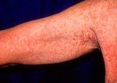
Itching of the skin is common. Stroking or rubbing the skin lesions causes urticaria and erythema around the lesion (Darier's sign); this reaction differs from dermographism, in which changes are seen on normal skin.
Systemic symptoms are very diverse. The most common are attacks of fever; severe are anaphylactoid reactions with syncope and shock. Other symptoms include epigastric pain due to peptic ulcer, nausea, vomiting, chronic diarrhea, arthralgia, bone pain, neuropsychiatric changes (irritability, depression, mood lability). Infiltration of the liver and spleen can lead to portal hypertension with subsequent ascites.
Forms
Mastocytosis can be cutaneous or systemic.
Cutaneous mastocytosis usually presents in children. Most patients present with urticaria pigmentosa, a localized or diffuse salmon-colored or brown maculopapular skin rash that results from multiple small collections of mast cells. Rarer forms include diffuse cutaneous mastocytosis, which is infiltrated skin with mast cells without discrete lesions, and mastocytoma, which has large, solitary collections of mast cells.
Systemic mastocytosis is more common in adults and is characterized by multifocal bone marrow lesions; other organs are often involved, including skin, lymph nodes, liver, spleen, and gastrointestinal tract. Systemic mastocytosis is classified as follows: painless, without organ dysfunction, and with a good prognosis; mastocytosis associated with other hematologic disorders (eg, myeloproliferative disorders, myelodysplasia, lymphoma); aggressive mastocytosis characterized by significant organ dysfunction; mast cell leukemia with more than 20% mast cells in the bone marrow smear, no skin lesions, multiorgan involvement, and a poor prognosis.
Diagnostics mastocytosis
A presumptive diagnosis is made based on clinical signs. Similar symptoms may be observed in anaphylaxis, pheochromocytoma, carcinoid syndrome, Zollinger-Ellison syndrome. The diagnosis is confirmed by biopsy of the affected skin areas and sometimes bone marrow. In patients with symptoms of peptic ulcer disease, the plasma gastrin level is measured to exclude Zollinger-Ellison syndrome; in patients with febrile attacks, the excretion level of 5-hydroxyindoleacetic acid (5-HIAA) is measured to exclude carcinoid. The level of mast cell mediators and their metabolites may be elevated in plasma and urine, but their detection does not allow a definitive diagnosis.
Treatment mastocytosis
Cutaneous mastocytosis. H2 blockers are effective as symptomatic therapy. Children with cutaneous mastocytosis do not require additional treatment, since most such cases resolve on their own. Adults with this form of mastocytosis are prescribed psoralen and ultraviolet irradiation or topical glucocorticoids 1 or 2 times a day. Mastocytoma usually undergoes spontaneous regression and does not require treatment. In children, the cutaneous form rarely progresses to the systemic form, but such cases can be observed in adults.
Systemic mastocytosis. All patients are given H1 and H2 blockers. Aspirin helps with fever but may increase leukotriene production, thus promoting symptoms related to the mast cells themselves; it is not given to children because of the high risk of Reye's syndrome. Cromolyn 200 mg orally 4 times a day [100 mg 4 times a day for children 2 to 12 years, not to exceed 40 mg/(kg x day)] is used to prevent mast cell degranulation. There are no treatments that can reduce the number of mast cells in tissues. Ketotifen 2-4 mg orally 2 times a day may be used but is not always effective.
In patients with severe forms, interferon a2b 4 million units subcutaneously once a week with a maximum dose of 3 million units per day is prescribed to relieve symptoms of bone marrow damage. Glucocorticoids (eg, prednisolone 40-60 mg orally once a day for 2-3 weeks) may be prescribed. In severe forms, splenectomy may improve quality of life.
Cytotoxic drugs (daunomycin, etoposide, 6-mercaptopurine) can be used in the treatment of mast cell leukemia, but their effectiveness has not been proven. The possibility of using imatinide (a receptor tyrosine kinase inhibitor) for the treatment of patients with c-kit mutations is being studied.