Medical expert of the article

New publications
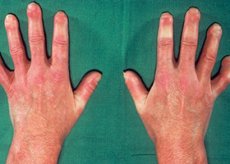
Eosinophilic fasciitis
Last reviewed: 04.07.2025

All iLive content is medically reviewed or fact checked to ensure as much factual accuracy as possible.
We have strict sourcing guidelines and only link to reputable media sites, academic research institutions and, whenever possible, medically peer reviewed studies. Note that the numbers in parentheses ([1], [2], etc.) are clickable links to these studies.
If you feel that any of our content is inaccurate, out-of-date, or otherwise questionable, please select it and press Ctrl + Enter.
Symptoms eosinophilic fasciitis
The onset of the disease is often observed in sedentary individuals, after performing heavy physical work (for example, after chopping wood). Pain, swelling, and inflammation of the skin and subcutaneous tissues develop, followed by their compaction, which leads to a characteristic "orange peel" skin change, most pronounced on the anterior surfaces of the extremities. The skin of the face and trunk are affected less often. Following induration and thickening of the fascia, limited range of motion in the joints of the upper and lower extremities develops; in addition, tendons, synovial sheaths, and muscles may be involved. Involvement of the fingers and toes is not typical of eosinophilic fasciitis. Muscle strength is usually not affected, but arthritis and myalgia, as well as carpal tunnel syndrome, may develop.
Fatigue and weight loss are characteristic. Aplastic anemia, thrombocytopenia and lymphadenopathy often develop.
Diagnostics eosinophilic fasciitis
Eosinophilic fasciitis should be suspected when a patient presents with typical manifestations. The skin changes should be differentiated from those of systemic sclerosis; however, the latter is usually characterized by Raynaud's phenomenon, involvement of the distal extremities, the appearance of telangiectasias, and visceral abnormalities (eg, esophageal atony), which are not observed in eosinophilic fasciitis.
The diagnosis is established by microscopic examination of biopsies of altered skin and fascia, with the biopsy material containing muscle fibers. The diagnosis is supported by the presence of fascial inflammation with or without eosinophils.
Blood tests are usually uninformative, but a complete blood count may reveal eosinophilia (especially in the active initial phase of the disease), and blood protein electrophoresis may reveal polyclonal hypergammaglobulinemia. Autoantibodies are usually not detected. MRI results, although not specific, can establish the presence of fascial thickening, accompanied by an increase in the signal intensity of the superficial muscles, correlating with the severity of inflammation.
 [ 15 ], [ 16 ], [ 17 ], [ 18 ], [ 19 ], [ 20 ], [ 21 ], [ 22 ]
Who to contact?
Treatment eosinophilic fasciitis
Most patients respond quickly to high-dose prednisolone (orally, 40-60 mg once daily, then tapered to 5-10 mg/day as symptoms subside). Low-dose glucocorticoids may be continued for 2 to 5 years. Despite the varying outcomes of the disease, eosinophilic fasciitis often resolves spontaneously without complications. However, due to the possibility of hematological disorders, monitoring of clinical blood test parameters is recommended.