Medical expert of the article

New publications
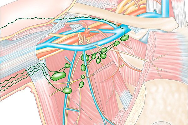
Mammary gland lymph nodes
Last reviewed: 04.07.2025

All iLive content is medically reviewed or fact checked to ensure as much factual accuracy as possible.
We have strict sourcing guidelines and only link to reputable media sites, academic research institutions and, whenever possible, medically peer reviewed studies. Note that the numbers in parentheses ([1], [2], etc.) are clickable links to these studies.
If you feel that any of our content is inaccurate, out-of-date, or otherwise questionable, please select it and press Ctrl + Enter.
The female mammary gland has a complex anatomy of structure. It lies on the large pectoral muscle and partly on the anterior serratus. The organ can easily move due to the connection with the underlying tissues of loose tissue. The anatomy of the structure also includes the lymph nodes of the mammary glands.
 [
[ Structure of the lymph nodes of the mammary glands
The paramammary lymph nodes are located on the pectoralis major muscle, along its outer edge. They are the main nodes of the first stage. The efferent vessels, which form the basis of the structure of the lymph nodes of the mammary glands, flow into the axillary lymph nodes. They, in turn, are the lymph nodes of the first stage. They are localized on the fourth tooth, sometimes called the Bartels node.
The main and largest group of lymph nodes are the axillary groups. Some of them are located on the surface, they are divided into three subgroups. These include the external, central and subscapular nodes. The external or lateral axillary nodes are located near the lateral thoracic artery. The central nodes are located along the axillary vein. They receive lymph from the outer quadrants of the mammary gland. The posterior axillary nodes have a slightly different location and run along the subscapular artery.
Another group of lymph nodes includes lymph nodes located under the collarbone. They are located in the area under the collarbone. They are referred to as first-stage nodes, which are included in the upper quadrants of the mammary glands. Lymphatic vessels flow into them. At the same time, they are referred to as first-stage nodes of the upper section of the mammary gland.
The parasternal lymph nodes are located along the internal mammary artery. They are located in the first to seventh intercostal spaces. The largest accumulation of them is observed within the second to fourth spaces. The parasternal lymph nodes are located in the second to fourth intercostal spaces. They are classified as the first stage nodes of outflow from the mammary gland. The second stage nodes include the efferent vessels of the axillary lymph nodes.
The glands of the lymphatic vessels from their base follow to the lymph nodes of the retromammary space. After that, they penetrate the pectoralis major muscle and pass into the interpectoral nodes. From this area, the lymph begins to flow into the central axillary lymph nodes.
Some of the vessels pass not only through the large but also the small pectoral muscles. Then through the ribs they penetrate to the parasternal lymph nodes, that is, the first stage. There are also non-permanent lymph nodes in the mammary gland. They are located in the area between the small and large pectoral muscles.
Regional lymph nodes of the mammary gland
The mammary gland is characterized by its complex structure. It "consists" of a lot of lymph nodes of different stages. The regional lymph nodes located in the mammary gland include a whole group of axillary nodes. It is divided into three main levels. The first level is the lower axillary nodes. They are located laterally to the lateral border with the pectoralis minor muscle. The second level is the middle axillary nodes. They are located in the area between the medial and lateral edges of the pectoralis minor muscle. The third level is represented by the apical axillary nodes. They are located in the center, relative to the medial edge of the pectoralis minor muscle. They include the subclavian and apical lymph nodes.
Regional lymph nodes also include internal ones. They are located on the affected side, i.e. they are most often affected by breast cancer. Moreover, they are even designated by a special symbol M.
Axillary lymph nodes in the mammary gland
Sometimes these lymph nodes are detected by mammography on the lateral part, which is located at the top of the quadrant of the mammary gland. It contributes to the appearance of a small round shadow. Due to this feature of the axillary lymph nodes in the mammary gland, areas of enlightenment can be seen. They are noticeable due to the accumulation of fat.
Axillary lymphadenopathy is a disorder of the axillary nodes. This process entails an increase in lymph nodes. When palpated, unpleasant pain is felt. This phenomenon can be a sign of the development of a serious disease. Including breast cancer, tuberculosis and many other diseases. Axillary lymph nodes suffer more often than others. Therefore, when they increase, it is necessary to begin to search for the cause that caused this process. The simplest method of examination in this case is a biopsy.
Intramammary lymph node of the mammary gland
Intramammary lymph nodes are coded as axillary or axillary lymph nodes. Due to their location, they most often suffer from serious diseases, including breast cancer. If there is a suspicion of damage to the mammary gland, the intramammary lymph node is examined first. The examination is carried out by means of mammography.
As mentioned above, the intramammary node is one of the axillary nodes. They, in turn, are divided into several levels, separated by their location. Detailed information about the axillary lymph nodes was presented above.
When the functionality of the intramammary lymph node is impaired, it significantly enlarges. In some cases, the process is accompanied by pain. An enlarged lymph node is a serious problem and requires immediate examination. A biopsy is used to examine this lymph node.
Examination of the lymph nodes of the mammary gland
Today, there are many methods for research. First of all, clinical diagnostics is carried out. This method of examining the lymph nodes of the mammary gland allows you to collect all the data on the patient, as well as conduct an examination and palpation. When collecting anamnesis, special attention should be paid to the duration of the disease, as well as the development of symptoms.
Physical examination involves inspection and palpation of the mammary gland. The procedure should be performed in daylight. Particular attention is paid to the symmetry of the mammary glands, the presence of deformations around the nipples and areolas. After inspection and palpation, morphological diagnostics are used. Verification of the diagnosis using this study is an important measure if there is a risk of developing a malignant process in the mammary gland.
- Cytological examination is most often used in oncology. It is characterized by diagnostic puncture using thin needles. The tip of the needle is used to determine the most dense place on the chest and puncture it. The material to be examined is collected with a syringe, after which it is transferred to glass. This method of examination will allow determining the nature of the process, as well as the degree of cell differentiation. The technique is not so common, it is used only to clarify the lymphogenous spread of cancer.
- Radiation diagnostics. Today, this type of research plays a leading role. The main methods of radiation diagnostics are considered to be X-ray and ultrasound examination. Other measures are also included here, such as computed tomography, magnetic resonance imaging, thermography and radionuclide method. They are used exclusively for special indications.
- X-ray examination. This method will allow you to take pictures of the affected area and understand the location of the pathological process by the darkening on them. In some cases, they resort to the help of X-rays in non-standard projections.
- Axillography. This method of examination allows to detect lesions of the lymph nodes. It gives a complete picture of what is happening.
- Ductography is performed if there is a suspicion of pathological secretion from the nipple. For this purpose, 0.5-2 moles of water-soluble iodine-based contrast agent are injected into the milk ducts of the mammary gland.
- Pneumocystography. This technique involves puncturing the cyst, which allows its outline to be seen.
- Ultrasound. This method of examination is the most common. It has a number of advantages and allows to accurately determine the presence of a pathological process. Moreover, the examination is harmless and safe.
- Thermography. This principle consists of remote registration of infrared radiation from the surface of the human body by thermal imagers. They reflect the degree of bioenergetic processes that occur in various areas of a particular organ. The result of the study can be obtained in the form of a thermogram.
- Computer tomography. This method of examination is used to detect metastases in breast cancer, as well as to assess the prevalence of the pathological process.
- Magnetic resonance imaging. This method of examination has modest capabilities. It is rarely used as a separate method of diagnosing the disease.
- Radionuclide examination of the mammary gland. This method is used as an additional one. It allows to determine the malignancy of the pathological process, as well as to evaluate the effectiveness of the prescribed antitumor treatment. This method of examination is highly effective.
Ultrasound of the mammary glands and lymph nodes
This method of examination is used due to its effectiveness and safety. It has no contraindications and is considered absolutely harmless. Today, ultrasound of the mammary glands and lymph nodes is used quite often. It is recommended for a general examination of a woman with complaints of soreness of the mammary glands. Ultrasound is safe even for pregnant women and young patients.
This method allows you to determine the exact cause of pain and monitor the condition of the mammary glands as a whole. In the early stages, it allows you to detect cancer and begin its timely treatment. It is necessary to resort to ultrasound in case of constant pain in the mammary glands, especially during the premenstrual phase. In case of discharge from the nipple, changes in skin color and enlargement of the axillary lymph nodes, an examination must be carried out. It is recommended to do an ultrasound at least 2 times a year. The procedure does not require special preparation. It is advisable to come for an examination in the first phase of the menstrual cycle.