Medical expert of the article

New publications
Lymph node biopsy
Last reviewed: 06.07.2025

All iLive content is medically reviewed or fact checked to ensure as much factual accuracy as possible.
We have strict sourcing guidelines and only link to reputable media sites, academic research institutions and, whenever possible, medically peer reviewed studies. Note that the numbers in parentheses ([1], [2], etc.) are clickable links to these studies.
If you feel that any of our content is inaccurate, out-of-date, or otherwise questionable, please select it and press Ctrl + Enter.
In order to understand the reasons for the enlargement of the lymph nodes, it is necessary to conduct a series of diagnostic studies. The most informative and widespread diagnostic method is currently considered to be a lymph node biopsy. This is a procedure during which a piece of biomaterial is removed for further examination.
In the human body, lymph nodes act as a kind of filtering station, attracting and neutralizing pathogenic microorganisms. When pathogens enter the node, it increases in size, which is also typical for inflammatory reactions or malignant processes. To understand what pathology is occurring in the lymphatic link, a procedure called biopsy is performed. [ 1 ]
What is the difference between a puncture and a biopsy of lymph nodes?
Histological analysis is prescribed for the diagnosis of many pathologies, since it successfully helps to determine the type of disease process, identify its phase, differentiate the tumor, etc. Often, it is this study that allows you to accurately establish a diagnosis and prescribe the correct treatment.
Lymph nodes are the main links of the body's immune system. They are "warehouses" of specific tissue that ensures the maturation of T and B lymphocytes, forms plasma cells that produce antibodies, and cleanses the lymph. Bacteria and foreign particles are filtered in the lymph nodes with the lymph flow. When they are in excess, the body's defense mechanism is activated, immunoglobulins are produced, and cellular memory is formed. All these reactions are an integral part of immunity, eliminating infectious and malignant agents.
Such protection normally always works, and the person himself may not even suspect that such reactions are occurring in his body. Only with a massive attack or a drop in immunity can the nodes increase in size, and pain may appear. However, for most patients, everything returns to normal within a few days.
If several groups of lymph nodes increase at once, the patient's health deteriorates sharply, the temperature rises, other painful symptoms appear, then in such a situation diagnostics are required, including a biopsy or puncture of the lymph node. Often these concepts are taken as synonyms, but this is not entirely true.
If we are talking about a puncture puncture, which involves collecting a liquid secretion with cells for further cytological examination, then the term "puncture" is usually used. Biopsy is mentioned if it means removing a large portion of biomaterial with subsequent histological analysis.
Puncture is a minimally invasive, fine-needle procedure that is virtually painless. A lymph node biopsy requires more traumatic intervention, often with the use of a scalpel. However, there is also the concept of "puncture biopsy", in which the node is punctured with a thicker needle device, allowing the necessary amount of tissue to be collected.
Indications for the procedure
When diagnosing lymphoproliferative and malignant pathologies, it is important not only to confirm the diagnosis morphologically, but also to detail it using cytology and histology. Such information can be obtained using a puncture and biopsy of the lymph node.
Puncture is used as an indicative diagnostic manipulation. Puncture is not suitable for determining lymphoproliferative pathology: a biopsy (either excisional or puncture) is required, with subsequent cytological and histological examination of the biopsy.
Indications for puncture may be:
- an enlarged single lymph node, without formed conglomerates and without signs of lymphoproliferative pathology;
- Ultrasound signs of fluid formation;
- the need to remove biomaterial for auxiliary examination after performing a biopsy.
A lymph node biopsy is a surgical procedure performed under local or general anesthesia. As a result of the procedure, a part of the lymph node or the entire node is obtained for further examination. Microscopic analysis is the key to accurate and correct diagnosis.
The basic indications for biopsy are:
- high risks of tumor development according to clinical information;
- lymphadenopathy of unknown origin (all diagnostic methods used did not help in making a diagnosis);
- lack of effectiveness of the therapy.
It is impossible to say exactly at what size of the lymph node a biopsy is mandatory. However, most specialists believe that a lymph node whose size exceeds 30 mm, and this is not associated with an infectious process, requires a biopsy.
Sometimes one biopsy is not enough: the patient is prescribed repeated procedures. This is possible if multiple histological changes were detected during the previous biopsy:
- lymph node necrosis;
- sinus histiocytosis;
- sclerosis;
- paracortical response with the presence of large numbers of macrophages and plasma cells.
Preparation
The preparatory stage before a lymph node biopsy may include consultation with a therapist, surgeon, endocrinologist, anesthesiologist, oncologist and hematologist. A general and biochemical blood test, and a study of the blood coagulation system are mandatory.
An ultrasound examination is prescribed to clarify the location of the pathological focus.
The doctor has a conversation with the patient in advance:
- clarifies the allergy status;
- receives information about the medications taken;
- in women, it specifies the phase of the menstrual cycle and excludes the possibility of pregnancy.
If the patient is taking blood thinning medications, they are discontinued 7-10 days before the biopsy.
If the procedure is to be performed using general anesthesia, then the preparation is carried out more thoroughly:
- It is forbidden to eat or drink on the day of the intervention;
- dinner the day before should be as light as possible, with the consumption of mainly easily digestible plant foods;
- 2-3 days before the intervention you should not drink alcohol, it is undesirable to smoke;
- The next morning, before going to the biopsy procedure, the patient should take a shower without using body lotions or creams.
Technique lymph node biopsy
A biopsy of shallow lymph nodes is usually short-lived – for many patients, the procedure is completed in about 20 minutes. Local anesthesia is usually used, although the puncture is generally considered painless. If ultrasound control is used, the doctor uses an ultrasound sensor to specify the location of the painful structure, puts a special mark, which is displayed on the monitor. The skin in the puncture area is treated with an antiseptic, and then with an anesthetic, or an injection of an anesthetic is given. The patient lies horizontally on the couch or is in a sitting position. If the biopsy is performed in the neck area, it is specially fixed, and the patient is explained about the need to temporarily refrain from swallowing. During the biopsy, the patient must remain completely still.
After taking the required amount of biological material, the puncture area is treated with an antiseptic. It may be recommended to apply a cold dry compress for half an hour.
There is no need for a long stay in the clinic or hospitalization of the patient: he can go home on his own if there are no other reasons to keep him. In the first period after the procedure, it is important to avoid physical activity.
If a biopsy is required from a deeply located node, general anesthesia may be required. In such a situation, the patient does not go home after diagnosis, but remains in the clinic - from several hours to 1-2 days.
An open biopsy requires a special set of instruments: in addition to a scalpel, these are clamps, a coagulating apparatus, and materials for suturing. This intervention lasts up to 60 minutes. The doctor selects the lymph node required for the biopsy, fixes it with his fingers, and then makes a 4-6 cm long incision in the skin. He dissects the subcutaneous fat layer, moves apart the muscle fibers, the network of nerves and vessels. If it is necessary to remove one or more nodes during the biopsy, the surgeon first ligates the vessels to prevent bleeding, lymph leakage, and the spread of tumor cells (if we are talking about a malignant process). After removing the lymph nodes, the doctor sends them for examination, revises the wound again, and sutures the incisions. In some cases, a drainage device is left, which is removed after 24-48 hours. The sutures are removed within a week.
How is a lymph node biopsy performed?
Taking a biopsy from a particular lymph node may have its own characteristics, which depend on the localization, depth of the structure, as well as the presence of vital organs and large vessels near the damaged link.
- A lymph node biopsy in the neck may be prescribed for otolaryngological and dental problems, which are the most common causes of lymphadenopathy. If the lymphadenopathy has an unclear origin, an ultrasound is prescribed first, and only after that, if necessary, a biopsy. Lymph nodes enlarge with malignant tumors, since cancer cells penetrate the lymph vessels that drain a separate area. These cells then settle in the filtering nodes as metastases and begin to develop. Often, with oncology, lymph node damage occurs "in a chain", which is easily determined by palpation. A biopsy in the neck can be performed either by a needle puncture with material removal, or by surgical access with complete removal of the link for histological analysis.
- Sentinel lymph node biopsy for melanoma is performed similarly to breast cancer biopsy. If there is information about metastasis to distant organs and lymph nodes, then performing a biopsy is considered useless. In the absence of metastases, sentinel node biopsy is completely justified. It is usually performed after removal of the melanoma itself. The lymph node can be visualized using various radiographic methods.
- Axillary lymph node biopsy is performed with the patient in a sitting position, raising his arm up and moving it back a little. Most often, this procedure is performed when the mammary gland is affected: lymph flows through the vessels to the nodes located in the armpit on the same side. Such lymph nodes form a kind of axillary-nodal chain. Its damage plays an important role in planning the treatment regimen for mammary gland pathology. The study is also appropriate for melanoma or squamous cell carcinoma of the upper limb, and lymphogranulomatosis.
- A biopsy of the inguinal lymph nodes is performed with the patient lying on a couch, the leg (right or left, depending on the side of the lesion) is moved to the side. This examination is most often prescribed when tumor processes are suspected (testicular cancer, external genitalia, cervix, prostate, bladder, rectum), or if it is not possible to determine the cause of lymphadenopathy by other means (for example, with lymphogranulomatosis or HIV infection).
- A biopsy of the supraclavicular lymph node is always caused by suspicions of quite serious pathologies: in many cases, these are tumors - metastases of cancer or lymphoma located in the chest or abdominal cavity. The supraclavicular lymph node on the right side can make itself known in tumor processes of the mediastinum, esophagus, lung. Lymph from the intrathoracic organs and abdominal cavity approaches the lymph node on the left side. Inflammatory diseases can also cause supraclavicular lymphadenopathy, but this happens much less often.
- Biopsy of the mediastinal lymph node is performed in the projection area of the upper third of the intrathoracic tracheal section, from the upper edge of the subclavian artery or pulmonary apex to the intersection of the upper border of the left brachiocephalic vein and the mid-tracheal line. The most common indications for biopsy of the mediastinal lymph nodes are: lymphoproliferative neoplasms, tuberculosis, and sarcoidosis.
- A biopsy of a lymph node in the lung is a common procedure for cancer, tuberculosis, and sarcoidosis. Lymphadenopathy is often the only sign of pathology, since many lung diseases are asymptomatic. In any case, before making a final diagnosis, the doctor must perform a biopsy and obtain histological information.
- Biopsy of the abdominal lymph nodes is prescribed if tumor processes in the gastrointestinal tract, female and male reproductive organs, and the urinary system are suspected. Enlarged lymph nodes of the abdominal cavity are often observed with hepatosplenomegaly. Biopsy is done for both primary and differential diagnostics. A large number of lymph nodes of the abdominal cavity are located parietal along the peritoneum, along the vessels, in the mesentery and along the intestine, near the omentum. They may enlarge if the stomach, liver, intestines, pancreas, uterus, appendages, prostate gland, and bladder are affected.
- A submandibular lymph node biopsy may be prescribed for pathologies of the teeth, gums, cheeks, larynx and pharynx, if the cause of lymphadenopathy cannot be found, as well as if metastases of a cancerous process or lymphoma are suspected.
- A biopsy for lymph node cancer is performed in the absence of metastases to distant organs and lymph nodes. Otherwise, the procedure is considered meaningless for the patient. If there is no distant metastasis, then they begin, first of all, to examine the first in the chain, the "sentinel" lymph node.
- Retroperitoneal lymph node biopsy is appropriate for malignant processes of the male and female reproductive system. Approximately 30% of patients already at the first stage of cancer have microscopic metastases in the lymph nodes, which cannot be determined using computed tomography or markers. Biopsy is usually performed on the side where the primary tumor was located. The procedure is usually performed as part of a retroperitoneal lymphadenectomy.
- Biopsy of the intrathoracic lymph nodes is a mandatory examination if lung cancer, esophagus, thymus, breast cancer, lymphoma and lymphogranulomatosis are suspected. Metastases from the abdominal cavity, pelvis, retroperitoneal space (kidneys, adrenal glands) in advanced stages can also spread to the mediastinal nodes.
- Biopsy of paratracheal lymph nodes is often performed in patients with oncological lung lesions. Paratracheal lymph nodes are located between the upper mediastinal and tracheobronchial nodes. In the case of a primary tumor process on the same side, they are classified as ipsilateral, and in the absence of a primary tumor, as contralateral.
Lymphatic fluid flows through the appropriate vessels. If cancer cells get into it, they end up, first of all, in the first lymph node of the chain. This first node is called a sentinel or sentinel node. If cancer cells are not found in the sentinel node, then the subsequent nodes should, in theory, be healthy.
Types of biopsies
There are several types of lymph node biopsy, depending on the technique of biomaterial extraction. Some types of procedures are performed in stages: first, a needle puncture is performed, and then an open intervention is performed if the puncture is not enough for diagnosis. An open biopsy is required if the cytology result is uncertain, questionable, or approximate.
- Open lymph node biopsy is the most complex and invasive option for such diagnostics. During the procedure, a scalpel is used, and the entire node is selected for examination, not just part of it. Such intervention is often the only correct one if malignant processes are suspected.
- Puncture biopsy of the lymph node is a relatively gentle and painless procedure that does not cause any particular discomfort to patients. During the diagnostics, a mandrin is used, which acts as a stylet. The mandrin is used to cut off and capture the required amount of biomaterial. Puncture biopsy involves the use of local anesthesia and does not require hospitalization of the patient.
- Excisional lymph node biopsy is a term often used to refer to an open biopsy, performed under general anesthesia, in which the affected node is removed through an incision.
- A trephine biopsy of a lymph node involves the use of a special large needle with notches that allow the removal of a tissue particle of the required size.
- Fine-needle biopsy of a lymph node is called aspiration: it involves the use of a thin, hollow needle device. Usually, the node is palpated and punctured: if this is not possible, ultrasound control is used. As a rule, fine-needle biopsy is prescribed when it is necessary to examine the submandibular or supraclavicular lymph nodes, when metastases of lymphoid structures are detected.
Ultrasound-guided lymph node biopsy
Currently, specialists consider the most acceptable technique for lymph node biopsy to be a targeted puncture procedure, or the so-called “biopsy under visual ultrasound control.”
This is the process of taking a sample of biomaterial, which is performed under ultrasound supervision: as a result, the placement and insertion of the puncture needle is carried out more accurately and safely. This is extremely important for the doctor, since often the suspicious lymph node is located in deep tissues near vital organs, or is small in size, which significantly complicates the procedure.
Ultrasound monitoring helps to insert the instrument precisely into the required place, without the risk of damaging nearby tissues and organs. As a result, the risk of complications is minimized.
The doctor determines the exact method by which the required area is visualized. An additional advantage of the technique is not only its safety, but also its low cost: no ultra-modern and expensive equipment is needed.
Biopsy with ultrasound is especially recommended if it is necessary to examine not only the affected structure, but also to find out the peculiarity of blood circulation near it. This approach will avoid injury to blood vessels and will prevent blood from leaking into the tissue.
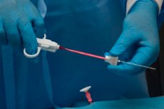
The procedure uses special needles with end sensors. This simple device helps to clearly monitor the position of the needle and its progress.
The recovery period after such an intervention is faster and more comfortable for the patient. [ 2 ]
Contraindications to the procedure
Before referring a patient for a lymph node biopsy, the doctor will prescribe a number of studies and tests that are necessary to rule out contraindications to this procedure. Basic preliminary diagnostics are a general blood test and an assessment of the quality of coagulation. Biopsy is not performed if there is a tendency to bleeding - for example, in patients suffering from hemophilia, since the intervention may injure the vessels.
Lymph node biopsy is contraindicated in case of purulent processes in the puncture area. It is not advisable to perform the procedure on pregnant or breastfeeding women, as well as during menstrual bleeding.
In general, experts highlight the following list of contraindications:
- disorders of the blood coagulation system (congenital disorders, acquired, or temporary - that is, associated with taking appropriate medications that thin the blood);
- platelet level below 60 thousand per µl;
- hemoglobin level less than 90 g/liter;
- INR greater than 1.5;
- prothrombin time exceeding the norm by 5 seconds;
- infectious and inflammatory processes in the area of biopsy;
- menstrual bleeding in women on the day of the procedure;
- decompensated chronic pathologies;
- treatment with nonsteroidal anti-inflammatory drugs during the last week.
Normal performance
Microscopic examination of a patient's lymph node biopsy is considered the most important in the diagnostic aspect of oncological pathologies and helps to assess the quality of drug therapy.
Lymph node histology is a minor surgical procedure in which a small piece of tissue is removed for further examination. With the help of a lymph node biopsy, specialists can study the features of its structure, detect painful deviations, and notice signs of an inflammatory reaction.
The lymph node is the basic link of the body's defense system, which is a connecting element between the lymphatic vessels. Lymph nodes help fight infectious invasion by producing leukocytes - specific blood cells. The node catches microbial and viral infections, malignant cells.
A lymph node biopsy helps to identify the presence of atypical cells, determine the specifics of the infectious inflammatory process, benign tumors, and purulent pathologies. Biopsy is most often performed in the inguinal, axillary, mandibular, and retroauricular areas.
A biopsy is prescribed to patients who need to determine the type of tumor process, especially if malignant pathology is suspected. Diagnostics are often prescribed to determine infectious diseases.
Lymph node biopsy results
After examining the biopsy (material obtained by biopsy of the lymph node) and detecting particles of pathology, specialists begin to count the cellular structures and derive a lymphadenogram. For this purpose, they use the immersion method of microscopic observation, which allows differentiating at least half a thousand cells and calculating their percentage presence.
Lymph node imaging data are extremely necessary and valuable for diagnosing non-specific forms of lymphadenitis.
Normal lymphadenogram results:
Contents of the corresponding cell types |
Percentage indicator |
Lymphoblasts |
From 0.1 to 0.9 |
Prolymphocytes |
From 5.3 to 16.4 |
Lymphocytes |
From 67.8 to 90 |
Reticular cells |
From 0 to 2.6 |
Plasmocytes |
From 0 to 5.3 |
Monocytes |
From 0.2 to 5.8 |
Mast cells |
From 0 to 0.5 |
Neutrophil granulocytes |
From 0 to 0.5 |
Eosinophilic granulocytes |
From 0 to 0.3 |
Basophilic granulocytes |
From 0 to 0.2 |
The biological material taken during a lymph node biopsy contains predominantly mature lymphocytes with prolymphocytes. Their total number can be from 95 to 98% of all cellular structures.
Reactive lymphadenitis is manifested by an increase in the number of reticular cells, the detection of macrophages and immunoblasts.
In acute lymphadenitis, an increase in the number of macrophages and neutrophils is observed.
Complications after the procedure
Usually, diagnostic lymph node biopsy is performed without any complications. Only in some cases complications develop:
- bleeding due to accidental vascular injury during biopsy;
- lymph discharge from the wound;
- paresthesia, impaired sensitivity in the area where the intervention is performed;
- infection associated with the entry of an infectious agent, in particular during a procedure;
- trophic disorders associated with mechanical injuries to nerve structures.
Some patients may experience impaired consciousness, dizziness, weakness. The condition should normalize within 1-2 days.
Dangerous symptoms requiring immediate medical attention:
- increased temperature, fever;
- the appearance of severe, throbbing, increasing pain in the area of the lymph node biopsy;
- discharge of blood or pus from the wound;
- redness, swelling at the biopsy site.
Consequences after the procedure
A lymph node biopsy is not performed if the patient has any contraindications. Otherwise, adverse effects may develop. For example, if a person suffers from disorders of the blood coagulation system, even a regular puncture biopsy may end in bleeding.
To prevent the occurrence of post-procedural problems, lymph node biopsy should be performed by a specialist, in compliance with all required conditions, aseptic and antiseptic rules.
In some cases, the following problems may arise:
- infection;
- bleeding from the wound;
- nerve damage.
However, the percentage of negative consequences is relatively small. But the information obtained during the biopsy is of great value to the doctor, allowing him to make a correct diagnosis and prescribe the appropriate effective treatment.
Care after the procedure
Usually, the lymph node biopsy procedure is not complicated and is well tolerated by patients. After the biomaterial is removed by aspiration or puncture, only the puncture site remains on the skin, which is treated with an antiseptic solution and sealed with a plaster. If an open biopsy was performed, the wound is sutured and bandaged. The sutures are removed within a week.
The wound after a lymph node biopsy should not be wetted. It is necessary to treat it with antiseptic solutions to prevent infection. If the body temperature suddenly rises, the intervention site swells, bleeds or bothers in any other way, then you need to urgently visit a doctor.
The appearance of short-term, mild pain after the procedure is acceptable.
What you should not do after a lymph node biopsy:
- take a bath;
- swim in pools and open water bodies;
- visit a bathhouse or sauna;
- practice vigorous physical exercise.
Such restrictions are in effect for approximately 2 weeks after the procedure, depending on the type and extent of such an intervention as a lymph node biopsy.