Medical expert of the article
New publications
Structure and function of the legs
Last reviewed: 04.07.2025

All iLive content is medically reviewed or fact checked to ensure as much factual accuracy as possible.
We have strict sourcing guidelines and only link to reputable media sites, academic research institutions and, whenever possible, medically peer reviewed studies. Note that the numbers in parentheses ([1], [2], etc.) are clickable links to these studies.
If you feel that any of our content is inaccurate, out-of-date, or otherwise questionable, please select it and press Ctrl + Enter.
The structure of the legs is a very complex design by nature. The bones that are inside the legs are the largest of the bones in the entire body. But nature designed it this way for a reason, because the legs bear the heaviest load of all body parts – they support the entire human mass. If a person is obese, the bones and joints of the legs receive a double load. More about the structure and role of bones and joints.
How do bones grow?
Girls' bones grow until they are 16 years old, and boys' bones grow until they are 17 years old. They gradually harden. When a child is small, his bones are soft and brittle, they are easy to break and damage, because bones are made mainly of cartilage. As a person grows, the cartilage hardens, they are more like bones, they are not so easy to break or damage.
When a person grows up, cartilage remains only in the joints. Without cartilage tissue in the joints, bones would not be able to slide easily, touching each other, and a person would not be able to bend arms, legs and other parts of the body where there are joints. For example, turn the neck. Thanks to the joints, bone tissue does not wear out, as it would without them.
Leg structure
They consist of the three largest bones of the pelvis - the ischium, ilium and pubis. These bones provide support for the torso and support the legs. By the age of 18, these bones fuse together in both boys and girls. This fusion of three bones is called the acetabulum.
The head of the femur bone is inserted into this cavity, like into a construction set. It rotates and thus allows a person to freely and easily rotate the limb. The femur bone is so strong that it can easily withstand a load in the form of the weight of a passenger car.
The knee joint has a cup that connects to the thigh bone, but is not connected to the shin bone. Therefore, the lower leg and the knee are connected by bone and joints, and this part of the leg is mobile thanks to the joints.
As for the knee joint, it is the most complex and durable structure of all the joints in the body.
 [ 7 ]
[ 7 ]
Structure of the foot
As we already wrote in the material about the structure of the foot, it consists of 26 bones - a huge number for such a small foot. The bones of the foot are divided into phalanges and metatarsal bones. The bones that are located in the foot make up two arches of the sole. They are located longitudinally. They allow the foot to be flexible and move dynamically in different directions. When walking, the foot acts as a spring. A person is diagnosed with flat feet if the spring function is impaired, that is, the arch of the foot is lowered in the same way as under the toes and heels.
 [ 8 ]
[ 8 ]
Why do we need cartilage?
They help the joints not to wear out and not to become inflamed when the joints rub against each other. Therefore, the bones outside the joints are covered with cartilage tissue, which is elastic and allows the heads of the bones to slide against each other. And the role of lubrication between the heads of the joints with cartilage on them is performed by synovial fluid. This fluid is produced by a membrane called synovial. As soon as the fluid begins to be produced insufficiently, the joints can no longer slide against each other, and therefore a person is greatly limited in movement.
Very rarely, but there are cases when the cartilaginous tissue begins to harden and become bone. Then the joints can no longer rotate and move, because the bones grow together. The person's leg becomes immobile, any movements in the direction of bending-unbending, turning cause pain. It is necessary to prevent the joints from growing into bones in advance, so as not to lose the mobility of the leg later.
The role of the ligaments of the legs
Ligaments have the property of being attached to the bones of the legs. Ligaments consist of connective tissue, it is quite strong. Ligaments are needed to fix the joints in a certain position, so that their movement, resting state and any other functions are stable, reliable.
Ligaments can tear (this is well known to athletes) if they are subjected to too much stress. When ligaments tear, it is very painful and takes a very long time to heal. If 21 days are given for the bones to heal, including rehabilitation, then it can take twice as long for torn ligaments to heal.
To prevent ligaments from tearing, it is important to exercise them: stretch them, warm them up with exercises.
If a person hardens his ligaments, then the joints work much easier and better. As for tendons, their structure is similar to the structure of ligaments, but they differ from ligaments in their role. Ligaments connect bones, and tendons connect bones and muscles.
Leg muscles
Legs need muscles to anchor bones and allow them to move. Muscles are divided into groups, and these groups are often multidirectional. This allows a person to move as he plans and to exclude movements that are in the opposite direction.
The part in front of the thigh consists of four muscles. They are the strongest of all the other bones in the human body. This is the most indicative group of muscles, which are collectively called the quadriceps. It has a very important role - it is responsible for bending the shin.
The so-called sartorius muscle is responsible for bending the shin and thigh. This gives the shin the ability to rotate only inward, while the thigh rotates outward. Other muscle groups – adductors and medial – allow the thigh to rotate inward, and thanks to them, the thigh can be moved away from the body and brought closer to it.
Muscles of the foot
The foot rises and falls thanks to the muscles of the lower leg, which make this possible. The muscles have the property of being attached by tendons to the bones that are located in the feet. Thanks to the two external muscles, the lower leg has the ability to lower the foot down, thanks to these muscles, the sole bends. The muscles that are located on the back of the lower leg help to raise the heel, as well as rise on tiptoes.
The foot has no more and no less than 11 muscles, small in size and volume. These muscles help to straighten and bend the toes, lift the foot off the floor, that is, walk. 11 muscles are not all, to enable a person to walk, a total of 38 muscles with different functions are needed.
Lazy muscles
If you do not train your leg muscles, they become flabby and become covered with fat deposits, which makes them perform their functions poorly. The fat from the hips is the last to go, even if a person is on a strict diet. It is important to constantly give strength training to the muscles, but calculate it correctly. Particular attention should be paid to the buttocks and thighs, training them. Then the legs will serve a person for a long time and efficiently.
Blood circulation in the legs

Blood moves through large arteries, small arteries and capillaries. In order for them to normally supply the legs with nutrients, the blood needs oxygen. And it needs to be enriched with oxygen.
There are different types of leg arteries: by location, they are called femoral, anterior and posterior tibial, popliteal, dorsal (serves to supply blood to the foot), lateral and medial (located on the sole). The blood flow in these arteries is very strong, so the movement of blood can be felt even by placing a finger on the skin above the artery.
The walls of the arteries depend on the size of these arteries. If the size is large, then the walls are thick, and the blood flows faster, since such an artery has a larger diameter. The composition of the walls is connective tissue. Smaller arteries have thinner walls, which consist of smooth muscle tissue. When the walls of the arteries contract, the blood flows through the arteries faster and more actively.
 [ 13 ]
Capillaries
The smallest and narrowest vessels of the leg (and the whole body) are called capillaries. Their walls are very thin, they are as thick as one cell of the body. Such walls are not made too thick so that the process of exchange of oxygen and nutrients in the capillaries proceeds faster. Capillaries are very sensitive to changes in heat and cold. If a person gets into cold conditions, the capillaries narrow, and then more heat is retained in the body. And if in hot temperatures, the capillaries expand. Then the body can regulate the temperature, lowering it.
Metabolic products enter the venules (small veins) from the blood capillaries, and are then transferred to the veins. These substances are transported through the bloodstream to the heart, and then to the lungs. There they are enriched with oxygen, giving up carbon dioxide.
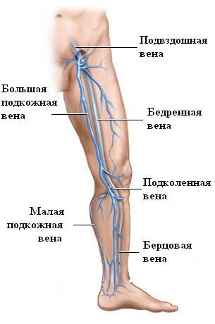
There are 8 main large veins in the legs. They converge into one femoral vein. These veins have special valves that help pump blood in the right direction. This blood moves with the help of the leg muscles, which pump it to the heart when the muscles contract. Because of this, to keep the heart healthy, doctors recommend walking and strolling, especially before bed.
Nerves located in the legs
All movements that our legs make are due to the motor nerves. They transmit commands from the brain. In addition to the motor nerves, there are also sensory nerves in the leg that transmit signals to the brain that a person has been injured, that the leg has hit ice or stepped on hot asphalt.
The nerves of the legs originate from the lumbar region and the sacrum (the same sections of the spine). The largest area of the thigh receives and transmits signals via the femoral nerve, the nerve of the perineum, as well as the tibial and subcutaneous nerves, are responsible for the impulses of the lower leg. The medial, gastrocnemius and lateral nerves control the sole of the foot.
Of course, these nerves do not exist on their own. They are interconnected, and an impulse transmitted by one nerve can be transmitted to others. This is why pain in one part of the body can be felt in another part. In addition, the interconnected system of nerves in different parts of the leg allows you to move your limbs as you wish.
Load on feet and their size
Previously, a person could use his toes the same way he uses his fingers now. With his toes, a person could grab a branch and hang on it or take some necessary object, for example, a stick. Now the functions of the foot have become less diverse, we limit our legs only to walking.
The foot has become much wider and larger than it was several centuries ago, because now people do not climb trees, but support their body weight with their legs. Accordingly, the load on them has increased. And it is always easier to lean on a larger area of the foot than on a smaller one. Therefore, the average minimum shoe size increases every year. This is indicated by statistics.
What foot is considered ideal?
Since the most important role of the foot is to bear the weight of the body, it must have an optimal shape. The shape, strength, elasticity and size of the feet depend on this, and therefore their health. How to check the shape of your own feet?
Prepare a clean white sheet of paper and a pencil. Place it on a hard surface. Stand with your feet on this sheet and trace the outline of your foot with a pencil. Now look at it carefully to determine visually how correct the structure of your feet is.
Pay attention to the big toe. The ideal toe is straight, larger than the other toes. The other toes point toward the big toe. Pay attention to the foot. It should not have a bump or ridge.
Look at the circumference of your heels. It should be round, uniform, without bumps or depressions. The heels themselves should mirror each other. Pay attention to the arch of your feet and the height of their rise. If the arch of your foot is too low, you should see an orthopedist – it could be flat feet.
Foot defects
If you find defects in your feet when examining their shapes, you should definitely consult a doctor. Flat feet can be caused by genetic changes, which is difficult to fix. But if you pay attention to the abnormal shape of your feet in childhood, you can still fix it. In childhood, the bones are still very soft and brittle, so you can combat defects with exercises and special orthopedic foot forms.
Some areas of the foot are more vulnerable. For example, deformation of the first toe (namely the metatarsal joint). This can also be the heel bone, as well as hammer toes.
Orthopedic devices will help to cope with this. You just need to consult a traumatologist or orthopedist at least once a year to avoid further development of foot deformation.
Timely treatment of legs
If you seek medical help in time, you can correct the foot deformity at its initial stage, when a person does not even suspect the abnormal development. Over time, if you do not pay attention to the abnormal development of the foot, the situation will worsen under the pressure of mechanical factors - walking, friction, pressure, increased loads.
Therefore, you should always pay attention to the most seemingly insignificant changes in the structure of the foot. For example, a bump on the heel, hair loss on the legs, a bone on the foot that grows or hurts, even calluses that were not observed before. And immediately consult a doctor about the health of the feet.

