Medical expert of the article
New publications
Intraocular pressure study
Last reviewed: 04.07.2025

All iLive content is medically reviewed or fact checked to ensure as much factual accuracy as possible.
We have strict sourcing guidelines and only link to reputable media sites, academic research institutions and, whenever possible, medically peer reviewed studies. Note that the numbers in parentheses ([1], [2], etc.) are clickable links to these studies.
If you feel that any of our content is inaccurate, out-of-date, or otherwise questionable, please select it and press Ctrl + Enter.

Orientation (palpation) examination
It is carried out with the patient's head in a motionless position and looking down. The doctor places the index fingers of both hands on the eyeball through the skin of the upper eyelid and presses on the eye one by one. The resulting tactile sensations (varying degrees of compliance) depend on the level of intraocular pressure: the higher the pressure and the denser the eyeball, the less mobility of its wall. The intraocular pressure determined in this way is designated as follows: Tn - normal pressure; T+1 - moderately elevated intraocular pressure (the eye is slightly dense); T+2 - significantly elevated (the eye is very dense); T+3 - sharply elevated (the eye is hard as a rock). When intraocular pressure decreases, three degrees of its hypotension are also distinguished: T-1 - the eye is somewhat softer than normal; T-2 - the eye is soft; T-3 - the eye is very soft.

This method of studying intraocular pressure is used only in cases where it is impossible to carry out its instrumental measurement: in case of injuries and diseases of the cornea, after surgical interventions with opening of the eyeball. In all other cases, tonometry is used.
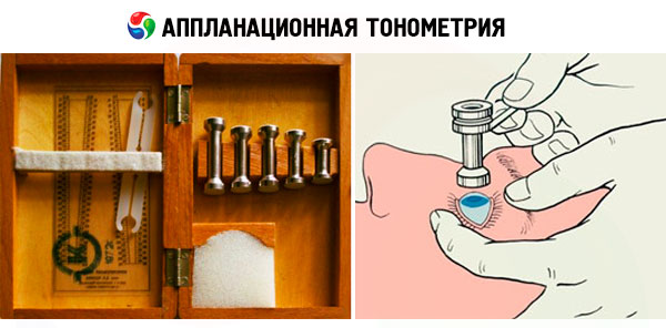
Applanation tonometry
In our country, this study is performed using the method proposed by A. N. Maklakov (1884), which involves placing a standard 10 g weight on the surface of the patient's cornea (after its drop anesthesia). The weight is a hollow metal cylinder 4 mm high, the base of which is widened and equipped with platforms made of milky-white porcelain 1 cm in diameter. Before measuring intraocular pressure, these platforms are covered with a special paint (a mixture of collargol and glycerin), and then, using a special holder, the weight is lowered onto the cornea of the patient's eye, which is wide open with the doctor's fingers, while the patient is lying on the couch.

Under the action of the weight's pressure, the cornea is flattened and the paint is washed off at the point of its contact with the weight's platform. A circle devoid of paint remains on the weight's platform, corresponding to the area of contact between the surface of the weight and the cornea. The resulting imprint from the weight's platform is transferred to paper pre-moistened with alcohol. The smaller the circle, the higher the intraocular pressure and vice versa.
To convert linear quantities into millimeters of mercury, S. S. Golovin (1895) compiled a table based on a complex formula.
Later, B. L. Polyak transferred this data to a transparent measuring ruler, with the help of which one can immediately obtain an answer in millimeters of mercury at the mark around which the imprint from the tonometer weight is inscribed.
The intraocular pressure determined in this way is called tonometric (P t ), since the ophthalmotonus increases under the effect of the weight on the eye. On average, with an increase in the tonometer mass by 1 g, the intraocular pressure increases by 1 mm Hg, i.e. the smaller the tonometer mass, the closer the tonometric pressure is to the true pressure (P 0 ). Normal intraocular pressure when measured with a 10 g weight does not exceed 28 mm Hg with daily fluctuations of no more than 5 mm Hg. The set includes weights weighing 5; 7.5; 10 and 15 g. Sequential measurement of intraocular pressure is called elastotonometry.
 [ 13 ]
Impression tonometry
This method, proposed by Schiøtz, is based on the principle of corneal indentation by a rod of constant cross-section under the influence of a weight of varying mass (5.5; 7.5 and 10 g). The magnitude of the resulting corneal indentation is determined in linear units. It depends on the mass of the weight used and the level of intraocular pressure. To convert the measurement readings into millimeters of mercury, the nomograms attached to the device are used.
Impression tonometry is less accurate than applanation tonometry, but is indispensable in cases where the cornea has an uneven surface.
Currently, the disadvantages of contact applanation tonometry have been completely eliminated due to the use of modern contactless ophthalmological tonometers of various designs. They implement the latest achievements in the field of mechanics, optics and electronics. The essence of the study is that from a certain distance, a portion of compressed air, dosed by pressure and volume, is sent to the center of the cornea of the eye being examined. As a result of its impact on the cornea, its deformation occurs and the interference pattern changes. The level of intraocular pressure is determined by the nature of these changes. Such devices allow measuring intraocular pressure with high accuracy without touching the eyeball.
Study of eye hydrodynamics (tonography)
The method allows obtaining quantitative characteristics of the production and outflow of intraocular fluid from the eye. The most important of them are: the coefficient of ease of outflow (C) of chamber fluid (normally not less than 0.14 (mm 3 -min)/mm Hg), the minute volume (F) of aqueous fluid (about 2 mm 3 /min) and the true intraocular pressure P 0 (up to 20 mm Hg).
To perform tonography, devices of varying complexity are used, including electronic ones. However, it can also be performed in a simplified version according to Kalf-Plyushko using applanation tonometers. In this case, the intraocular pressure is initially measured using sequentially weights weighing 5; 10 and 15 g. Then, a weight weighing 15 g is placed with a clean area on the center of the cornea for 4 minutes. After such compression, the intraocular pressure is measured again, but the weights are used in the reverse order. The resulting flattening circles are measured with a Polyak ruler and two elastic curves are constructed based on the established values. All further calculations are made using a nomogram.
Based on the results of tonography, it is possible to differentiate the retention (reduction of fluid outflow pathways) form of glaucoma from the hypersecretory (increased fluid production) form.

