Medical expert of the article

New publications
Examination of cranial nerves. Pair V: trigeminal nerve (n. trigeminus)
Last reviewed: 07.07.2025

All iLive content is medically reviewed or fact checked to ensure as much factual accuracy as possible.
We have strict sourcing guidelines and only link to reputable media sites, academic research institutions and, whenever possible, medically peer reviewed studies. Note that the numbers in parentheses ([1], [2], etc.) are clickable links to these studies.
If you feel that any of our content is inaccurate, out-of-date, or otherwise questionable, please select it and press Ctrl + Enter.
The motor branches of the trigeminal nerve innervate the muscles that provide movement of the lower jaw (masticatory, temporal, lateral and medial pterygoid; mylohyoid; anterior belly of the digastric); tensor tympani muscle; tensor veli palatini muscle. Sensory fibers supply the main part of the scalp (facial skin and fronto-parietal part of the scalp), the mucous membrane of the nasal and oral cavities, including the frontal and maxillary sinuses; part of the auditory canal and tympanic membrane; the eyeball and conjunctiva; the anterior two-thirds of the tongue, teeth; the periosteum of the facial skeleton; the dura mater of the anterior and middle cranial fossae, the tentorium cerebelli. The branches of the V nerve are the ophthalmic, maxillary and mandibular nerves.
Sensation in the face is supplied by both the trigeminal nerve and the upper cervical spinal nerves.
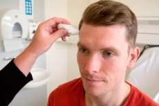
Pain, tactile and temperature sensitivity are tested sequentially in the innervation zones of all three branches of the V pair on both sides (using a pin, a soft hair brush, a cold surface of a metal object - a neurological hammer, a dynamometer). Synchronously touch symmetrical points in the forehead area (I branch), then the cheek (II branch), chin (III branch).
Dissociated sensory disturbance of the face, i.e. disturbance of pain and temperature sensitivity with preservation of tactile sensitivity, indicates damage to the nucleus of the spinal tract of the trigeminal nerve (nucl. tractus spinalis n. trigemini) with preservation of the main sensory nucleus of the trigeminal nerve located in the dorsolateral part of the pontine tegmentum (nucl. pontinus n. trigemini). Such a disorder most often occurs with syringobulbomyelia, ischemia of the posterolateral parts of the medulla oblongata.
Trigeminal neuralgia is characterized by sudden, short, and very intense, repeated attacks of pain, so short-lived that they are often described as a shooting pain or electric shock. The pain spreads to the innervation zones of one or more branches of the trigeminal nerve (usually in the area of the II and III branches, and only in 5% of cases - in the area of the I branch). With neuralgia, there is usually no loss of sensitivity on the face. If trigeminal pain is combined with disturbances of superficial sensitivity, trigeminal neuralgia-neuropathy is diagnosed.
The corneal reflex is examined using a piece of cotton wool or a strip of newspaper. The patient is asked to look at the ceiling and, without touching the eyelashes, the cotton wool is lightly touched to the edge of the cornea (not to the sclera) from the lower outer side (not above the pupil!). The symmetry of the reaction on the right and left is assessed. Normally, if the V and VII nerves are not damaged, the patient flinches and blinks. The preservation of corneal sensitivity in the presence of paralysis of the facial muscles is confirmed by the reaction (blinking) of the contralateral eye.
To assess the motor portion of the trigeminal nerve, the symmetry of opening and closing the mouth is assessed, noting whether there is any displacement of the lower jaw to the side (the jaw shifts towards the weakened pterygoid muscle, and the face appears distorted).
To assess the strength of the masticatory muscle, the patient is asked to clench his teeth tightly and the m. masseter is palpated on both sides, and then an attempt is made to unclench the patient's clenched jaws. Normally, the doctor is unable to do this. The strength of the pterygoid muscles is assessed by moving the lower jaw to the sides. The detected asymmetry may be caused not only by paresis of the masticatory muscles, but also by malocclusion.
To elicit the mandibular reflex, the patient is asked to relax the facial muscles and slightly open the mouth. The doctor places the index finger on the patient's chin and delivers light blows with a neurological hammer from top to bottom on the distal phalanx of this finger, first on one side of the lower jaw, then on the other. In this case, the masseter muscle on the side of the blow contracts and the lower jaw rises upward (the mouth closes). In healthy people, the reflex is often absent or elicited with difficulty. An increase in the mandibular reflex indicates bilateral damage to the pyramidal tract (corticonuclear tracts) above the middle sections of the bridge.
 [
[