
Medical expert of the article

New publications
Hydrops fetalis: causes, diagnosis, treatment, and prognosis
Last updated: 30.03.2026

All iLive content is medically reviewed or fact checked to ensure as much factual accuracy as possible.
We have strict sourcing guidelines and only link to reputable media sites, academic research institutions and, whenever possible, medically peer reviewed studies. Note that the numbers in parentheses ([1], [2], etc.) are clickable links to these studies.
If you feel that any of our content is inaccurate, out-of-date, or otherwise questionable, please select it and press Ctrl + Enter.
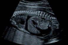
Hydrops fetalis is not a distinct disease, but a severe syndrome in which abnormal fluid accumulates in at least two fetal anatomical compartments. Typically, this includes ascites, pleural effusion, pericardial effusion, and generalized cutaneous edema. Placentomegaly and polyhydramnios are common, but are not included in strict diagnostic criteria. [1]
Modern clinical practice divides fetal hydrops into immune and non-immune. The immune form is associated with hemolytic disease of the fetus, most often due to erythrocyte alloimmunization. The non-immune form is now significantly more common, accounting for approximately 85-95% of all cases, primarily due to the widespread use of anti-D immunoglobulin and the decline in the incidence of classical Rh isoimmunization in countries with available prophylaxis. [2]
From a clinical perspective, hydrops fetalis is always a concerning situation, as it reflects decompensation of the intrauterine condition. It is the final phenotype of a wide variety of disorders: severe anemia, heart failure, lymphatic dysfunction, chromosomal abnormalities, infection, metabolic disease, congenital malformations, and complications of multiple pregnancies. Therefore, after hydrops is identified, the primary goal is not only to confirm the syndrome itself but also to quickly identify its cause, especially if it is potentially treatable in utero. [3]
Crucially, the prognosis for fetal hydrops is extremely variable. In some cases, intrauterine blood transfusion or arrhythmia treatment can save the pregnancy and the fetus. However, in other situations, hydrops reflects a severe genetic or structural pathology with an extremely unfavorable outcome. Therefore, modern management requires a tertiary center, a multidisciplinary team, and simultaneous assessment of fetal and maternal risks. [4]
Below is a brief table of the syndrome. The data is based on current guidelines and reviews. [5]
| Parameter | Brief description |
|---|---|
| What is this? | Pathological accumulation of fluid in the fetus in at least 2 compartments |
| Main manifestations | Ascites, pleural effusion, pericardial effusion, cutaneous edema |
| Main forms | Immune and non-immune |
| Which form is more common today? | Non-immune |
| The main clinical task | Quickly determine the cause and understand whether treatment can be carried out in utero |
| Why is this dangerous? | This is a sign of fetal decompensation and a high risk of intrauterine death. |
Code according to ICD 10 and ICD 11
In the International Classification of Diseases, 10th revision, the coding of hydrops fetalis depends on its nature. For immune hydrops, codes from the section on hemolytic disease of the fetus and newborn are used: P56.0 for hydrops fetalis due to isoimmunization and P56.9 for hydrops fetalis due to another and unspecified hemolytic disease. For the non-immune form, code P83.2 is used - hydrops fetalis not associated with hemolytic disease. In obstetric documentation, code O36.2 is additionally used - pregnancy management for hydrops fetalis. [6]
In the International Classification of Diseases, 11th revision, the coding became more precise. For immune hydrops, the KA85 block is used, where KA85.0 denotes hydrops fetalis due to isoimmunization, KA85.Y denotes other specified forms of hydrops fetalis due to hemolytic disease, and KA85.Z denotes unspecified hemolytic hydrops. For the nonimmune form, the code KC41.1 is used – hydrops fetalis not associated with hemolytic disease. [7]
Below is a practical coding table. [8]
| Classification | Code | Meaning |
|---|---|---|
| ICD 10 | P56.0 | Hydrops fetalis due to isoimmunization |
| ICD 10 | P56.9 | Hydrops fetalis due to other or unspecified hemolytic disease |
| ICD 10 | P83.2 | Hydrops fetalis not associated with hemolytic disease |
| ICD 10 | O36.2 | Pregnancy management with hydrops fetalis |
| ICD 11 | KA85.0 | Hydrops fetalis due to isoimmunization |
| ICD 11 | KA85.Y | Other specified hydrops fetalis due to hemolytic disease |
| ICD 11 | KA85.Z | Hydrops fetalis due to hemolytic disease, unspecified |
| ICD 11 | KC41.1 | Hydrops fetalis not associated with hemolytic disease |
Epidemiology
Hydrops fetalis remains a rare but very severe syndrome. According to modern reviews, the overall incidence of hydrops fetalis ranges from approximately 1 case per 1,500 to 3,800 births, but the exact estimate depends on whether only live births or also intrauterine deaths and terminations are taken into account. Larger population-based registries from the United States and Sweden provide a lower estimate of approximately 1.6-2.5 cases per 10,000 live births. [9]
Non-immune hydrops is currently the dominant form. According to reviews and current educational resources, it accounts for over 90% of cases in countries where Rh alloimmunization prophylaxis is available. Non-immune hydrops occurs in approximately 1 in 1,700-3,000 pregnancies, but only about 1 in 4,000 live births, as intrauterine death and termination of pregnancy remain common in severe forms. [10]
The etiology of non-immune hydrops varies across regions. One modern review identifies the most common causes as cardiovascular disorders, lymphatic dysplasia, hematological causes, and chromosomal abnormalities. However, the profile may shift regionally; for example, in regions with a high incidence of alpha thalassemia, hematological causes play a greater role. This is a very important practical point: the etiology of hydrops depends not only on the "textbook" but also on the population in which the pregnancy is being managed. [11]
An accurate prenatal diagnosis is not always possible. According to clinical guidelines from tertiary centers, prenatal etiological diagnosis is achievable in approximately 50-60% of cases, and with the addition of postnatal testing, placental examination, and autopsy, diagnostic accuracy increases to 75-85%. This explains why some cases, even with modern testing, remain idiopathic. [12]
Below is a summary table of epidemiological landmarks.[13]
| Indicator | Approximate data |
|---|---|
| Overall incidence of fetal hydrops | Approximately 1 in 1,500-3,800 births |
| Frequency in large live birth registries | About 1.6-2.5 per 10,000 live births |
| The proportion of non-immune hydrops among all cases | Approximately 85-95% and more |
| Non-immune hydrops during pregnancy | About 1 in 1,700-3,000 pregnancies |
| Non-immune hydrops in live births | About 1 in 4,000 live births |
| Prenatal etiological diagnostics | Approximately 50-60% of cases |
| Prenatal and postnatal etiological diagnostics together | Approximately 75-85% of cases |
Reasons
The causes of fetal hydrops are conveniently divided into immune and non-immune. Immune hydrops develops against the background of severe hemolytic disease of the fetus, when maternal antibodies destroy fetal red blood cells. In countries with good access to anti-D prophylaxis, this form has become significantly less common, but has not completely disappeared. It still occurs in cases of Rh isoimmunization and other red blood cell antigen conflicts. [14]
Non-immune hydrops is considerably more diverse. Current reviews and the 2026 Society of Maternal-Fetal Medicine guidelines emphasize that it can result from genetic diseases, congenital malformations, intrauterine infections, fetal arrhythmias, placental tumors, complications of monochorionic twins, and a host of other disorders. The most common major etiologic factors are cardiovascular causes, lymphatic dysplasia, hematological disorders, and chromosomal abnormalities. [15]
Cardiovascular causes include structural heart defects, cardiomyopathies, severe tachyarrhythmias and bradyarrhythmias, cardiac tumors, arteriovenous malformations, and severe cardiac dysfunction. Hematological causes include severe fetal anemia due to fetomaternal hemorrhage, parvovirus infection, hemoglobinopathies, and glucose-6-phosphate dehydrogenase deficiency. Infectious causes include primarily parvovirus B19, cytomegalovirus, syphilis, toxoplasmosis, and other congenital infections. [16]
Genetic and metabolic diseases constitute a separate, large block. Current recommendations increasingly include microarray analysis in the algorithm, and in the case of a negative result and the absence of an obvious cause, exome or genomic sequencing, as a significant portion of non-immune hydrops is associated with monogenic diseases, RAS pathway syndromes, metabolic defects, and lymphatic disorders. This is one of the most notable diagnostic evolutions of recent years. [17]
The causes of dropsy can be conveniently summarized in a table. [18]
| Group of reasons | Examples |
|---|---|
| Immune | Rhesus isoimmunization and other forms of hemolytic disease of the fetus |
| Cardiovascular | Heart defects, cardiomyopathy, arrhythmia, cardiac tumors |
| Hematological | Severe anemia, fetomaternal hemorrhage, hemoglobinopathies |
| Infectious | Parvovirus B19, cytomegalovirus, syphilis, toxoplasmosis |
| Genetic | Aneuploidies, microdeletions, monogenic syndromes |
| Lymphatic | Lymphatic dysplasia, cystic hygroma |
| Placental and multiple births | Complications of monochorionic twins, placental tumors |
| Metabolic | Lysosomal storage diseases and other inborn errors of metabolism |
Risk factors
Risk factors are determined primarily by the underlying cause, not the syndrome itself. For immune hydrops, the main risk factor is maternal alloimmunization to fetal red blood cell antigens. The risk is particularly high in patients without adequate anti-D immunoglobulin prophylaxis or with an existing antibody response from previous pregnancies, transfusions, or invasive procedures. [19]
For non-immune hydrops, risk factors are more varied. Important factors include a family history of genetic diseases, known chromosomal abnormalities, early detection of cystic hygroma or severe nuchal pathology, structural cardiac abnormalities, multiple monochorionic pregnancies, maternal infections, autoimmune antibodies in fetal bradyarrhythmia, and ethnic population risk factors for hemoglobinopathies. [20]
Infectious risks should be emphasized separately. During periods of increased parvovirus B19 activity, pregnant women with symptoms of viral infection, confirmed exposure, or suspected fetal anemia require special vigilance. The Society of Maternal-Fetal Medicine (SMF) in 2024 emphasized that serologic testing should be considered for symptoms, suspected fetal anemia, or existing nonimmune hydrops. [21]
The timing factor is also clinically important. The earlier in pregnancy hydrops develops, the higher the likelihood of a severe chromosomal, genetic, or lymphatic cause, and the worse the prognosis, on average. With late onset, anemia, infections, arrhythmias, and some potentially treatable causes are more often detected. This is not an absolute rule, but it is very useful in prenatal counseling. [22]
Pathogenesis
From a pathophysiological perspective, fetal hydrops is the result of an imbalance between fluid entry into the interstitium and its removal by the lymphatic system. Modern reviews describe four main mechanisms: increased hydrostatic pressure in capillaries, decreased plasma oncotic pressure, impaired lymphatic outflow, and damage to the capillary wall. In reality, several mechanisms are often present in a single fetus. [23]
In severe anemia, high-output cardiac failure becomes the leading mechanism. The fetal heart attempts to compensate for hypoxia by increasing its output, but this gradually leads to decompensation, venous congestion, increased hydrostatic pressure, and fluid leakage into tissues and serous cavities. This is why parvovirus anemia and severe hemolytic disease so often lead to hydrops. [24]
In structural cardiac pathology and arrhythmias, the mechanism is similar, but the primary cause is no longer anemia, but pump failure or electrical destabilization of the heart. In lymphatic dysplasia, fluid is unable to be removed from the tissues. In hypoproteinemia and a number of metabolic diseases, oncotic fluid retention in the vascular bed decreases. Intrauterine infections additionally affect vascular permeability, the myocardium, and the liver of the fetus. [25]
It's important to understand that hydrops is a stage of decompensation, not an early marker. That is, once it has already appeared, the fetus's compensatory capabilities are significantly reduced. This is why modern guidelines consider hydrops a condition requiring urgent, and sometimes emergency, follow-up evaluation at a tertiary care center, especially if intrauterine therapy is potentially possible. [26]
Below is a pathogenetic diagram in tabular form. [27]
| Pathogenetic mechanism | What's happening | Typical reasons |
|---|---|---|
| Increase in hydrostatic pressure | Fluid leaks from the vessels into the tissues | Heart failure, arrhythmia, anemia |
| Decrease in oncotic pressure | Water is retained less well in the vascular bed | Hypoproteinemia, liver dysfunction |
| Violation of lymphatic drainage | Fluid is not removed from the interstitium | Lymphatic dysplasia, cystic hygroma |
| Increased capillary permeability | The vascular wall allows fluid to pass through more easily | Infections, inflammation, ischemia |
| Combined mechanism | Several paths operate simultaneously | Chromosomal syndromes, severe systemic diseases |
Symptoms
In the pregnant woman, hydrops fetalis often causes no specific symptoms in the early stages and is first detected during an ultrasound examination. Therefore, speaking of "maternal symptoms" in the literal sense is not entirely accurate. However, the patient may notice a rapid increase in abdominal size, a feeling of tension, shortness of breath, discomfort, and a decrease or change in fetal movements if hydrops is associated with polyhydramnios or severe fetal decompensation. [28]
The main "symptoms" of hydrops are ultrasound findings in the fetus. These include ascites, pleural effusion, pericardial effusion, and generalized cutaneous edema. A thickened placenta and polyhydramnios are often detected simultaneously. However, pleural effusion or ascites alone do not necessarily indicate hydrops unless there is a second abnormal fluid compartment, although modern experts recommend initiating diagnostic testing as soon as even one fetal effusion appears. [29]
Indirect signs depend on the cause. In severe anemia, an increase in peak systolic blood flow velocity in the middle cerebral artery may be seen. In a cardiac cause, cardiomegaly, impaired contractility, signs of heart failure, or arrhythmia may be seen. In case of infection, additional markers of congenital damage may be present, such as intracranial or hepatic calcifications, ventriculomegaly, or hepatosplenomegaly. [30]
Another important condition for the mother is mirror syndrome, or Ballantyne's syndrome. This is a rare but potentially dangerous complication in which the pregnant woman develops edema, hypertension, oliguria, hypoproteinemia, hemodilution anemia, thrombocytopenia, and sometimes acute pulmonary edema. It is called "mirror syndrome" because the edema occurs in both the fetus and the mother. [31]
Classification, forms and stages
The primary classification of hydrops fetalis remains etiologic. Immune hydrops fetalis is associated with hemolytic disease of the fetus. Non-immune hydrops fetalis includes all other causes. This distinction is crucial because the immune and non-immune forms have different pathogenesis, different diagnostic procedures, and different prognoses. The first step after identifying hydrops fetalis should always be to exclude or confirm alloimmunization. [32]
Within the non-immune form, it's practical to divide cases by the dominant mechanism: anemic, cardiac, genetic, lymphatic, infectious, metabolic, placental, and multiple pregnancy-related. This classification is more convenient for the clinician than trying to remember dozens of rare diagnoses, because it immediately suggests which tests are needed first. [33]
In terms of clinical dynamics, one can speak of an early compensated phase and a late decompensated phase, although this is not formally an international staging scale. In the early phase, there may be limited effusions and a potentially reversible condition. In the late phase, there is massive hydrops, placentomegaly, polyhydramnios, severe fetal heart failure, and a high risk of intrauterine death. The later the intervention, the less reserves the fetus has. [34]
Ultrasound classification is also important for practice: minimal hydrops with two fluid compartments, severe generalized hydrops, and hydrops with additional unfavorable features, such as significant placentomegaly, severe cardiac dysfunction, severe anemia, multiple malformations, or early gestational onset. These categories do not formally replace the etiologic diagnosis but assist in family counseling and management decisions. [35]
The classification can be conveniently summarized as follows. [36]
| Approach to classification | Options |
|---|---|
| By origin | Immune and non-immune |
| By leading mechanism | Anemic, cardiac, lymphatic, infectious, genetic, metabolic, placental |
| By time of detection | Early debut and late debut |
| By severity | Limited, severe, generalized decompensated |
| By treatment potential | Potentially reversible and extremely unfavorable with little curability |
Complications and consequences
For the fetus, the main complications include intrauterine hypoxia, severe heart failure, progression of anemia, hemodynamic compromise, intrauterine death, and the need for emergency delivery. For certain causes, such as severe anemia and certain arrhythmias, intrauterine intervention can significantly improve the prognosis. However, if the underlying cause is unavoidable, hydrops becomes a marker of a very high risk of fetal death or severe postnatal instability. [37]
For the neonate, complications include respiratory failure due to pleural effusions and generalized edema, the need for immediate drainage procedures, severe anemia, heart failure, hypoproteinemia, the need for resuscitation, and prolonged intensive care. Postnatal treatment largely depends on the cause and whether the fetus's condition was stabilized before delivery.[38]
For the mother, the most serious complication is mirror syndrome. According to clinical guidelines, it can be accompanied by edema, hypertension, proteinuria, oliguria, and deterioration of liver and kidney function, with acute pulmonary edema reported in approximately 20% of cases. This is one of the key reasons not to indefinitely delay a decision on treatment if hydrops progresses and the underlying cause is untreatable. [39]
Long-term consequences depend on the cause and the success of treatment. Tertiary care guidelines cite a risk of neurodevelopmental delay of approximately 10% among survivors, but this rate varies greatly depending on the underlying etiology, gestational age, the severity of intrauterine hypoxia, and the need for intensive care. Therefore, families should not be promised a uniform outcome "based on a diagnosis of hydrops" without clarifying the cause. [40]
When to see a doctor
If hydrops fetalis is already diagnosed, further management should not be carried out through routine observation, but rather in a center with access to advanced prenatal diagnostics, fetal echocardiography, invasive procedures, and consultations with a geneticist, neonatologist, and maternal-fetal medicine specialist. Current guidelines emphasize that such a patient requires a multidisciplinary approach and a rapid diagnostic pathway. [41]
Reasons for urgent re-admission include decreased fetal movements, rapid abdominal enlargement, increasing shortness of breath, edema, headache, increased blood pressure, decreased urine output, and any signs of possible mirror syndrome. Although hydrops itself is diagnosed in the fetus, the danger extends to the mother as well. [42]
Special attention should be given to any confirmed cases of exposure to parvovirus B19, the appearance of viral infection symptoms in a pregnant woman, the detection of fetal tachyarrhythmia or bradyarrhythmia, signs of severe fetal anemia, or rapidly increasing polyhydramnios. In some of these situations, time directly impacts the possibility of effective intrauterine intervention. [43]
Diagnostics
Diagnosis begins with ultrasound confirmation of the syndrome itself. Traditionally, diagnosis requires the presence of at least two abnormal fluid compartments: ascites, pleural effusion, pericardial effusion, or generalized cutaneous edema. After this, the first mandatory step is to differentiate between the immune and non-immune forms using maternal blood group, Rh factor, indirect Coombs test, and a search for irregular antibodies. [44]
If an immune cause is excluded, the current algorithm requires an expanded etiologic search. The Society of Maternal-Fetal Medicine (SMF) recommends fetal diagnostic testing for all pregnancies with one or more fetal effusions in 2026, including chromosomal microarray analysis with or without karyotyping. If uncertainty persists after microarray analysis and there is no obvious cause, exome or genomic sequencing should be offered, and if there is a high probability of a monogenic disease, it is reasonable to consider such testing even in parallel with microarray analysis. [45]
An ultrasound examination should be as detailed as possible. It includes a search for structural defects, especially cardiac and thoracic ones, an assessment of the placenta, the amount of amniotic fluid, the biophysical profile, and Doppler ultrasound. One of the key indicators is the peak systolic blood flow velocity in the middle cerebral artery. A value greater than 1.5 times the median for the gestational age has a high prognostic value for severe fetal anemia. [46]
Fetal echocardiography is essential because it helps identify both structural anomalies and functional disorders: arrhythmias, cardiomegaly, tumors, myocardial dysfunction, and venous drainage patterns. If anemia is suspected, especially if the gestational age allows, cordocentesis is performed, with blood prepared in advance for immediate intrauterine transfusion. In advanced hydrops, even normal blood flow velocity in the middle cerebral artery does not always rule out anemia, so the clinical context is more important than a single number. [47]
Infectious testing is performed as indicated, but if there is an appropriate differential diagnosis, it should include molecular tests. Guidelines list parvovirus B19, cytomegalovirus, toxoplasma, syphilis, and other agents. Genetic and infectious testing can be performed simultaneously during amniocentesis, and chorionic villus sampling can be considered early on. In some cases, fetomaternal hemorrhage, hemoglobinopathies, glucose-6-phosphate dehydrogenase deficiency, diabetes, antibodies to Ro and La antigens, and biochemical markers of congenital metabolic diseases are also assessed. [48]
Postnatal and sometimes postmortem diagnosis are also very important. If a prenatal diagnosis is not established, the liveborn infant should undergo further genetic and clinical testing. In the case of intrauterine death or termination of pregnancy, autopsy and placental examination significantly increase the chances of establishing the cause and accurately assessing the risk of recurrence in future pregnancies. [49]
The step-by-step diagnostic algorithm can be conveniently presented as follows. [50]
| Step | What are they doing? | For what |
|---|---|---|
| 1 | Confirm at least 2 fluid compartments on ultrasound | Confirm the fact of dropsy |
| 2 | Alloimmunization is excluded | Separate the immune and non-immune forms |
| 3 | A detailed anatomical ultrasound examination is performed | Find defects and indirect signs of the cause |
| 4 | Assesses the blood flow velocity in the middle cerebral artery | Detect severe fetal anemia |
| 5 | Fetal echocardiography is performed | Exclude structural and functional cardiac causes |
| 6 | Perform genetic testing | Find chromosomal or monogenic etiology |
| 7 | Conduct an infectious search according to indications | Find a treatable infectious cause |
| 8 | Perform invasive procedures as indicated | Confirm the diagnosis and carry out treatment |
| 9 | Postnatal or postmortem examination will be arranged if the cause is unclear | Clarify the etiology and risk of relapse |
Differential diagnosis
First and foremost, it's important to distinguish between true fetal hydrops and conditions that can mimic it. Isolated pleural effusion, isolated ascites, large cysts, localized edema, massive cystic hygroma without generalized fluid changes, and pronounced placentomegaly do not necessarily indicate hydrops according to classical criteria. However, current guidelines emphasize that even a single fetal effusion requires a thorough etiologic investigation. [51]
The next level involves distinguishing between immune and non-immune hydrops. This requires blood type, Rh factor, an indirect Coombs test, and a search for irregular antibodies. Without this, it is impossible to correctly interpret subsequent findings and choose the right path—for example, from alloimmunization to intrauterine transfusions or from the non-immune form to genetic and infectious testing. [52]
The differential diagnosis then revolves around four main axes: anemia, heart, genetics, and infection. Increased blood flow velocity in the middle cerebral artery, reticulocytopenia, and parvovirus markers support an anemic cause. Cardiomegaly, myocardial dysfunction, and arrhythmia support a cardiac cause. Early detection, multiple anomalies, cystic hygroma, and a negative infectious workup support a chromosomal or monogenic cause. Calcifications, hepatosplenomegaly, and serologic or molecular findings support a congenital infection. [53]
Maternal conditions that may be associated with hydrops fetalis should be considered separately. Ro and La antibodies in fetal bradyarrhythmia, poorly controlled diabetes, fetomaternal hemorrhage, and certain infectious syndromes can guide the diagnosis. Therefore, the differential diagnosis of hydrops fetalis involves not only a fetal examination but also a comprehensive assessment of the mother, placenta, and obstetric context. [54]
Treatment
Treatment of hydrops fetalis is always determined by the underlying cause. There is no universal "therapy for hydrops." The main principle of modern fetal medicine is to treat not the ultrasound phenotype itself, but the underlying pathogenic mechanism that led to it. Therefore, the first task is to quickly determine whether the case is potentially treatable in utero. [55]
If severe fetal anemia is the cause, intrauterine blood transfusion becomes the key treatment. This applies to both immune alloimmune anemia and a number of non-immune causes, including parvovirus infection, fetomaternal hemorrhage, some hemolytic conditions, and anemia of unknown origin with compelling evidence of its severity. Current guidelines emphasize that if anemia is suspected, cordocentesis and readiness for immediate transfusion should be considered early, rather than after the entire diagnostic workup has been completed. [56]
When fluid accumulates in the chest or abdomen, the treatment strategy depends on the cause and volume of the effusion. In selected cases, aspiration of the pleural effusion or ascites is performed, and in cases of recurrent and hemodynamically significant effusions, shunt placement is performed. This approach is especially important for large pleural effusions, lymphatic disorders, and certain cystic or pulmonary lesions. The goal of treatment is to reduce compression of the lungs and heart and give the fetus a chance to maintain hemodynamic stability until delivery or until further intrauterine intervention. [57]
If hydrops is caused by fetal arrhythmia, treatment may be intravascular or maternal, depending on the type of rhythm disturbance. For tachyarrhythmias, maternal antiarrhythmic therapy is considered, while for severe bradyarrhythmia with maternal antibodies to Ro and La antigens, the strategy is determined by the type of block, timing, severity of heart failure, and the center's experience. Here, treatment success directly depends on the accuracy of fetal echocardiography and the speed of therapy initiation. [58]
For infectious causes, treatment should also be targeted. Syphilis in both mother and fetus requires specific antibacterial therapy. If fetal parvovirus anemia is suspected, intrauterine transfusion, rather than antiviral therapy, remains the primary treatment. For cytomegalovirus, toxoplasmosis, and other infections, decisions are more complex and depend on the timing, confirmation of the diagnosis, and the center's local protocol. The most important general principle is this: laboratory confirmation of infection should lead to a reconsideration of the treatment plan and not simply remain a nice line in the report. [59]
Genetic and metabolic causes often lack a simple intrauterine solution, but new opportunities are emerging. Recent reviews indicate growing interest in targeted strategies for certain RAS pathway syndromes and some lymphatic disorders, including the use of mitogen-activated protein kinase inhibitors in selected pediatric cases after birth. While this is not yet a universal or routine approach for the fetus, it is no longer purely theoretical. For some lysosomal storage diseases, enzyme replacement therapy is possible after birth, which changes prenatal counseling even when intrauterine intervention is limited. [60]
In severe hydrops, it's crucial to remember maternal safety. If mirror syndrome develops, the approach is twofold: symptomatic care for the mother with diuretics, antihypertensive therapy, and strict monitoring, as well as a decision on whether to continue the pregnancy. If the cause of the hydrops is eliminated and the hydrops regresses, the mother's condition may also improve. If this is not possible, the definitive treatment for mirror syndrome is termination of the pregnancy. Therefore, expectant management is only permissible after a very clear discussion of the maternal risks. [61]
The timing of delivery is decided on an individual basis. The Society of Maternal-Fetal Medicine (SMF) recommends against using preterm delivery as an automatic response to hydrops. Preterm delivery should be reserved for specific obstetric indications: preeclampsia, mirror syndrome, premature rupture of membranes, preterm labor, worsening hydrops, or situations where the risk of continuing the pregnancy already exceeds the risk of preterm delivery. This is crucial, as preterm delivery itself dramatically increases neonatal morbidity. [62]
The route of delivery also depends not only on the diagnosis but on the entire context. Current guidelines recommend that cesarean section be performed for standard obstetric indications if active resuscitation and intensive care are planned for the newborn. In cases of massive pleural effusions, intrauterine thoracentesis immediately before birth can help re-inflate the lungs. In cases of extremely unfavorable prognosis and risk of dystocia, palliative drainage procedures are permitted to facilitate vaginal delivery. [63]
After birth, treatment continues in the neonatal intensive care unit. Depending on the cause and condition, the baby may require drainage of effusions, blood transfusions, respiratory support, inotropic therapy, treatment for heart failure, genetic testing, infection screening, and long-term observation. This further emphasizes that hydrops fetalis is not a "single ultrasound diagnosis," but a complex syndrome that begins in utero and often continues into the neonatal period. [64]
The therapeutic possibilities can be conveniently summarized in a table. [65]
| Cause or mechanism | Basic treatment approach |
|---|---|
| Severe fetal anemia | Cordocentesis and intrauterine blood transfusion |
| Large pleural effusion or ascites | Fluid aspiration, sometimes a shunt |
| Fetal tachyarrhythmia | Antiarrhythmic therapy through the mother or other specialized tactics |
| Bradyarrhythmia in maternal antibodies | Individualized specialized treatment |
| Infectious cause | Etiotropic therapy, if it exists, and correction of consequences |
| Syphilis | Specific antibacterial therapy |
| Mirror syndrome | Intensive monitoring of the mother, symptomatic care, decision on termination of pregnancy |
| Extremely severe progression without a curable cause | Individual counseling, discussion of all acceptable treatment options |
| After birth | Neonatal resuscitation, drainage of effusions, transfusions, treatment of the underlying cause |
Prevention
Prevention of immune hydrops fetalis is based primarily on preventing maternal alloimmunization. The widespread use of anti-D immunoglobulin has led to a dramatic reduction in the incidence of classical Rh-associated hydrops in countries with accessible pregnancy monitoring systems. Therefore, adherence to standards for the prevention of Rh isoimmunization remains one of the most successful strategies for preventing severe fetal pathology in obstetrics. [66]
Prevention of non-immune hydrops is much more complex because it depends on the cause. This includes prenatal screening for infections when indicated, prompt identification and treatment of syphilis, targeted testing for parvovirus B19 in cases of symptoms or exposure, genetic counseling for families with a history of the disease, appropriate management of multiple pregnancies, and early evaluation of suspicious ultrasound findings such as cystic hygroma, cardiomegaly, or significant effusion. [67]
Early and high-quality ultrasound monitoring plays a significant preventative role. The earlier a potentially treatable cause is identified, the greater the chance of intervening before the development of fully decompensated hydrops. This is why modern reviews increasingly emphasize the importance of consistent prenatal phenotyping, Doppler ultrasound, fetal echocardiography, and genetic diagnostics, not only as a therapeutic but also as a preventative measure. [68]
Forecast
The prognosis for hydrops fetalis remains generally grave. According to clinical guidelines from tertiary centers, the overall mortality rate for non-immune hydrops can reach approximately 50-70%, although it varies greatly depending on the cause, time of detection, and the availability of intrauterine treatment. Particularly unfavorable are early onset, chromosomal abnormalities, and major structural defects. [69]
The best prognosis is observed for potentially reversible causes: severe anemia, some arrhythmias, some isolated effusions, and certain infectious conditions, if they are diagnosed early and managed in a specialized center. With available intrauterine therapy, the question is no longer "is there hydrops or not?" but rather "will we be able to intervene before irreversible decompensation occurs?" [70]
The maternal prognosis is assessed separately. In the absence of mirror syndrome and severe obstetric complications, the maternal prognosis is usually favorable. However, if mirror syndrome develops, the risks increase significantly and may include pulmonary edema, intensive care, and the need for early delivery. This is one reason why the management of pregnancies with fetal hydrops cannot be considered solely a fetal problem. [71]
Below is a brief prognostic table. [72]
| Prognostic factor | What does it mean? |
|---|---|
| Early detection of a treatable cause | Improves chance of survival |
| Possibility of intrauterine therapy | Improves fetal prognosis |
| Chromosomal or severe structural pathology | Usually worsens the prognosis |
| Severe fetal cardiac dysfunction | Increases the risk of death |
| Early gestational onset | More often associated with a more severe cause |
| Mirror syndrome in mother | Worsens the obstetric prognosis and changes tactics |
FAQ
Are hydrops fetalis and polyhydramnios the same thing?
No. Polyhydramnios often accompanies hydrops fetalis, but is not included in its mandatory diagnostic criteria. Diagnosis typically requires pathological fluid accumulation in at least two fetal compartments. [73]
Can hydrops fetalis be cured?
Sometimes, yes, if the underlying cause can be eliminated. This is especially true for severe anemia, some arrhythmias, and some isolated effusions. However, with severe genetic and structural causes, treatment options are significantly limited. [74]
Does hydrocele always indicate a severe chromosomal abnormality?
No. Chromosomal abnormalities are an important, but not the only, cause. Hydrocele can be associated with anemia, infection, cardiac pathology, lymphatic dysplasia, complications of multiple pregnancy, and other conditions. Therefore, without extensive diagnostics, it is impossible to draw a definitive conclusion about the cause from a single ultrasound. [75]
Should all pregnant women with hydrops fetalis undergo amniocentesis?
Current guidelines increasingly support invasive diagnostic testing if it can change management and prognosis. The Society of Maternal-Fetal Medicine recommends genetic testing for all pregnancies with one or more fetal effusions, and tertiary care centers often include amniocentesis in their basic algorithm for non-immune hydrops fetalis. [76]
Is hydrops fetalis dangerous for the pregnant woman?
Yes, in some cases. The most significant maternal complication is mirror syndrome, which can resemble severe preeclampsia and be accompanied by pulmonary edema, hypertension, and organ dysfunction. [77]
Is it possible to simply wait for labor and not intervene?
This depends on the cause, the gestational age, and the condition of the mother and fetus. Current recommendations do not automatically support either immediate delivery or indefinite waiting. The approach should be strictly individualized and take into account the possibility of intrauterine treatment, the risks of prematurity, and maternal complications. [78]
Key points from experts
Teresa N. Sparks, MD, a maternal-fetal medicine specialist, is a co-author of the 2026 Society of Maternal-Fetal Medicine consultation document on non-immune hydrops fetalis. The key practical implication of her work is that non-immune hydrops fetalis requires a modern, stepwise search for a cause, with mandatory genetic testing, and in cases of negative microarray analysis and the absence of an obvious cause, exome or genomic sequencing should be actively considered, because the precise etiology directly affects management and prognosis. [79]
Mary E. Norton, MD, one of the most renowned international experts in prenatal diagnosis and co-author of previous and current guidelines on non-immune hydrops fetalis. Her position emphasizes another important principle: hydrops fetalis is not a single diagnosis, but the end point of multiple pathogenetic pathways. Therefore, the crucial factor is not the diagnosis of hydrops, but the earliest possible identification of treatable causes, especially anemia, arrhythmias, and certain infectious forms. [80]
Asma Khalil, MD, professor of fetal medicine, is a researcher of early and late non-immune hydrops fetalis. Her work emphasizes that the timing of hydrops detection provides independent clinical information: early forms are more often associated with severe genetic and lymphatic causes, while later forms are associated with anemia, infection, and some potentially treatable conditions. This is of great importance for family counseling, starting as early as the first ultrasound visit. [81]