Medical expert of the article

New publications
Giant biped
Last reviewed: 06.07.2025

All iLive content is medically reviewed or fact checked to ensure as much factual accuracy as possible.
We have strict sourcing guidelines and only link to reputable media sites, academic research institutions and, whenever possible, medically peer reviewed studies. Note that the numbers in parentheses ([1], [2], etc.) are clickable links to these studies.
If you feel that any of our content is inaccurate, out-of-date, or otherwise questionable, please select it and press Ctrl + Enter.
The giant fluke is a worm that parasitizes the human body mainly in the liver, causing acute and chronic liver dysfunction, as well as other organs. This parasite is widespread in Africa and Asia, but cases of infection are also possible in Russia and Ukraine. It is necessary to know some features of its cycle in order to predict not only the course of symptoms, but also possible methods of prevention at different stages.
Structure giant bilharzia
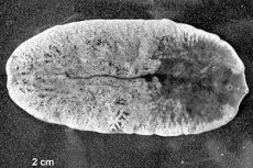
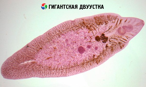
The giant fluke or Fasciola gigantica is a parasite that belongs to the flukes or flatworms of the genus Trematoda. The features of their life cycle and structure allow them to be combined into one genus with other parasites - schistosomes, opisthorchis.
The structural features of the giant fluke are such that these parasites can be the largest in their class. The length of an adult giant fluke can reach about seven centimeters. Their body is elongated, in the shape of a tulip leaf - narrowing at the ends. The color of the worm can be from pale pink to gray, depending on the conditions. These parasites got their name "flukes" because they have suckers on the front part of the abdominal end. Between these suckers is the oral end, through which food enters. The digestive system of the fluke is a closed type, that is, there is a digestive tube where the main processes of digestion of food occur. Then this food moves along the intestines along the entire length of the body, and after digestion is thrown back out through the mouth. Such features allow them to parasitize for a long time in closed spaces without access to oxygen. This localization is also explained by the incompletely developed hematopoietic and respiratory systems, which allows them to remain without oxygen for a long time and migrate through human vessels, feeding on red blood cells and other blood cells.
The giant fasciola reacts to movement and changes in body shape thanks to a branched nervous system. It begins near the oral sucker in the form of a ring of nerve tissue, from which a nerve ganglion extends along the entire length of the body. Thus, all organs are innervated from this ganglion, and the reaction of analyzers is also ensured.
The reproduction of the parasite is complex, since the giant fluke is a hermaphrodite. There are female and male individuals. For reproduction, favorable conditions must be in place, and some time passes for the fertilization of the eggs. Then, the peculiarities of the change of hosts allow the fasciola to undergo successive stages of development.
Life cycle giant bilharzia
The life cycle of the giant fluke begins with the main host, which is cattle and small cattle - goats, sheep, cows, bulls, buffalo. These worms are localized in the intestines of cattle, and then persisting for some time they mature and become sexually mature individuals. In this state, they are able to migrate through the intestinal wall and enter the portal vein system. So the parasite gets to the liver, where its final location is. There the parasite reproduces and releases eggs, which can get back into the intestines through the bile duct system and be excreted with feces. So with feces are excreted eggs that are not pathogenic for humans until they are fully mature. Then the eggs get into fresh water bodies, where warm water is needed for further development. In water, the larva grows and develops for two days, then it is necessary for it to get into the body of the mollusk. There, the fluke develops further, reaching the larval stage, which is invasive to humans.
The ways in which people are infected with the giant fluke are limited to indirect ways, when a person accidentally encounters an area where the parasite is found. At the same time, one can become infected through food when eating vegetables, fruits, and greens that have fasciola larvae on their surface. Infection is also possible when accidentally drinking water in which these parasites swim. These features of the life cycle must be taken into account in order to know the main ways of transmission and ways to prevent the disease.
Symptoms
The characteristic localization of the parasite in the body of the final host contributes to the same localization in the human body. Therefore, there are some specific symptoms of fascioliasis that are characteristic of the defeat of this group of flukes.
When entering the human intestine, the eggs of the giant fasciola develop, grow, then at the larval stage they penetrate the submucosal layer and are absorbed into the blood. With the blood flow through the portal vein system, the parasite enters the liver, where it is actualized. There, further growth of the larvae occurs, their activation - in this state, they are able to move along the ducts and enter the gallbladder, in the process of which the normal location of the ducts and their relationship are disrupted. The function of bile outflow is disrupted first of all, and as a secondary process, bile stagnation and disruption of the function of the liver itself occur.
The incubation period of the disease lasts from several days to five to seven weeks. In this case, a person may not even remember the fact of infection, which makes diagnosis very difficult. This period lasts from the moment of entry into the intestine until activation in the liver and disruption of its function.
The acute stage of the disease develops with a single massive lesion of the liver by a significant number of parasites. In this case, the symptoms are very pronounced. Jaundice appears, which makes patients see a doctor. It is accompanied by itching of the skin, since the release of bile acids into the blood is pronounced. In this case, symptoms of pain in the right side or right hypochondrium appear, the severity of the pain syndrome increases with the use of fatty foods. The pain can also be dull, weakly expressed. An allergic rash is often an accompanying symptom. This symptom is often observed due to the ability of helminths to cause increased allergization of the body, which is often manifested by a diffuse rash all over the body with itching of the skin. Also, in the acute course, dyspeptic phenomena can be observed - bitterness in the mouth, nausea, vomiting, abdominal pain and stool disorders such as diarrhea.
But such a detailed clinical picture is less common than a latent course. Often, with an insignificant number of parasites, mild symptoms are observed, there may only be asthenovegetative syndrome, which cannot be explained. In this case, a chronic form is formed, which is characterized by a slow constant release of eggs into the intestinal lumen, and then reinfection. In this case, there may be no symptoms from the liver, only changes in allergic reactivity and the formation of a predisposition to stone formation and chronic cholecystitis in the gallbladder are expressed.
Diagnostics
The diagnosis of this pathology should be complete and timely, because at the initial stage of development it is easier to act on a small number of worms. First, it is necessary to carefully collect anamnesis, finding out possible etiological factors of infection. Considering the incubation period, it is necessary to find out the anamnesis for the last two months. Then it is necessary to examine the patient and detail the complaints. During the examination, positive cystic symptoms, pain in the right hypochondrium can be determined, but the liver should not increase.
Instrumental diagnostic methods are more informative in terms of diagnosing not only fasciola parasitism, but also in terms of assessing the condition of the bile ducts and liver. Ultrasound of the liver and bile ducts reveals dilation of the ducts, formation of echo-positive shadows in the projection of these ducts, impaired bile outflow, and a reactive gallbladder. Based on this, one can suspect the presence of a parasite.
Laboratory blood tests are not specific, but they may also show changes in the form of eosinophilia, which can confirm the etiology of helminthic invasion. In case of severe jaundice, the patient needs a biochemical blood test. An increase in the bilirubin level is determined due to its direct fraction, as well as an increase in alkaline phosphatase, as a sign of cholestasis and intraductal parasitism of the fluke. The most specific and sensitive method for diagnosing giant fluke is blood testing and polymerase chain reaction. In this case, the qualitative and quantitative presence of the worm in the body is determined in the form of its DNA. This allows you to identify antibodies or the antigen itself in the human body and accurately determine the pathogen.
These are the main diagnostic methods for this pathology, which must be used at the initial symptoms of the disease to prevent the chronic course of the pathology.
 [ 10 ]
Treatment
Treatment of any helminthic invasion should be carried out only in combination with other means that prepare the gastrointestinal tract for deworming. Therefore, it is necessary to start with a diet that cleanses the intestines. It is necessary to completely limit sweet, starchy foods for the duration of treatment. It is necessary to eat porridge and cooked vegetables that stimulate intestinal peristalsis. After this, it is advisable to take a course of laxative therapy. To do this, it is necessary to take a single course with the use of laxatives. It is better to take herbal preparations with a laxative effect. Then it is recommended to use a course with treatment with sorbents by taking them for three days. You can use Sorbex, White Coal, Polysorb. After such a course of cleansing therapy, they proceed to treating the helminthic invasion itself. Anthelmintic drugs are used that have a predominant effect on flatworms and their larval forms.
- Hexihol is a drug that is especially active in localizing parasitic worms in the liver. It is available in powder form. The treatment regimen with this drug can be three-day, five-day, or ten-day. The three-day regimen is the most effective, since it allows for the maximum concentration of the drug to be created in the shortest possible time. In this case, the drug is prescribed in a daily dose of 0.2 milligrams per kilogram of the patient's body weight. The drug is taken three times a day. In this case, the first dose should be taken after a light breakfast, dissolving the powder in a glass of warm milk. After three days of treatment, it is necessary to adhere to the diet for at least a week, which will maintain the result and improve the body's response to the drug. When treating with this drug, it is necessary to monitor not only the dynamics of clinical symptoms, but also a biochemical analysis with the level of bilirubin and transaminases.
- Thiabendazole is a broad-spectrum anthelmintic that is active against not only adult worms, but also larvae. This drug is available in the form of 500-milligram tablets, the dosage of the drug is two tablets twice a day with a three-day course of treatment. Thus, the maximum dose of the drug for one course should not exceed 6 grams. Side effects are possible during the administration of the drug with severe helminthic invasion - nausea, abdominal pain, itching of the skin, as well as pronounced intoxication symptoms with enlarged lymph nodes, dizziness and subfebrile temperature. It is not recommended for children under five years of age to use this drug, and it should not be used during pregnancy.
Considering the predominant liver damage and the disruption of intrahepatic bile outflow, it is recommended to use hepatoprotectors and drugs to improve bile outflow. For this purpose, it is recommended to use Ursofalk to improve bile outflow, which normalizes the function of the ducts and relieves the symptoms of jaundice. From the group of hepatoprotectors, you can use Enerliv, Livker, Gepabene, Geptral. Together with improving liver function, it is necessary to normalize bowel function after a cleansing course, which will help eliminate the parasite more quickly. Therefore, probiotics are also used in complex therapy.
Prevention giant bilharzia
Prevention of infection with giant fluke can be non-specific and specific. Non-specific methods of prevention are very simple - you need to follow the rules of hygiene, wash vegetables and fruits before eating, and avoid drinking water from untreated sources. Specific prevention can be carried out with any antiparasitic drug twice a year in the spring and autumn using prophylactic doses of the drug.
The giant fluke is a parasite from the group of flatworms, which is localized in the liver and bile ducts with a violation of the function of bile outflow and the development of clinical symptoms. Human infection does not occur often, since the final host is cattle. Symptoms of the pathology can be hidden or obvious, which requires proper diagnosis. Treatment of the giant fluke should be aimed at eliminating the parasite, restoring liver and intestinal function.
[ 11 ]