Medical expert of the article

New publications
Fibrous hypertrophic gingivitis
Last reviewed: 29.06.2025

All iLive content is medically reviewed or fact checked to ensure as much factual accuracy as possible.
We have strict sourcing guidelines and only link to reputable media sites, academic research institutions and, whenever possible, medically peer reviewed studies. Note that the numbers in parentheses ([1], [2], etc.) are clickable links to these studies.
If you feel that any of our content is inaccurate, out-of-date, or otherwise questionable, please select it and press Ctrl + Enter.
Periodontal diseases and their consequences often become a reason for patients to visit dentists. Among periodontal pathologies, hyperplastic processes in the gingival tissues occupy a significant share. Fibrous hypertrophic gingivitis is a chronic inflammatory disease that is accompanied by reactive growth of fibrous connective tissue elements and basal structures of the gingival epithelium without violating the integrity of the gingival attachment. The causes of such a phenomenon are multiple - both local and general. Pathology can occur as an independent disease, or as a sign of relapse of generalized periodontitis. Treatment is complex, involving specialists of different profiles - in particular, a general dentist, periodontist, orthodontist, physiotherapist. [1]
Epidemiology
According to the World Health Organization, among all diseases of the oral cavity the most common is dental caries, but the second place is confidently occupied by gum disease. It is important to note that the safety and health of teeth largely depends on the condition of the gums, as gum lesions entail the destruction of the periodontium. As a consequence - the appearance of unpleasant odor, unsightly appearance, loosening and loss of teeth.
The most common causes of fibrotic hypertrophic gingivitis in both adults and children are poor oral hygiene, the presence of incorrect implants and fillings, as well as hormonal changes (more typical for adolescents and women). Statistics show that pathology in early childhood can occur in only 1-2% of cases, and older people get sick much more often. The risks of the disease increase significantly when active biological processes in the body begin to occur: hormonal changes, loss and growth of teeth. These cyclical changes create the basis for the development of the malfunction. An additional "contribution" is made by an incorrect bite, the use of special devices to correct the dentition. An important unfavorable factor is stomatitis.
The highest incidence of fibrotic hypertrophic gingivitis occurs at age 13 years.
Among adult patients, the most common illnesses are:
- Pregnant women;
- Diabetics;
- HIV-positive.
Doctors to see for fibrotic hypertrophic gingivitis: dentist, periodontist.
Causes of the fibrotic hypertrophic gingivitis.
General and local factors may be involved in the development of fibrous hypertrophic gingivitis. Among the local causes, the most common are bite disorders, individual dental defects (overcompletion, deformity, crowding, etc.), dental deposits (plaque, calculus), undersized frenulum, improper fillings or prosthetics, poor oral hygiene, etc.
Among the common causes, the picture of the hormonal background is of particular importance. It is known that fibrous hypertrophic gingivitis often occurs in adolescents during puberty, as well as in women during pregnancy or menopause. Other pathological causes can be endocrine pathologies (thyroid disease, diabetes mellitus), long-term treatment with certain medications (hormones, anticonvulsants, immunosuppressors, calcium channel blockers), as well as vitamin deficiencies and leukemia.
- Pathogenic and opportunistic microflora inhabit virtually the entire oral cavity, however, in itself it does not pose a threat: its development and growth are controlled by the immune system, both local and general. Microorganisms are able to provoke the start of the inflammatory process and fibrotic hypertrophic gingivitis only in the presence of favorable conditions for them.
- Improper or insufficient oral care leads to the steady appearance of dental plaque, which becomes an excellent breeding ground for pathogenic flora, which contributes to the development of pathological processes.
- If hygienic rules of oral care are ignored for a long time, plaque thickens and "stiffens". This factor in most cases leads to trauma and gingival prolapse, due to which the inflammatory process takes over deeper tissues develops fibrous hypertrophic gingivitis.
- Gingivitis can be a consequence of improper installation of dentures and fillings, avid smoking, hypovitaminosis, endocrine and digestive pathologies, failures of immune defense. Hereditary predisposition to such diseases is not excluded.
Risk factors
Factors that can provoke the development of fibrotic hypertrophic gingivitis are divided into two categories: endogenous and exogenous. To endogenous factors may include weakening of immunity, hormonal changes, metabolic disorders and so on. Exogenous factors can be divided into such groups:
- Physical (mucosal trauma, burns, etc.);
- Biological (caused by the influence of pathogenic flora);
- Chemical (caused by the influence of aggressive solutions and substances);
- Iatrogenic (related to a previously traumatic medical manipulation).
The most common factor is considered to be a biological one, primarily related to poor oral hygiene. Food particles accumulate in the gum zone, plaque builds up, calculus forms, and favorable conditions are created for the growth and development of bacterial flora.
Risk groups for the incidence of fibrotic hypertrophic gingivitis include the following individuals:
- Patients with bite disorders, with orthodontic devices (corrective plates, braces), with poorly placed fillings and implants;
- Heavy smokers;
- People who do not take proper care of their mouths, or who do so improperly;
- Patients with salivary problems, suffering from increased dryness of mucous membranes;
- Long-term sick people with weakened immune system;
- Adolescents during active puberty;
- Women who are pregnant, menopausal, or taking hormonal contraceptives;
- Patients with somatic diseases (diabetes, hypovitaminosis, digestive, endocrine or nervous pathologies);
- Long-term users of hormonal drugs, immunosuppressors, anticonvulsants, calcium channel blockers);
- Oncology patients;
- Children in the period of active growth and change of teeth, with bite anomalies and "adenoid" (mouth) breathing;
- Patients with blood diseases (leukemia, myeloleukemia, leukemic reticulosis, etc.).
Pathogenesis
Among the main causes of fibrotic hypertrophic gingivitis is the prolonged presence of dental plaque containing predominantly Gram-negative microorganisms. Epithelial tissue in the dentoalveolar junction is a kind of semipermeable membrane in which the exchange between the external and tissue environment takes place. Extensive microflora deposited on the epithelial surface interact with the subepithelial tissues. A special negative role is played by a dense subgingival plaque containing anaerobic pathogenic bacteria (actinobacilli, bacteroidetes, porphyromonas, compilobacteria, peptostreptococci, eubacteria, streptococci, spirochetes, etc.).
The bacterial microflora in the oral cavity, on the one hand, inhibits the development of microorganisms that enter the mouth from the outside. But, on the other hand, it is a potential source of autoinfection. Thus, with an increase in the number of microbes against the background of poor oral hygiene, the fall of immune defense, the bacterial flora from saprophytic transforms into pathogenic, providing the start of most gingivitis and periodontitis.
Poor hygiene, the presence of food debris on the teeth form an excellent breeding ground for microorganisms that begin to multiply and produce substances that contribute to the formation of tartar.
Even a short absence of oral hygiene care (3-4 days) leads to an increase in bacterial growth by 10-20 times, and the thickness of the microbial layer on the gingival surface can reach 0.4 mm. At the same time, the composition of the plaque is transformed and becomes more complex: aerobic gram-positive bacilli and filamentous bacteria are added to the coccal flora. Starting from the fifth day of lack of hygienic care, there is an increase in the number of anaerobes, spirochetes and vibrios. In some areas of gums periodontal reaction changes, migration of neutrophils and macrophages increases, gingival fluid secretion increases. Histologically there is a picture of acute inflammatory process.
The initial lesion may persist for months or even years. The gingival tissue transforms into fibrotic tissue.
According to morphological signs in fibrous hypertrophic gingivitis there is overgrowth of connective elements of gingival papillae, expansion of vessels, swelling of collagen fibers, lymphoplasmocytic infiltration. Transition of edematous form to fibrous form is accompanied by reduction of edema, signs of proliferation of fibroplasts, coarsening of collagen fibers.
Symptoms of the fibrotic hypertrophic gingivitis.
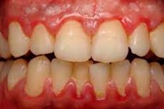
Fibrous hypertrophic gingivitis in most cases develops gradually, for a long time, "quietly", without special symptoms. In some cases there is discomfort, slight soreness (an uncharacteristic symptom), slight bleeding during brushing and eating. A closer look can trace the enlargement of interdental papillae, more saturated or, conversely, pale color of the gum.
During the dental examination, one notices gingival overgrowth, presence of plaque (calculus). The dento-gingival junction remains intact (no pockets).
The first signs of fibrous overgrowth are usually complaints of enlarged gums, their thickening, unaesthetic appearance. Often patients indicate the appearance of difficulties in chewing food. The mucous membranes in the area of the lesion change their color, the surface becomes uneven, bumpy. On examination, soft and hard dental layers are visualized.
The pathological fibrous hypertrophic focus can be located locally (on a limited area of the gingiva) or generalized (over the entire surface).
Stages
Depending on the overgrowth of gingival tissues, such stages of hypertrophic gingivitis are distinguished:
- Mild stage - is represented by hypertrophic processes at the base of the gingival papillae, and the enlarged gingival margin covers the dental crown by one third;
- Middle stage is accompanied by dynamically increasing enlargement and dome-shaped change in the configuration of gingival papillae, and gingival growth leads to closure of the dental crown by 50%;
- Severe stage is characterized by obvious hyperplastic processes in the papillae and gingival margin, and the crown of the tooth is more than half closed.
Forms
According to the spread of the pathological process, localized (local, within 1 to five teeth) and generalized (more than five teeth) fibrous hypertrophic gingivitis are distinguished. In some cases, localized shallow types of the disease are counted as a separate pathology, such as papillitis.
According to the variation of hyperplasia gingivitis is edematous (inflammatory) and granulating (fibrous). Edematous gingivitis is represented by swelling of connective tissue of gingival papillae, dilated vessels, limoplasmocytic infiltration of gingival tissues. Fibrous gingivitis is characterized by proliferative changes in connective tissue structures of gingival papillae, thickening of collagen fibers, signs of parakeratosis. Swelling is weakly expressed, inflammatory infiltrate is minimal.
Complications and consequences
Without the necessary treatment, the hypertrophic form of gingivitis transforms into an atrophic form, which poses a danger in terms of periodontitis and complete loss of teeth.
It is important to prevent the development of fibrotic hypertrophic gingivitis, and if this has happened, all efforts should be directed at eliminating the pathology. Experts note that fibrous overgrowths require a longer, more complex and expensive treatment, involving not only a direct impact on the pathological focus, but also strengthening immunity and health of the body as a whole, stabilizing metabolic processes and hormonal balance.
The development of adverse effects can be avoided if you visit the dentist in a timely manner and follow other important recommendations:
- Brushing your teeth regularly;
- Choose the right toothbrush and change it every 2-3 months;
- Eat right, do not ignore the consumption of solid vegetables and fruits;
- To stop smoking.
It is mandatory to visit the dentist twice a year for preventive purposes - for timely diagnosis of disorders.
Diagnostics of the fibrotic hypertrophic gingivitis.
The main method of diagnosis of fibrous hypertrophic gingivitis is clinical examination. One can notice lumpy, thickened gums, which grow and prevent the patient from eating normally and even talking.
Instrumental diagnosis consists of a gingival sulcus bleeding test (detection of hidden bleeding areas with a periodontal probe), as well as radiography to assess the root cause and severity of the pathology. Fibrous gingivitis is often accompanied by osteoporosis of the tip of the interdental septa, which is determined radiologically.
Other possible procedures include:
- Oral hygiene index;
- Periodontal index;
- Papillary-marginal-alveolar index;
- Schiller-Pisarev test (iodine reaction, staining of gingival glycogen);
- Less often - biopsy, morphologic analysis of tissues.
Laboratory tests are nonspecific, can be prescribed by specialized specialists (endocrinologist, hematologist) in the framework of determining the root causes of the hypertrophic process and background diseases. [2]
Differential diagnosis
Differential diagnosis of fibrous hypertrophic gingivitis is carried out with epulis and gingival fibromatosis.
|
Epulis |
Gingival fibromatosis |
|
A benign growth on the gums, formed from the alveolar process and consisting of epithelial tissue. It has the appearance of a bump, sometimes with a pedicle attaching the formation to the interdental space. Fibrous epulis does not have a pedicle. The overgrowth enlarges slowly, it is painless, but it is uncomfortable during chewing and speech activity. The treatment is surgical. |
Hereditary disease with predominantly dominant type of inheritance. It occurs more often in the first and tenth year of life. The presumed pathologically responsible gene is SOS1. The gingiva is thickened, it is painless, pale pink in color. The predominant localization is on the cheek side. Not uncommon in patients with Down syndrome. The treatment is surgical. |
In addition to epulis and fibromatosis, gingival overgrowths of other origin are possible in the oral cavity (especially in children). The fact is that the gums in children are characterized by high reactivity, so a chronic inflammatory reaction in the area of permanent or deciduous teeth often occurs against a background of strong tissue changes - for example, hypertrophy of fistulas or hyperplasia of the marginal gingiva. In most cases, such changes are soon overcome after the disappearance of the irritating factor or removal of the diseased tooth - the focus of chronic periodontitis.
Treatment of the fibrotic hypertrophic gingivitis.
The treatment of patients with fibrous hypertrophic gingivitis depends on the origin of the disease, its clinical presentation, and the degree of gingival connective tissue overgrowth. The therapeutic strategy is discussed with the family physician (if the patient requires ongoing medication support - e.g. Anticonvulsants or hormones), endocrinologist (if there are hormonal disorders), hematologist (if hyperplastic gingivitis is a consequence of blood diseases) or other specialized specialists, depending on the situation. For example, in the case of medically caused hypertrophic gingivitis, the active drug should be replaced - in particular, Phenytoin is replaced with Gabapentin or Topiramate, and Cyclosporine A - with Tacrolimus. However, drug substitution is relevant and effective only in cases when the provoking medication has been taken for only a few months (up to six months). If the provoking medication has been taken for a long time, its replacement is ineffective.
At the initial stage of therapy to reduce the swelling of hypertrophic gingiva recommended gargling - daily for 15-20 days. Use herbal preparations based on St. John's wort (you can take ready pharmacy Novoimanin), chamomile or calendula, oak bark or sage. These plants have an astringent and anti-inflammatory effect, create a protective coating on the mucosal surface, protecting the gums from irritation and reducing pain.
After the reduction of inflammatory swelling and disappearance of bleeding use special biogenic stimulants with sclerosing and keratolytic properties. For this purpose is perfectly suitable Befungin: it is applied applicatorily up to three times a day for a month, previously diluted with boiled water in equal proportions. A similar effect is demonstrated by Maraslavin - a herbal remedy based on clove color, wormwood, pepper and wine vinegar.
Often and successfully practiced physiotherapy - in particular, electrophoresis of heparin, Lidase, Ronidase, potassium iodide 5%, calcium chloride 10% (daily or once every two days for three weeks). If there is no bleeding, vacuum massage can be prescribed, and after the inflammatory reaction is suppressed - darsonvalization.
It is important to identify and eliminate the factors that provoked the development of fibrotic hypertrophic gingivitis. Thus, many patients are recommended to have a professional oral cleaning, correction of a chafing implant or filling.
If the initial therapeutic course becomes successful, then further invasive manipulations are sharply limited and the patient is dynamically monitored until the etiologic factor of gingivitis development is completed, for example, until the end of puberty and so on.
If the therapy has not led to the expected result, then sclerosing procedures are prescribed with the drug Orthochrom, which contains sulfuric acid and chromic anhydride. Orthochrom has a cauterizing ability with a limiting effect (up to 6 seconds). Injection of 50% glucose solution, Lidase and lidocaine, hydrocortisone emulsion (0.1-0.2 ml up to eight times with an interval of 24-48 hours) into the papillary apex is also used. In recent years, more preferable is the introduction of Longidase - a modern drug that inhibits the processes of connective tissue hyperplasia and inhibits the inflammatory response of gingivitis.
If conservative treatment is ineffective, fibrous gingivitis is operated on by gingivectomy: excised gingival tissues are removed, root surfaces are cleaned and polished. In some cases, perform modeling of the gingiva with special scissors or electrotome. Finally, the wound is cleaned from dead tissue and blood clots, treated with antiseptic solutions and covered with medicinal periodontal dressings.
In some cases (e.g., hematologic diseases or patients after chemotherapy), gingivectomy is performed using cryodestruction, diathermocoagulation, high-frequency or laser surgery. [3]
Prevention
The absence of decayed teeth is not an absolute indicator of a healthy oral cavity. The condition of the gums is also important, because gum pathologies pose a danger not only directly to the oral cavity, but also to the body as a whole. What to do to prevent the development, in particular, such a disease as fibrous hypertrophic gingivitis?
Gum disease (also called periodontal disease) is most often provoked by microorganisms that inhabit plaque and tartar. Other provoking factors in the development of gingivitis include smoking, self-treatment with certain medications, hormonal disorders, and genetic predisposition.
The most common are such gingival pathologies as gingivitis and periodontal disease. In general, to avoid the development of a pronounced pathology, it is necessary to pay attention to the following symptoms in a timely manner:
- Redness, bleeding, swollen gums;
- Bad breath;
- Dental mobility;
- Excessive tooth sensitivity;
- Tooth loss;
- The appearance of persistent plaque on the enamel.
If the above signs appear, you should definitely visit your dentist.
To prevent the development of fibrotic hypertrophic gingivitis, the following recommendations should be followed:
- Brush your teeth regularly twice a day (in the morning after breakfast and in the evening before going to bed);
- Practice proper brushing techniques and try to remove plaque before it begins to harden;
- Use toothpastes with fluoride: they better cope with pathogens and gently clean the oral cavity;
- In addition to brushing, floss regularly to clean the spaces between the teeth that are inaccessible to brush lint;
- Rinse your mouth thoroughly after each meal (you can use plain warm water or a special mouthwash);
- Visit the dentist in a timely manner (even if you think your teeth are fine - make preventive visits).
An urgent visit to the dentist is necessary if:
- Gums bleed when brushing or eating hard foods;
- Mucous membranes have become overly sensitive or swollen;
- There's some pus on the gums;
- There's a bad taste in your mouth;
- The bad odor does not disappear even after brushing your teeth;
- The spaces between the teeth have gotten smaller or larger, the teeth have become loose.
It is important to realize that fibrous hypertrophic gingivitis can lead not only to dental problems, but also to general diseases of the body. To prevent complications, it is necessary to carefully follow all the recommendations of specialists.
Forecast
Unfortunately, despite the fact that people are sufficiently informed about the need for oral hygiene and the availability of the widest range of personal care products for teeth and oral mucosa, cases of fibrous hypertrophic gingivitis are quite common. The greatest therapeutic effect in this pathology is the surgical procedure involving excision of the hypertrophic areas and stabilization of the occlusal relationship. Some particularly complex cases require the help of other medical specialists - for example, correction of the hormonal balance in the body.
Juvenile hypertrophic gingivitis and similar disease in pregnant women can often be limited to conservative therapy: normalization of hormonal balance indicators, as well as successful childbirth in patients lead to a decrease in the manifestations of the pathological process, or even to its elimination. It is important to understand that fibrous hypertrophic gingivitis has a tendency to exacerbations, so it is necessary to pay enough attention to the elimination of any potential provoking factors.
To prevent the development of exacerbations should be as far as possible to exclude possible physical damage to the gums, regularly observe sanitary-hygienic rules, if necessary, perform professional cleaning of the oral cavity, timely eliminate all dental problems. It is equally important to treat endocrine pathologies in a timely manner, competently approach the intake of certain drugs.
Given that fibrous hypertrophic gingivitis may have a different etiopathogenetic origin, the prognosis may differ. The impact of systemic factors is complemented by poor oral hygiene. To date, medicine has a large arsenal of conservative and surgical therapeutic techniques, which, if competently used, help to achieve good results and prevent the development of destruction of gum tissues in the future.
Literature
Dmitrieva, L. A. Therapeutic stomatology: national guide / edited by L. A. Dmitrieva, Y. M. Maksimovskiy. - 2nd ed. Moscow: GEOTAR-Media, 2021.