Medical expert of the article

New publications
Excimerlaser correction of refractive anomalies
Last reviewed: 06.07.2025

All iLive content is medically reviewed or fact checked to ensure as much factual accuracy as possible.
We have strict sourcing guidelines and only link to reputable media sites, academic research institutions and, whenever possible, medically peer reviewed studies. Note that the numbers in parentheses ([1], [2], etc.) are clickable links to these studies.
If you feel that any of our content is inaccurate, out-of-date, or otherwise questionable, please select it and press Ctrl + Enter.
Under the influence of excimer laser radiation, a lens of a given optical power is formed from the cornea’s own substance.
S. Trokel et al. (1983) demonstrated the possibility of dosed evaporation of the cornea with micron accuracy using an excimer laser.
The priority in conducting excimer laser operations for the purpose of correcting refractive errors in Russia belongs to the ophthalmological school of Academician Svyatoslav Fedorov (1984), and abroad - to T. Seiler (Germany, 1985) and L'Esperance (USA, 1987).
Laser radiation with a wavelength of 193 nm breaks interatomic and intermolecular bonds in the surface layers of the cornea with an accuracy of up to tenths of a micron. Clinically, this phenomenon is manifested in layer-by-layer evaporation of the cornea - photoablation.
The operations are performed according to individual programs created on the basis of complex mathematical calculations. The construction and implementation of the program for changing the corneal refraction is carried out using a computer. The operation does not have a negative impact on other structures of the eye - the lens, vitreous body, retina.
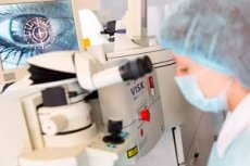
Each ophthalmological excimer laser system includes an excimer laser (a source of ultraviolet radiation), a forming optical system, the purpose of which is to transform the structure of the laser beam and deliver it to the surface of the cornea; a control computer, an operating microscope, a surgeon's chair and an operating table for the patient.
Depending on the type of the forming system, which determines the capabilities and features of the corneal evaporation technology, all installations are divided into homogeneous (diaphragm and mask), scanning, semi-scanning and spatial. Thus, when using the principle of laser diaphragm, the radiation hits the diaphragm or system of diaphragms in a wide beam, gradually opening or closing with each new pulse. In this case, a thicker layer of tissue evaporates in the center of the cornea than at its edges, as a result of which it becomes less convex and refraction decreases. In other installations, the radiation hits the cornea through a special mask of uneven thickness. Through a thinner layer in the center, evaporation occurs faster than at the periphery.
In scanning systems, the surface of the cornea is treated with a laser beam of small diameter - "flying spot" technology, and the beam moves along such a trajectory that a lens of a given optical power is formed on the surface of the cornea.
The "Profile" system developed by S. N. Fedorov is a spatial type of laser. The basic idea of the spatial distribution of laser energy in the "Profile-500" system is that the radiation hits the cornea with a wide beam with a Gaussian, i.e. parabolic, profile of laser energy distribution. As a result, in the same unit of time, in places where the energy of greater density was applied, the tissue evaporates to a greater depth, and in places where the energy density was lower, to a lesser depth.
The main refractive excimer laser surgeries are photorefractive keratectomy (PRK) and laser intrastromal keratomileusis (LASIK).
Indications for refractive excimer laser surgeries are primarily intolerance to contact and spectacle correction, myopia, hyperopia and astigmatism of varying degrees of severity, as well as the professional and social needs of patients at least 18 years of age.
Contraindications to photorefractive keratectomy include glaucoma, retinal conditions preceding detachment or detachment, chronic uveitis, eye tumors, keratoconus, decreased corneal sensitivity, dry eye syndrome, diabetic retinopathy, ectopia pupillae, severe allergic status, autoimmune pathology and collagenoses, severe somatic and mental illnesses. In the presence of cataracts, photorefractive keratectomy is inappropriate, since immediately after cataract extraction, the refraction of the eye can be corrected using an artificial lens.
Photorefractive keratectomy is performed on an outpatient basis under local anesthesia. The technique of performing the operation on foreign installations includes two stages: removal of the epithelium and evaporation of the corneal stroma. At the first stage, scarification of the epithelium in the central zone of the cornea is performed mechanically, chemically or by laser. The duration of this stage of the operation depends on the type of laser and can vary from 20 seconds to several minutes, after which evaporation of the corneal stroma is performed.
During the first day, pain syndrome, lacrimation, and photophobia may be observed. From the first day after the operation, the patient is prescribed instillations of an antibiotic solution until complete epithelialization of the cornea (48-72 hours). Then a course of corticosteroid therapy is carried out according to the scheme lasting 1-2 months. In order to prevent steroid hypertension, beta-blockers are used simultaneously 1-2 times a day.
The described technology allows for effective and safe correction of myopia up to 6.0 D and astigmatism up to 2.5-3.0 D. The technology of performing photorefractive keratectomy with a transepithelial approach (without preliminary scarification of the epithelium) on the domestic installation "Profile-500" allows for immediate correction of myopia up to 16.0 D in combination with complex myopic astigmatism up to 5.0 D without any additional interventions.
Patients with hyperopia and hyperopic astigmatism undergo photorefractive keratectomy less frequently, which is explained by the need for de-epithelialization of a large area of the cornea and, accordingly, its long healing (up to 7-10 days). With hyperopia greater than 4.0 D, LASIK surgery is usually performed.
The change in refraction depends on the thickness of the evaporated cornea. The residual thickness of the cornea in the thinning zone should not be less than 250-300 μm to prevent postoperative deformation of the cornea. Therefore, the limit of the method's capabilities is determined by the initial thickness of the cornea.
Early postoperative complications of photorefractive keratectomy include long-term (more than 7 days) non-healing corneal erosion; postoperative keratitis (dystrophic, infectious); severe epitheliopathy accompanied by edema and recurrent erosions; coarse subepithelial opacities within the entire corneal evaporation zone.
Late postoperative complications include subepithelial corneal opacities; overcorrection; myopization; irregular astigmatism; dry eye syndrome.
The formation of subepithelial opacities is usually associated with a large volume of corneal evaporation with high degrees of correctable refractive errors. As a rule, due to the implementation of resorption therapy, it is possible to achieve complete disappearance or significant regression of opacities. In cases of development of persistent irreversible corneal opacities, repeated photorefractive keratectomy can be performed.
The LASIK operation is a combination of surgical and laser treatment. It consists of three stages: formation of a superficial corneal flap (valve) on a stalk with a microkeratome; laser evaporation of the deep layers of the cornea under the flap; placing the valve back in its original place.
Mild pain sensations (a "speck" in the eye) are usually noted in the first 3-4 hours after surgery. Tearing usually stops after 1.5-2 hours. Drug therapy is limited to instillations of antibiotics and steroids for 14 days after the intervention.
In cases of myopia correction by performing the "LASIK" operation, the maximum refractive effect is determined by the anatomical features of the patient's cornea. Thus, given that the thickness of the valve is usually 150-160 μm, and the residual thickness of the cornea in the center after laser ablation should not be less than 250-270 μm, the maximum possible correction of myopia with the "LASIK" operation does not exceed 15.0-17.0 diopters on average.
"LASIK" is considered to be an operation with fairly highly predictable results in cases of mild to moderate myopia. In more than 80% of cases, the postoperative refractive result is within 0.5 D of the planned one. Visual acuity of 1.0 is observed on average in 50% of patients with myopia up to 6.0 D, and visual acuity of 0.5 and higher - in 90%. Stabilization of the refractive result, as a rule, occurs 3 months after the "LASIK" operation. In cases of high degrees of myopia (more than 10.0 D), in 10% of cases there is a need for repeated operations to further correct residual myopia, which are usually performed within 3 to 6 months. During repeated operations, the corneal flap is lifted without repeated cutting with a microkeratome.
When correcting hyperopia, a refractive result within 0.5 D of the planned value can be achieved only in 60% of patients. Visual acuity of 1.0 can be achieved only in 35-37% of patients, visual acuity of 0.5 and higher is noted in 80%. The achieved effect in 75% of patients remains unchanged. The incidence of complications during LASIK surgery ranges from 1 to 5%, with complications most often occurring at the stage of corneal flap formation.
It is quite obvious that technical progress in the near future will lead to the emergence and widespread clinical use in medicine, in particular ophthalmology, of new generation lasers, which will allow contactless and non-opening refractive surgeries. Laser energy, focused at one point, can destroy intermolecular bonds and evaporate corneal tissue at a given depth. Thus, the use of femtosecond systems already now makes it possible to correct the shape of the cornea without damaging its surface. Excimer laser refractive surgery is one of the most dynamically developing high-tech areas in ophthalmology.
 [
[