Medical expert of the article

New publications
Dracunculiasis: causes, symptoms, diagnosis, treatment
Last reviewed: 05.07.2025

All iLive content is medically reviewed or fact checked to ensure as much factual accuracy as possible.
We have strict sourcing guidelines and only link to reputable media sites, academic research institutions and, whenever possible, medically peer reviewed studies. Note that the numbers in parentheses ([1], [2], etc.) are clickable links to these studies.
If you feel that any of our content is inaccurate, out-of-date, or otherwise questionable, please select it and press Ctrl + Enter.
Dracunculiasis is a biohelminthiasis. Mature individuals are localized in the subcutaneous tissue, most often in the lower extremities.
The life cycle of dracunculiasis
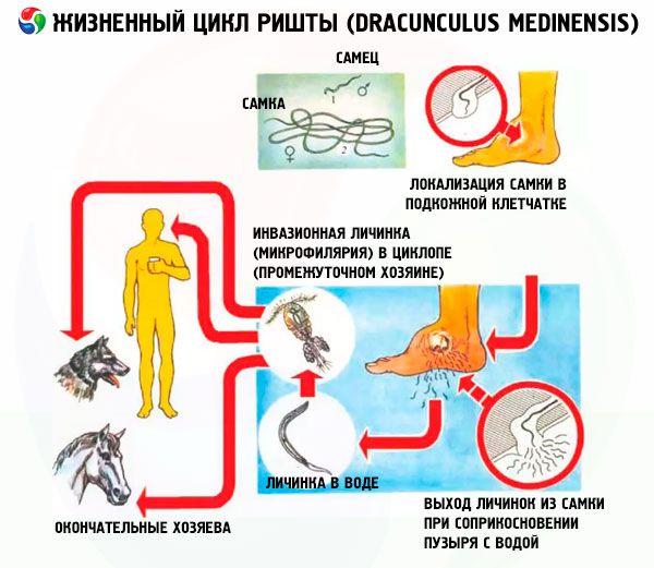
Dracunculiasis is a biohelminthiasis. The final host is a human, sometimes animals: dogs, monkeys. Intermediate hosts are freshwater crustaceans of the genus Cyclops or Eucyclops.
A person becomes infected with dracunculiasis by swallowing cyclops with water, infested with mature larvae (microfilariae). In the gastrointestinal tract, the cyclops are digested. The larvae penetrate the intestinal wall and migrate along the connective tissues towards the lower extremities. Three months after the invasion, the female is fertilized. After this, the female migrates to the subcutaneous tissue of the lower extremities, grows, reaching a length of 75-100 cm. About a year after the larvae penetrate the human body, up to 3 million larvae are formed in her uterus. The head end of the female reaches the skin, causing the formation of a bubble on it up to 5-8 cm in diameter, filled with liquid. The larvae exit the female's genital tract through a rupture in the uterus and the wall of the helminth's body near its anterior end. They are excreted from the body of the final host through an opening formed in the skin by the secretion of special glands located at the front end of the female helminth. Small rhabditiform larvae with a long threadlike end are 0.5-0.75 mm long and 15-25 µm wide.
When in contact with water, the bubble bursts. The front end of the female sticks out of it. The larvae are ejected from the body of the guinea worm due to the contraction of its muscles when in contact with water, which is possibly due to the cooling of the front end of the helminth under the influence of water. Over the course of 2-3 weeks, the female "gives birth" to 3 million larvae into the water. After this, the females die. They are absorbed or calcified.
Larvae that fall into the water live in it for 3-6 days and are swallowed by cyclops, in their bodies they grow, develop, molt twice and at a temperature of 25-30 °C after 12-14 days reach the invasive stage.
The maximum lifespan of a parasite in the human body is less than 18 months.
Epidemiology of dracunculiasis
Dracunculiasis is common in countries with hot and dry climates, in tropical regions of Africa, in the south of the Arabian Peninsula, in the south of Iran, in Pakistan, India, China, and South America.
Foci of dracunculiasis are formed in areas where the population drinks raw water from small artificial or natural stagnant reservoirs, which residents enter barefoot (at this time, the female guinea worm gives birth to larvae in the water). The development of parasites occurs synchronously in all infested people. Females become capable of giving birth to larvae simultaneously in almost all carriers of the helminth. This achieves a sharp increase in the probability of infection of a huge number of cyclops, and then the final hosts within a short period of time. This feature of the development cycle has an adaptive value in areas with arid climates and rare rainy periods. In foci of dracunculiasis, a large number of people infected with this helminth are detected within a short time interval.
Infection with dracunculiasis occurs as a result of accidental ingestion of cyclops when drinking water from stagnant open water bodies. In the human body, the parasite develops very slowly. The epidemiological incubation period (the period from the moment of infection to the moment of release of larvae into the external environment) for dracunculiasis is very long and amounts to 12 months or more. The infested final host becomes a source of invasion only a year after infection.
The main source of invasion is an infected person.
Dracunculiasis is spread due to unsanitary conditions, poor water supply, lack of running water and sewerage. Dracunculiasis is prevalent in poor families living in poorly maintained houses and drinking raw dirty water, using feces to fertilize gardens.
A major role in polluting water with guinea worm larvae is played by water carriers who enter stagnant water bodies barefoot to collect water, as well as religious people who perform ritual ablutions in water bodies. As a result of the large number of larvae entering water bodies, the presence of many cyclops, and the habit of the population to drink raw water, the intensity of transmission of the invasion in dracunculiasis foci is high.
The pathogenic effect of guinea worm is associated with the sensitization of the body by the metabolic products of the helminth, mechanical damage to tissues and the addition of a secondary infection.
What causes dracunculiasis?
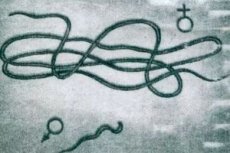
The causative agent of dracunculiasis is Dracunculus medinensis, a guinea worm with clearly expressed sexual dimorphism. The thread-like female is a large nematode 30-129 cm long and 0.5-1.7 mm wide, the male is 12-30 mm long and 0.2-0.4 mm wide.
The posterior end of the male is bent toward the ventral side. It has 4 pairs of preanal and 6 pairs of postanal papillae, 2 dark brown spicules 0.49-0.73 mm long, and a gubernaculum 0.2 mm long. On the rounded anterior end of the female there is a quadrangular cuticular elevation with 4 double marginal papillae and amphids located behind them. The mouth is triangular in shape, the short esophagus consists of muscular and glandular sections, separated by a narrowing located at the level of the nerve ring. The esophagus passes into a cylindrical intestine, ending in an anal opening near the posterior end of the body. The caudal end of the female ends in a subulate appendage facing ventrally. The vagina, located in the middle of the body, leads to two uteri lying one behind the other. The oviducts, coming from the tubular ovaries, open into them. Females are viviparous.
During the long development in the body of the final host (11-13 months), the primary cavity of the female is almost completely filled with uteruses filled with embryos. The vaginal opening, rectum and vulval opening atrophy. The remaining part of the intestinal tube shrinks and is pushed aside. The larvae exit through ruptures in the uterus and cuticle at the anterior end of the body.
Symptoms of Guinea worm disease
Patients learn about the presence of guinea worm several months after infection, 8-10 days before the blister forms on the skin. The first symptoms of dracunculiasis are accompanied by severe allergic reactions. Itching, urticaria, nausea, vomiting, asthmatic symptoms, fever, swelling of the joints near which the helminths are located occur.
Soon after the rupture of the blister, allergic reactions cease. The further course of the invasion is determined by the absence or presence of a secondary infection.
Specific signs of this disease are erythema, thickening of the skin, formation of blisters and ulcers at the site of the helminth's exit to the surface. The first symptoms of dracunculiasis are the formation of a small capsule that turns into a blister. The blister is filled with a transparent yellowish liquid that contains guinea worm larvae, leukocytes, lymphocytes and eosinophils. The formation of the blister is accompanied by itching and burning pain, which can be relieved by cold water. The blister ruptures upon contact with water, and the front end of the female sticks out of it. An ulcer appears at the site of the blister, surrounded by a ridge of edematous skin and covered with a white necrotic mass, which is rejected after a few days. In uncomplicated cases, the ulcer heals quickly. If there is only one helminth in the body, clinical manifestations last no more than 4-6 weeks and end in recovery. Local lesions are localized mainly on the shins and ankles (90%), sometimes found on other parts of the body: on the back, abdomen, scrotum, buttocks, mammary glands, tongue, shoulders.
The symptoms of dracunculiasis depend on the localization of the parasite. A more severe course of dracunculiasis is observed when the worm is localized in the area of large joints, when the parasite dies, with secondary bacterial infection, or a combination of these. Single lesions occur more often, but there are known cases of parasitism in one person up to 50 helminths. The process is painful and deprives the patient of the ability to work for a long time. Sometimes the helminth dies before the larvae hatch. In these cases, there are no symptoms of dracunculiasis.
Complications of dracunculiasis
When the guinea worm is localized in the joint area, acute arthritis develops, which in 1% of cases ends in ankylosis. Other large joints and muscles may be involved in the process. Penetration of a bacterial infection into the parasite localization site can cause purulent abscesses, phlegmon, sometimes gangrene, epididymitis, orchitis, and sepsis. Cases of tetanus registered in areas endemic for dracunculiasis occur as a result of previous guinea worm invasion. In the absence of complications, the prognosis is favorable.
 [ 5 ]
[ 5 ]
Diagnosis of dracunculiasis
Diagnostics of dracunculiasis in endemic foci with characteristic skin manifestations is not difficult. A cord-like formation can be felt under the skin. At the site of the rupture of the bladder, the anterior end of the guinea worm and its larvae can be found. Calcified parasites are detected by X-ray examination.
Outside endemic foci, differential diagnostics of dracunculiasis from furunculosis, abscess, phlegmon is necessary, and the patient must be asked about the possibility of his being in foci of dracunculiasis.
Who to contact?
Treatment of dracunculiasis
Traditional treatment for dracunculiasis is winding the helminth's body on a stick, several centimeters a day, avoiding its breakage. If allergic reactions develop, antihistamines are prescribed. Metronidazole is used at a dose of 250 mg x 3 x 10 days, for children - 25 mg/kg in three doses, the total daily dose should not exceed the adult dose. The drugs do not destroy the helminth, but facilitate its extraction in the traditional way.
How to prevent dracunculiasis?
The Global Guinea Worm Eradication Programme includes:
- providing the population with safe drinking water;
- allocation of special reservoirs for the collection of drinking water and protection from pollution;
- filtration of water from open stagnant reservoirs to prevent the penetration of cyclops;
- identification and treatment of patients;
- preventing the insemination of water bodies with helminth larvae by applying a bandage to the guinea worm bladder.