Medical expert of the article

New publications
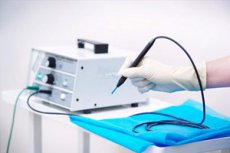
Diathermocoagulation in gynecology, dentistry and dermatology
Last reviewed: 29.06.2025

All iLive content is medically reviewed or fact checked to ensure as much factual accuracy as possible.
We have strict sourcing guidelines and only link to reputable media sites, academic research institutions and, whenever possible, medically peer reviewed studies. Note that the numbers in parentheses ([1], [2], etc.) are clickable links to these studies.
If you feel that any of our content is inaccurate, out-of-date, or otherwise questionable, please select it and press Ctrl + Enter.
Among the various procedures that can stop bleeding and slow down pathological tissue overgrowth, diathermocoagulation, which is an effective method based on the use of electric current, is particularly widespread. High-frequency alternating current helps to quickly coagulate tissue proteins, which helps to accelerate the treatment of neck erosion, gingival hyperplasia and other pathologies. Diathermocoagulation can be successfully combined with physiotherapy, the use of drugs. [1]
Indications for the procedure
The effect of the current on the tissue involves heating it to a certain temperature, at which coagulation (folding) of protein structures occurs. At the same time with coagulation, vessels are "sealed", which leads to a stop of bleeding. In addition, the development of inflammatory reaction is blocked, which improves the recovery of damaged tissue. The possibility of deep exposure to high-frequency current allows to treat both superficially located pathologies and deeper structural lesions (in particular, some gynecological disorders).
Diathermocoagulation may be prescribed:
- To eliminate some neoplasms (in particular, on the skin, in the oral cavity), which can not be removed by any other method;
- For cervical erosion, endocervicitis;
- Papillomas or bladder ulcers (in these situations, thin electrodes are used, which are inserted into the bladder through a catheterized cystoscope);
- For closed bone tuberculosis foci;
- For skin lesions caused by lupus erythematosus;
- For cutaneous leishmaniasis, warts, skin papillomas;
- For retinal detachment;
- For inflammation of the dental pulp, etc.
Diathermocoagulation is actively used to stop bleeding - particularly during surgical interventions. To stop bleeding, the damaged vessel is fixed with a hemostatic clamp, which is connected to an active electrode. For the same purpose, diathermocarbonization (fulguration) is sometimes used, which is a method of charring by a spark arising from the active electrode to the vessel at a distance of 1-2 mm.
Most often diathermocoagulation is used in gynecological and dermatological practice, which is explained by the high efficiency of high-frequency current on skin and mucous tissues.
In dentistry, thermal exposure is used in pulpitis (for pulp coagulation), periodontitis (for coagulation of root canal contents), benign mucosal lesions of the oral cavity (hemangioma, papilloma, epulis, fibroma), as well as for coagulation of granulations in periodontal pockets.
Common indications for diathermocoagulation include:
- The need for treatment of prolonged non-healing erosive and inflammatory processes;
- Getting rid of ectopic foci, areas of hyperkeratosis, leukoplakia, pathologic growths of benign nature.
This type of treatment is actively used to eliminate acne, telangiectasia, rosacea, to remove benign neoplasms (including atheromas, angiomas, scars). The method can be used in dentistry, gynecology, cosmetology, general surgery, veterinary medicine and other medical fields.
Preparation
Preparation for cervical diathermocoagulation is performed with mandatory preliminary diagnostic examination and preoperative treatment.
Before the procedure, the woman is thoroughly examined, using generally accepted tactics. The attending physician establishes a clinical diagnosis and treats existing inflammatory pathologies accordingly.
Both indications and possible contraindications to performing diathermocoagulation must be determined. This condition must be met to improve the prognosis of the disease and avoid errors in the discrepancy of clinical diagnoses. It is necessary to warn the doctor about existing chronic, inflammatory-infectious and systemic pathologies, about disorders of the cardiovascular and respiratory system, about possible allergies, about malfunctions of the blood coagulation system, about complications after surgery and anesthesia.
On the day of cervical diathermocoagulation, you should abstain from eating and drinking six hours before the procedure. It is necessary to take a shower and shave off the hair on the external genitalia. The patient should take with her the results of such studies: a general blood test, a test for hepatitis B and C, Wasserman reaction, the presence of antibodies to HIV. The results of electrocardiography with a description are also required.
Before performing diathermocoagulation on other parts of the body any special preparation is not required, except for preliminary diagnosis of the body for contraindications to the procedure. If thermal manipulations in the oral cavity are supposed to be performed, the patient should clean teeth well, remove plaque and calculus, treat inflammatory diseases (including oropharynx).
Technique of the diathermocoagulation
Diathermocoagulation is the "cauterization" of tissue by means of alternating high-frequency current from lamp-electronic generating devices. The technique is based on local heating of the tissue to approximately 80 to 100°C, which entails the folding of protein fractions.
The main merits of the methodology are:
- Tissues treated with the electrode become sterile at the same time;
- Under the influence of elevated temperature in the coagulated area is cauterized and thrombosed vessels, which blocks the entry of infection, toxic substances and tumor structures into the circulatory system;
- Nerve endings are also cauterized, so post-procedure pain is usually low.
Strong currents are not used, because the tissue treated with the electrode is quickly dehydrated, resulting in an increase in the level of resistance and a decrease in the current in the circuit. As a result, coagulation does not occur, and under the influence of strong current increases the risk of rupture of the vascular wall to the point of thrombus formation. This contributes to the development of bleeding, which is even more aggravated when the electrode adheres to the surface of the vessel. Against the background of bleeding diathermocoagulation becomes impossible: coagulated blood on the electrode needle prevents the cauterization process, and the bleeding blood, being an excellent conductor, "takes" the vast majority of the current. In such a situation, the treated area should be well dried and only then continue the procedure.
Two main methods of diathermocoagulation are known:
- Monopolar, with only one generator pole connected;
- Bipolar, with two generator poles connected.
Taking into account the size of the electrode area, monoactive and bi-active methods of diathermocoagulation are distinguished. The most popular is the bipolar monoactive method, when one passive electrode (lead plate with dimensions of 200-300 cm²) is applied to the lumbar region, the outer thigh surface or another area remote from the heart, from the places of passage of large vessels and nerves. A second small active electrode is placed in an insulated clamp (holder), which may have an additional mechanism for interrupting the current supply. The active electrode can be different in shape: needle, disk-shaped, spherical, loop, etc., which depends on the characteristics of the area to be treated.
The active electrode is applied closely, but not aggressively, to the body surface and the current is applied for the required period of time (usually a few seconds), until the tissue lightens slightly. Then the current supply is stopped and proceed to the treatment of the next area. If deep coagulation is required, the procedure is performed layer by layer, with each coagulated layer removed with tweezers. If the electrode becomes contaminated with adhering particles of coagulated tissue, it should be cleaned immediately, as the contamination will interfere with the procedure.
The biactive technique involves placing two electrodes close to each other.
Papilloma diathermocoagulation
Diathermocoagulation is widespread in both medicine and cosmetology. It can be used to easily get rid of small imperfections on the skin, such as papillomas. Diathermocoagulation is also considered an optimal technique for removing warts and tattoos, to eliminate pink acne. Most often the procedure copes with its task in one go: the problem is solved quickly, almost painlessly and effectively.
A papilloma is a benign skin growth that develops during overgrowth of the upper epidermal layer of the skin. It has the appearance of a skin growth with the size of 1-7 mm, sometimes more. The shape of the formation is round, the color is from light beige to dark brown. There may be a single localization of the growth, or multiple overgrowths of the papillomatosis type.
The appearance of papillomas is associated with the activity of the human papillomavirus (HPV). The growths usually occur against a background of weakened immunity, after prolonged illness or regular overwork, with frequent courses of medication or changes in hormonal balance - in particular, unpleasant growths often appear during pregnancy, with the onset of menopause or during the period of active sexual development in adolescents.
It is not possible to get rid of a papilloma with medication. If a neoplasm has appeared, it must be removed. It should be remembered: weak immunity and improper lifestyle can provoke the reappearance of growths. Removal can be performed by different methods, and one of them is diathermocoagulation.
The high-frequency electric current delivered by the coagulator leads to a volumetric thermal burn of the tissue in the area of exposure. A crust is formed on the treated area, which peels off after some time almost without trace. In case of large and deep papillomas may remain a small trace in the form of a light spot: after a few months, it smoothes out and also becomes imperceptible.
Diathermocoagulation, as a method of eliminating skin neoplasms, has a number of advantages over other methods. It is effective, safe and affordable. With this method of removal minimizes the likelihood of infection in the wound, and the development of bleeding after the procedure is completely excluded. This fact makes diathermocoagulation one of the most popular procedures in practical dermatology and cosmetology. [2]
Diathermocoagulation of cervical erosion
Cervical erosion is one of the most frequent gynecologic diseases. Such a diagnosis is made if there is an epithelial erosive defect on the vaginal part of the cervix. Specialists categorize erosions into true and pseudoerosions, or ectopias. True erosion is said if the mucosa of the uterine cervix in a certain area is marked by the absence of part of the epithelium in the form of a wound surface. Such pathology can occur after mechanical injuries, childbirth, infections, hormonal disorders. Pseudoerosion, or ectopia, is characterized by changes in the epithelium due to inflammatory gynecological diseases.
Cervical erosion is often accompanied by almost no symptoms. Only occasionally you may find the appearance of bloody discharge after sexual intercourse or vaginal examination. Some women experience pulling discomfort in the lower abdomen.
Even despite the lack of pronounced symptoms, cervical erosion requires mandatory treatment - first of all, to prevent the entry of infection into the wounds, which can turn into an inflammatory process, as well as to prevent malignant degeneration of pathology.
To date, gynecologists use different techniques to treat erosion. Among them is diathermocoagulation, which is a proven and reliable way to get rid of the problem. The procedure involves the use of a pair of electrodes and local anesthesia. One ball-shaped electrode is inserted intravaginally. The second electrode is placed under the lumbar region and the current is passed: under the spherical electrode the tissue is heated and coagulated. The duration of the treatment session is about 20-25 minutes, and the effectiveness of the technique is estimated at 70-80%. Neck tissue is fully restored after 8-12 weeks.
Diathermocoagulation is used to treat cervical erosions as often as other similar methods such as cryodestruction, laser photocoagulation, radiofrequency therapy, etc. However, thermocoagulation is not prescribed for unborn patients of childbearing age who plan to have children in the future. However, thermocoagulation is not prescribed for unborn patients of childbearing age who plan to have children in the future.
Diathermocoagulation of the cervix for cervical cancer
Cervical cancer is one of the most dangerous female diseases. Its appearance can be prevented by early detection and treatment of precancerous lesions. In particular, secondary prevention involves the detection and elimination of precancerous conditions during systematic examination. Thus, epithelial dysplasias and preinvasive carcinoma require special attention - pathologies accompanied by changes in multilayer squamous epithelial tissue. Such disorders can be provoked by various reasons, such as early sexual activity, promiscuity, childbirth at a young age, infectious diseases (including human papillomavirus).
The pathology can be diagnosed by cytologic and histologic examination. The doctor chooses the type of treatment individually, taking into account not only the pathology, but also the age of the patient and her desire to have children in the future.
If intraepithelial carcinoma or microinvasive cancer is detected, the cervix is removed with a surgical scalpel: a so-called knife conization or amputation is performed. In epithelial dysplasia, it is possible to use not diathermocoagulation, but a procedure with a similar name - diathermoconization, which involves the use of a specific lancet-like electrode. The pathologically altered tissue is excised in a cone-shaped manner, with the tip of the cone "looking" into the area of the internal pharynx.
Removal of the uterine cervix with a surgical scalpel is considered to be the more preferred method, which is due to the absence of tissue damage in the type of charring "cone" outlines, which in some cases prevents adequate assessment of the nature of pathologic changes.
In case of moderate dysplasia of the epithelium in patients under 40 years of age, diathermocoagulation is possible, but after 40 years of age, amputation, neck diathermoconization with mandatory evaluation of the state of the slices of the removed element of the organ are performed. If concomitant pathology (cancer, myoma) is detected, the operation can be extended to complete amputation of the uterus. Always at the stage of preparation for treatment (diathermocoagulation, diathermoconization), the doctor must accurately establish the diagnosis and exclude the presence of invasive cancer. The main treatment method for microinvasive carcinoma is surgery. Young patients undergo organ-preserving intervention using a scalpel, laser. If a woman is in the menopausal period, it is recommended to perform uterine extirpation.
Cervical diathermocoagulation for leukoplakia
Leukoplakia is a lesion of the mucous membrane of the uterine cervix, which is manifested by the formation of a milky-white half transparent film or lightened zones on the epithelial surface. The disease may occur in a simple form, with thickening and dying off of the upper layer of the epithelium, or in a proliferative form, in which all layers of the epithelium are affected, including the basal and parabasal layers.
Leukoplakia is dangerous, first of all, due to the increased risk of degeneration into dysplasia and cancer. Therefore, the disease should be detected and treated in a timely manner.
As for the procedure of diathermocoagulation, it is often the cause of leukoplakia, along with hormonal disorders and various infectious-inflammatory processes. But it is recommended to treat leukoplakia in two main ways: laser or radio wave method.
- Laser cauterization is a virtually painless and safe procedure that rapidly cleans and heals the tissue. If the leukoplakia is extensive, several treatments may be required.
- The radio wave method involves the use of a radioscalpel, which is used to "vaporize" pathological tissues. Treatment is painless and there is no risk of bleeding.
In simple leukoplakia can be used therapeutic tactics, including the correction of hormonal disorders. In the absence of positive dynamics, the focus is removed by laser-destructive method or cryodestruction. It is also possible to use electric current, but not in the form of diathermocoagulation, but in the form of diathermoconization. The choice of treatment technique is based on the results of the examination, as well as on the age of the patient, her desire to preserve fertility, etc.
Diathermocoagulation in dentistry
Dentists have been using diathermocoagulation since about the middle of the 20th century. Today practicing doctors use electric high-frequency coagulation to eliminate pathological formations on the mucous tissues of the oral cavity and on the skin, as well as for endodontic treatment of root canals, removal of gingival hypertrophy, ingrowths in the cavity of caries, etc. There are known successful cases of application of diathermocoagulation for treatment of periodontitis, maxillary odontogenic sinusitis and for zaapical therapy. The disadvantage of the method is the difficulty of dosing of exposure, which in certain situations can lead to the development of complications. If electrocoagulation is used irrationally, the adverse effects may include pain, gingival necrosis or osteomyelitis with alveolar sequestration.
Given this, diathermocoagulation, which is effective in other areas, is not often used in practical endodontics. The most common bipolar diathermocoagulators are not used in root canal treatment because of the risk of periodontal overheating.
Bipolar electrocoagulators are equipped with a pair of electrodes. One of them has a special retainer that holds the electrode: special tools required by the doctor are placed in it. The other electrode plays a passive role and is placed on the patient's body. The standard current frequency used is no more than 1000 kHz. The efficiency of diathermocoagulation is higher in the presence of moisture, but for endodontic therapy bipolar coagulators are not used, because the presence of blood and exudative secretions in the root canal under the influence of excessive theproduction can damage the periodontium and alveolar bone tissue.
Monopolar electrocoagulators have only one electrode and a fixation holder. There is no passive second electrode. The procedure is performed with an alternating current frequency of more than 2000 kHz. If the environment is intensely humid, the quality of coagulation suffers, so it is necessary to periodically dry the treated tissues with gauze or cotton swabs. This type of diathermocoagulation is used for removal of formations, gingival coagulation, root canal treatment.
In dentistry, it is very important to correctly adjust the frequency of the applied current and the output impedance. If this is not done, coagulation will either not occur or will be excessive, resulting in burns to the periodontium and bony alveolus.
During diathermocoagulation of soft tissues, the treated blood and lymphatic vessels and interstitial spaces are thrombosed. This helps to reduce the absorption of metabolic products and toxic substances, prevents infectious spread, and stops bleeding.
Monopolar diathermocoagulation is used in endodontic therapy to help coagulate the root canal filling, block bleeding, and disinfect tissue for subsequent dental manipulations.
However, experts point out that the full potential of this thermal treatment method has not yet been fully explored.
Diathermocoagulation of the gingiva
The features of gingival diathermocoagulation consist in the excision of mucosal tissues. Cauterization is performed with an electrocoagulator or a medical laser. Instrumentation heated to a certain temperature cuts the neoplasm and simultaneously coagulates small vessels, so bleeding during the procedure is completely excluded.
The patient feels almost no pain, but for greater comfort, the doctor performs local anesthesia beforehand. The risk of infection in the wound is practically nil, as the tissues are cauterized and treated with antiseptic solutions.
To date, two variations of thermal gum treatment have been utilized:
- Monopolar variant, which is suitable for getting rid of large growths, in particular those that are localized deep in the tissues. For the procedure, a return plate and an electrode are used, through which the electric current passes through the desired area of tissue. This method of treatment is quite effective and is suitable for the removal of tumor processes.
- Bipolar variant is used for therapy of gingival diseases and local inflammatory processes with minimal risk of complications.
The most optimal method of diathermocoagulation is chosen by the doctor, based on individual indications and limitations. It is possible to apply the procedure:
- For the removal of gingival neoplasms;
- To eliminate mucous overgrowths, inflammation of gum pockets;
- In periodontal disease, periodontitis, pulpitis, gingivitis, neck carious processes.
The most common use of diathermocoagulation is associated with gingival papilla overgrowth: periodontal volume increases, interdental spaces are formed, and soft tissue overgrowth occurs and fills the resulting voids. Mucosal overgrowth can be provoked by mechanical damage.
Before starting the procedure, the doctor removes plaque and calculus from the patient. Before coming to the clinic, the patient is advised to eat well, because after the procedure of diathermocoagulation he will have to give up food for at least three hours.
At the end of treatment, the patient is released home: complete healing of the gum will occur in 2-4 weeks. To accelerate recovery, it is recommended to use special antiseptic solutions and medications prescribed by the doctor (most often these are drugs of non-steroidal anti-inflammatory series). For a month after the procedure, it is not recommended to traumatize the oral mucosa with hard toothbrushes, rough and hot food.
Diathermocoagulation of dental pulp
In the process of diathermocoagulation of dental pulp, an alternating electric current with high frequency (within 1-2 MHz), low voltage and sufficient strength (up to 1-2 A) is used. The residual pulp tissue is cauterized under thermal influence, which is the result of the transformation of electricity into thermal energy: temperature values rise between 40 and 90°C, which causes the protein fractions of blood and tissue to curdle.
The undeniable "plus" of diathermocoagulation is the following:
- Elimination of residual pulp is not accompanied by bleeding, because the lumen of the vessels are "sealed";
- Infection spread from the canal to the vasculature is excluded.
The procedure is carried out as follows:
- The dental cavity is cleaned of blood;
- The active root electrode is placed in the tooth canal, not bringing it one and a half to two millimeters to the apex;
- Are applied with electric current with exposure of 2-3 seconds for each channel, with output power from 6 to 8 W;
- Eliminate residual pulp tissue.
If there are lateral pulp branches, a so-called gradual diathermocoagulation is performed:
- The active electrode needle is placed in the canal orifice and gradually moved to the root apex;
- Without turning off the coagulator, the electrode is slowly withdrawn from the canal;
- -exposure is 3-4 seconds;
- When bleeding is completely stopped, begin to treat the canals with instrumental and medication.
The procedure is performed under local injection anesthesia.
Diathermocoagulation for pulpitis
Chronic hypertrophic pulpitis involves the use of a loop electrode or a special thermocouter for pulp amputation. Removal is performed according to the technology described above. If there is bleeding from the pulp stump, then inject a hemostatic agent, dry the canal and again perform diathermocoagulation.
Chronic gangrenous pulpitis and periodontitis require direct layer-by-layer diathermocoagulation. The needle electrode is placed one third of the canal depth and coagulated for 2 seconds, after which it is moved one third deeper and coagulated again for 2 seconds. Then move the electrode to the apex and again coagulate for 1-2 seconds. Using pulpoextractor clean the root canal, treat with antiseptic solution and put a filling. To prevent the entry of infection into the canal after the completion of coagulation, salivary fluid is not allowed to enter the canal, and treatment is carried out with sterile turundas.
Diathermic exposure has anesthetic and hemocoagulating properties. The heat formed in the treatment area destroys toxic products of tissue decay, and the protein clot inhibits the absorption of infectious agents and toxins into the bloodstream. Around the treated area is formed an area of diathermization, in which there is increased lymph and blood circulation, optimizes metabolism, which contributes to rapid tissue repair and stop the inflammatory process.
According to specialists, immediate and distant adverse effects after such a procedure are not observed.
Diathermocoagulation of eyelashes
The procedure of diathermocoagulation of eyelashes involves their removal: sometimes it is necessary if there are relevant indications - for example, trichiasis. This is a peculiarity of eyelash growth in which the hairs sprout not outward and upward, but inward and downward, which leads to uncomfortable sensations and eye irritation. Trichiasis can be congenital, or it can be a consequence of traumatic injuries or diseases affecting the eyelid margin.
Diagnosis of pathology is quite simple: visually noticeable is the incorrect location of the eyelashes, and the patient himself complains of constant irritation of the eyeball. Diathermocoagulation is prescribed by a doctor.
It would seem that improperly grown eyelashes can simply be removed in the usual way. However, in this case, they will re-grow with a violation. To eliminate the problem, the hair should be removed together with the follicle, which is possible surgically or with diathermocoagulation.
Since the treatment area is small, the procedure is performed using a microscope. The specialist gets rid of only the wrongly growing, unfolded hairs, while the rest of the normal cilia remain intact.
After the procedure is completed, it is recommended to drip antiseptic ophthalmic drops or put bactericidal eye ointments in the eyes for several days.
Diathermocoagulation of warts
Diathermocoagulation is a suitable technique for removing warts and other similar skin defects. The unsightly growth is removed with the help of a special electric device called electrocoagulator. Working electrodes of the device for a few seconds heated to the required temperature under the influence of electric current, thanks to which the defect is removed. After the procedure, a crust is formed in the area of exposure, which disappears for several days.
An undoubted "plus" diathermocoagulation - it is possible to get rid of several warts at once in one session. And if necessary, you can send the removed neoplasm for histological analysis. In general, the choice of treatment depends on the location and degree of spread of rashes.
Common warts are often eliminated using diathermocoagulation, as this method is both effective and inexpensive. But flat warts located in cosmetically significant areas (eg, on the face) is not recommended to remove using such destructive methods, as these types of neoplasms often grow deep into the tissue, and after the procedure can be left quite an impressive trace.
Getting rid of warts with diathermocoagulation can be performed in most clinical centers or dermatology departments, and even in many cosmetic salons. However, when choosing a place to perform the procedure, you should always pay attention to the quality of the equipment and the qualifications of the staff - especially the specialist who will perform the removal. If everything is done competently and correctly, then soon there will be no trace of the former wart at all.
Diathermocoagulation of the vessel
Vascular diathermocoagulation is used not only during surgical interventions to stop bleeding, but also in cases of blood loss and vascular damage in the nasal cavity, pharynx, upper digestive tract - using endoscopic methods.
A prerequisite for performing endoscopic bleeding arrest is good access to the injured vessel.
Diathermocoagulation refers to universal, effective and proven hemostatic methods. Monopolar, bipolar and multipolar coagulation of the bleeding site with high-frequency current is commonly used, which causes rapid tissue heating, thrombosis of the bleeding vessel or thickening of the previously formed thrombus. At the same time, there is a coagulation damaging effect on other tissues, which may pose a threat of perforation of hollow organs. The risk of such a complication increases, depending on the type of bleeding source, current power, duration of exposure and the qualifications of the specialist who performs the treatment.
In monoactive coagulation, the passive electrode (plate electrode) is applied to the outside of the patient's femoral surface, and the active electrode is brought through the instrument channel of the endoscopic device to the area to be treated. Bipolar and multipolar techniques involve bringing all electrodes to the distal end of the probe. The current affects the tissue located between the electrodes, without spreading it to the depth of the structures and the patient's body.
Using coagulation instruments and an endoscope, the physician first clamps the vessel and then performs the coagulation action. The duration of continuous coagulation is no more than 2-3 seconds. After that, the doctor evaluates the effectiveness of the effect, rinses the surface and, if necessary, repeats the current treatment again.
Based on clinical practice, the monoactive method is more suitable for stopping bleeding of chronic ulcers. The biactive method is used for bleeding caused by ruptures of mucous tissues of the stomach and esophagus, acute ulcers, erosions and other lesions that are not accompanied by pronounced scarring and sclerotic tissue changes, or in cases where there is no need (or possibility) to perform deep coagulation.
If the bleeding cannot be stopped in this way, or the vessel is damaged again, then most often the patient is prescribed emergency surgery. By the way, such a development is rare.
Contraindications to the procedure
Like any medical manipulation, diathermocoagulation has its own list of contraindications:
- Individual intolerance to electric current;
- Severe pathologies of the cardiovascular system, including disturbed heart rhythm, atherosclerotic cardiosclerosis on the background of a pronounced disorder of coronary circulation, cerebral sclerosis and circulatory disorders in the brain, aortic aneurysm, insufficient blood circulation of the 2nd or 3rd degree;
- Nervous pathologies associated with hyperexcitability of the nervous system;
- Blood diseases;
- Hyperthyroidism;
- Severe pulmonary emphysema;
- Renal failure;
- Malignant tumor processes;
- Severe course of diabetes mellitus in the stage of decompensation or unstable compensation;
- For women - inflammatory-infectious pathologies of genital organs, the fourth degree of vaginal cleanliness, pregnancy, suspected malignant processes.
In dental practice, diathermocoagulation is not prescribed for baby teeth in children in the period of resorption of their root system, with unformed roots of permanent teeth, as well as in completely impassable canals.
Thermocoagulation treatment is allowed only after the diagnosis of malignancy of the lesion to be removed has been completely excluded. For example, before sending a patient for therapy of cervical erosion, a preliminary biopsy is performed. [3]
Complications after the procedure
After cervical diathermocoagulation, patients may experience reproductive problems. And under certain circumstances, the possibility of conception may become jeopardized.
Under no circumstances should thermocoagulation treatment be performed during pregnancy. Any interference with the mucosal tissue may lead to spontaneous abortion.
The loss of elasticity of the cervical tissues due to diathermocoagulation will have a negative impact on the quality of their extensibility during labor: the risk of rupture increases significantly, so it is recommended that such women do not plan natural childbirth, and immediately prepare for cesarean section.
In the postoperative recovery period, patients often complain of pulling pain in the area of the procedure (in cervical diathermocoagulation, pain is noted in the lower abdomen and lumbar area). Women may have a short disorder of the menstrual cycle, the appearance of vaginal discharge (watery or bloody), which indicates the rejection of necrotic tissue and the beginning of the period of wound healing. If recovery is delayed, and negative symptoms are present for more than 1-2 weeks, it is necessary to consult the attending doctor.
A reason to see a doctor should be such unfavorable signs:
- The transformation of a dry wound into a wet one;
- Bleeding wounds;
- A rise in temperature;
- Purulent discharge;
- Severe redness and swelling of tissues in the area of exposure lasting several days, with increasing negative dynamics.
Possible consequences that do not require a mandatory doctor's visit:
- Formation of a light spot (hypopigmentation) at the site of exposure, which occurs after deep penetration of the current into the tissues and takes about two years;
- Repeated formation of pathological growths (papillomas, warts) - does not pose a threat to health, but if desired, removal can be performed again;
- The appearance of a depression (fossa) in the area of exposure, which does not require intervention and disappears on its own within a few years.
The probability of complications after diathermocoagulation depends largely on the literacy of the procedure, the level of training of medical professionals, the quality of equipment, compliance with all the rules of preparation and completeness of preliminary diagnostic measures.
The timing of tissue repair also depends on various factors:
- From the peculiarities of the main and background diseases of the patient, from the individual state of the organism and the quality of immune defense;
- Depending on the age of the patient;
- From the quality of hormonal balance and metabolic processes;
- On the degree of compliance with all doctor's recommendations and prescriptions.
"Minus" diathermocoagulation is considered that during the procedure it is necessary to very carefully control the zone of exposure. If even a little beyond the pathological focus, healthy tissue will be affected, which can also affect the development of complications. In addition, the period of tissue recovery is relatively long, and during it the patient must carefully follow all the doctor's instructions and even come to the appointment for a checkup. This will help prevent the appearance of unpleasant consequences.
As alternative methods, the doctor can always offer other, more modern and affordable treatment options - for example, laser therapy or cryodestruction. Laser treatment is considered to be particularly safe, after which the tissue recovers quite quickly.
Care after the procedure
After performing a diathermocoagulation session, the patient is advised to adhere to all medical recommendations:
- Avoid excessive physical activity;
- Do not strain the damaged area, do not lift heavy weights, and when treating the cervix - do not have sexual intercourse until the wound is completely healed.
In addition, it is necessary to support and strengthen the immune system in every possible way, which will avoid complications and speed up the recovery process.
If thermal removal of external defects (warts, papillomas) was performed, then during the first few days after the intervention use special antiseptic and drying agents, for example:
- Diamond green solution, fucorcin;
- An intense solution of potassium permanganate;
- Chlorhexidine;
- Miramistin.
To ensure comprehensive stimulation of recovery, it is additionally recommended to take multivitamin preparations and immunomodulating agents. If the affected area is swollen, it should not be alarming: the swelling will subside within a few days (sometimes up to one week).
After the crust comes off, the wound is treated with regenerating ointments. Panthenol, Actovegin, Levomekol, etc. Will do a great job.
During the first three days it is not recommended to wet the wound, do not apply cosmetics on it, do not expose it to sunlight. For 4 weeks, do not visit swimming pools, baths, baths, bathing in public water bodies.
Alcohol is not recommended throughout the healing period, as it promotes vascular dilation, which can lead to the development of bleeding.
If the simplest rules are followed, the recovery phase will be quick and comfortable.
Testimonials
The majority of patients who have undergone diathermocoagulation left mostly positive feedback about the procedure, calling it effective, affordable and fast - both in terms of execution and tissue healing. Pain during the recovery phase is mild and short-lived, and does not cause any particular discomfort.
The actual procedure cannot be called pleasant, because thermocoagulation is a burn of the skin or mucosa, although it is used for good purposes. Pain when performing the treatment is not strong, but they are present: first of all, painful are the contractions of the uterus at each application of current, if the therapy of neck erosion is performed. Another nuance is an unpleasant odor of "burnt meat" emitted during cauterization. Some particularly impressionable patients are advised to wear a gauze bandage to cover the respiratory system.
A longer healing period is noted when eliminating skin and gynecological problems by diathermocoagulation. For example, with erosion of the uterine cervix, thermal treatment is effective, but the duration of tissue regeneration is quite long. Doctors advise: if there is an opportunity to use another, more modern method, it is better to be insured and choose new technologies - for example, laser therapy. But it is still better to consult with your doctor: take into account the degree of neglect of the primary pathology, and the presence of background diseases, as well as the age and general state of health of the patient.
Currently, in all clinical centers and hospitals, doctors use a variety of highly effective and modern treatment methods. The choice of the optimal treatment procedure is left to a highly qualified specialist with extensive therapeutic experience. Therefore, the patient should definitely consult with the doctor about which treatment method will be the most suitable for him - whether it is diathermocoagulation, or other therapeutic effects.
Literature used
Practical skills in obstetrics and gynecology, Textbook for students of medical universities, clinical interns and residents, doctors of obstetrics and gynecology. Edited by Prof. L. I. Trubnikova, Ulyanovsk 2015
Dentistry. Endodontics. 2nd ed., per. And ext. Textbook for universities. Britova A. A., 2023
Dermatology. Textbook in two parts. 3rd edition. Part 1. Edited by V. G. Pankratov. Minsk BGMU, 2012