Medical expert of the article

New publications
Diagnosis of diabetes mellitus
Last reviewed: 04.07.2025

All iLive content is medically reviewed or fact checked to ensure as much factual accuracy as possible.
We have strict sourcing guidelines and only link to reputable media sites, academic research institutions and, whenever possible, medically peer reviewed studies. Note that the numbers in parentheses ([1], [2], etc.) are clickable links to these studies.
If you feel that any of our content is inaccurate, out-of-date, or otherwise questionable, please select it and press Ctrl + Enter.
In accordance with the definition of diabetes mellitus as a syndrome of chronic hyperglycemia proposed by WHO in 1981, the main diagnostic test is the determination of blood glucose levels.
The level of glycemia in healthy people reflects the state of the insular apparatus of the pancreas and depends on the method of blood sugar testing, the nature of the blood sample taken for testing (capillary, venous), age, previous diet, time of food intake before the test and the influence of certain hormonal and medicinal drugs.
For the purpose of studying blood sugar, the Somogyi-Nelson, orthotoluidine, and glucose oxidase methods allow determining the true glucose content in the blood without reducing substances. Normal glycemia values are 3.33-5.55 mmol/l (60-100 mg%). (To convert the blood sugar value expressed in mg% or mmol/l, use the formulas: mg% x 0.05551 = mmol/l; mmol/l x 18.02 = mg%.)
The level of basal glycemia is affected by food intake at night or immediately before the study; a diet rich in fats, intake of glucocorticoid drugs, contraceptives, estrogens, diuretics of the dichlorothiazide group, salicylates, adrenaline, morphine, nicotinic acid, dilantin can contribute to some increase in blood sugar levels.
Hyperglycemia can be detected against the background of hypokalemia, acromegaly, Itsenko-Cushing's disease, glucosteroma, aldosteroma, pheochromocytoma, glucagonoma, somatostatinoma, toxic goiter, brain injuries and tumors, febrile diseases, chronic liver and kidney failure.
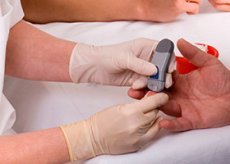
For mass detection of hyperglycemia, indicator paper impregnated with glucose oxidase, peroxidase and compounds that are colored in the presence of glucose is used. Using a portable device - a glucometer, operating on the principle of a photocalorimeter, and the described test paper, it is possible to determine the glucose content in the blood within the range from 50 to 800 mg%.
A decrease in blood glucose levels relative to the norm is observed in diseases caused by absolute or relative hyperinsulinism, prolonged fasting and heavy physical exertion, and alcoholism.
Oral tests used to determine glucose tolerance
The most widely used are the standard oral glucose tolerance test with a 75 g glucose load and its modification, as well as the test with a test breakfast (postprandial hyperglycemia).
The standard glucose tolerance test (STT), in accordance with the WHO recommendation (1980), is a study of glycemia on an empty stomach and every hour for 2 hours after a single oral load of 75 g of glucose. For the children being examined, a glucose load of 1.75 g per 1 kg of body weight (but not more than 75 g) is recommended.
A necessary condition for the test is that the patient consumes at least 150-200 g of carbohydrates per day with food for several days before the test, since a significant reduction in the amount of carbohydrates (including easily digestible ones) helps to normalize the sugar curve, which complicates the diagnosis.
Changes in blood parameters in healthy individuals, patients with impaired glucose tolerance, as well as questionable results when using a standard glucose tolerance test are presented in the table.
Blood glucose content during oral (75 g) glucose tolerance test, mmol/l
Research conditions |
Whole blood |
Venous blood plasma |
|
Venous |
Capillary |
||
Healthy |
|||
On an empty stomach |
<5.55 |
<5.55 |
<6.38 |
2 hours after exercise |
<6.70 |
<7.80 |
<7.80 |
Impaired glucose tolerance |
|||
On an empty stomach |
<6.7 |
<6.7 |
<7.8 |
2 hours after exercise |
>6.7-<10.0 |
>7.8-<11.1 |
>7.8-<11.1 |
Diabetes mellitus |
|||
On an empty stomach |
>6.7 |
>6.7 |
>7.8 |
2 hours after exercise |
>10.0 |
>11.1 |
>11.1 |
Since the blood sugar level 2 hours after the glucose load is of greatest importance in assessing glycemic indices during the oral glucose tolerance test, the WHO Committee of Experts on Diabetes Mellitus proposed a shortened version for mass studies. It is carried out similarly to the usual one, but the blood sugar test is performed only once 2 hours after the glucose load.
A carbohydrate load test can be used to study glucose tolerance in a clinical or outpatient setting. The subject must eat a test breakfast containing at least 120 g of carbohydrates, 30 g of which must be easily digestible (sugar, jam, preserves). Blood sugar is tested 2 hours after breakfast. The test indicates impaired glucose tolerance if glycemia exceeds 8.33 mmol/l (pure glucose).
Other glucose load tests do not have any diagnostic advantages, according to WHO experts.
In diseases of the gastrointestinal tract accompanied by impaired glucose absorption (post-resection gastric syndrome, malabsorption), a test with intravenous administration of glucose is used.
Methods of diagnosing glucosuria
The urine of healthy people contains very small amounts of glucose - 0.001-0.015%, which is 0.01-0.15 g/l.
When using most laboratory methods, the above amount of glucose in urine is not determined. Some increase in glucosuria, reaching 0.025-0.070% (0.25-0.7 g / l), is observed in newborns during the first 2 weeks and in elderly people over 60 years. Glucose excretion in urine in young people depends little on the amount of carbohydrates in the diet, but can increase 2-3 times compared to the norm against the background of a high-carbohydrate diet after prolonged fasting or a glucose tolerance test.
In mass screening of the population to detect clinical diabetes, methods are used to quickly detect glucosuria. The indicator paper "Glukotest" (produced by the Reagent plant, Riga) has high specificity and sensitivity. Similar indicator paper is produced by foreign companies under the names "test-type", "clinistics", "glukotest", "biofan" and others. The indicator paper is impregnated with a composition consisting of glucose oxidase, peroxidase and ortholidin. A strip of paper (yellow) is dipped in urine; if glucose is present, the paper changes color from light blue to blue after 10 s due to the oxidation of ortholidin in the presence of glucose. The sensitivity of the above types of indicator paper ranges from 0.015 to 0.1% (0.15-1 g / l), while only glucose without reducing substances is determined in urine. To detect glucosuria, it is necessary to use daily urine or urine collected within 2-3 hours after a test breakfast.
Glucosuria detected by one of the above methods is not always a sign of the clinical form of diabetes mellitus. Glucosuria can be a consequence of renal diabetes, pregnancy, kidney disease (pyelonephritis, acute and chronic nephritis, nephrosis), Fanconi syndrome.
Glycated hemoglobin
Methods that allow detecting transient hyperglycemia include determining glycosylated proteins, the period of presence of which in the body varies from 2 to 12 weeks. By binding to glucose, they accumulate it, representing a kind of memory device that stores information about the glucose level in the blood (Blood glucose memory). Hemoglobin A in healthy people contains a small fraction of hemoglobin A 1c, which includes glucose. The percentage of glycosylated hemoglobin (HbA 1c ) is 4-6% of the total amount of hemoglobin. In patients with diabetes mellitus with constant hyperglycemia and impaired glucose tolerance (with transient hyperglycemia), the process of glucose incorporation into the hemoglobin molecule increases, which is accompanied by an increase in the HbA 1c fraction. Recently, other small fractions of hemoglobin have been discovered - A 1a and A 1b, which also have the ability to bind to glucose. In patients with diabetes mellitus, the total content of hemoglobin A 1 in the blood exceeds 9-10% - a value characteristic of healthy individuals. Transient hyperglycemia is accompanied by an increase in the levels of hemoglobin A 1 and A 1c for 2-3 months (during the life of the erythrocyte) and after normalization of the blood sugar level. To determine glycosylated hemoglobin, the following methods are used: column chromatography or calorimetry.
Determination of fructosamines in blood serum
Fructosamines belong to the group of glycosylated proteins of blood and tissues. They arise in the process of non-enzymatic glycosylation of proteins during the formation of aldimine, and then ketoamine. An increase in the content of fructosamine (ketoamine) in the blood serum reflects a constant or transient increase in the level of glucose in the blood for 1-3 weeks. The final product of the reaction is formazan, the level of which is determined spectrographically. The blood serum of healthy people contains 2-2.8 mmol/l of fructosamines, and in cases of impaired glucose tolerance - more.
 [ 8 ], [ 9 ], [ 10 ], [ 11 ], [ 12 ], [ 13 ], [ 14 ], [ 15 ]
Determination of C-peptide
Its level in the blood serum allows to evaluate the functional state of the β-cell apparatus of the pancreas. C-peptide is determined using radioimmunological test kits. Its normal content in healthy individuals is 0.1-1.79 nmol/l, according to the test kit of the company "Hoechst", or 0.17-0.99 nmol/l, according to the company "Byk-Mallin-crodt" (1 nmol/l = 1 ng/ml x 0.33). In patients with type I diabetes mellitus, the level of C-peptide is reduced, in type II diabetes mellitus it is normal or increased, and in patients with insulinoma it is increased. The level of C-peptide can be used to judge the endogenous secretion of insulin, including against the background of insulin therapy.
[ 16 ], [ 17 ], [ 18 ], [ 19 ]
Determination of immunoreactive insulin
The study of immunoreactive insulin (IRI) allows to judge the secretion of endogenous insulin only in patients who do not receive insulin preparations and have not received them before, since antibodies are formed to exogenous insulin, distorting the result of determining immunoreactive insulin. The content of immunoreactive insulin in the serum of healthy people is 0-0.29 μU/ml. Type I diabetes mellitus is characterized by a reduced, and type II - by a normal or increased basal insulin level.
[ 20 ], [ 21 ], [ 22 ], [ 23 ], [ 24 ], [ 25 ], [ 26 ], [ 27 ]
Tolbutamide test (according to Unger and Madison)
After fasting blood sugar testing, the patient is given 20 ml of 5% tolbutamide solution intravenously and blood sugar is tested again after 30 minutes. In healthy individuals, blood sugar decreases by more than 30%, and in patients with diabetes - less than 30% of the initial level. In patients with insulinoma, blood sugar drops by more than 50%.
[ 28 ], [ 29 ], [ 30 ], [ 31 ], [ 32 ]
Glucagon
The content of this hormone in the blood is determined by the radioimmunological method. Normal values are 0-60 ng/l. The level of glucagon in the blood increases with decompensated diabetes mellitus, glucagonoma, starvation, physical exertion, chronic liver and kidney diseases.
If the disease developed in childhood or adolescence and was compensated for by insulin administration for a long period, then the question of the presence of diabetes type I is not in doubt. A similar situation arises in the diagnosis of diabetes type II, if compensation for the disease is achieved by diet or oral hypoglycemic drugs. Difficulties usually arise when a patient who was previously classified as suffering from diabetes type II needs to be transferred to insulin therapy. About 10% of patients with diabetes type II have autoimmune damage to the islet apparatus of the pancreas, and the question of the type of diabetes can only be resolved with the help of a special examination. The method that allows in this case to establish the type of diabetes is the study of C-peptide. Normal or increased values in the blood serum confirm the diagnosis of type II, and significantly reduced values - type I.
Methods for detecting potential impaired glucose tolerance (IGT)
The group of persons with potential NTG is known to include children of two parents with diabetes, a healthy twin from a pair of identical twins if the second one has diabetes (especially type II), mothers who have given birth to children weighing 4 kg or more, as well as patients with the presence of a genetic marker of type I diabetes. The presence of diabetogenic HLA histocompatibility antigens in the subject in various combinations increases the risk of developing type I diabetes. Predisposition to type II diabetes can be expressed in facial flushing after taking 40-50 ml of wine or vodka, if it is preceded (12 hours before - in the morning) by taking 0.25 g of chlorpropamide. It is believed that in people predisposed to diabetes, under the influence of chlorpropamide and alcohol, activation of enkephalins and dilation of skin vessels occurs.
The potential impairment of glucose tolerance should apparently also include the "syndrome of inappropriate insulin secretion", which is expressed in periodically occurring clinical manifestations of spontaneous hypoglycemia, as well as (an increase in body weight of patients, which may precede the development of IGT or clinical diabetes by several years. The GTT indicators in subjects at this stage are characterized by a hyperinsulinemic type of sugar curve.
To detect diabetic microangiopathy, methods of vital biopsy of skin, muscles, gums, stomach, intestines, kidneys are used. Light microscopy allows to detect proliferation of endothelium and perithelium, dystrophic changes of elastic and argyrophilic walls of arterioles, venules and capillaries. Using electron microscopy, it is possible to detect and measure thickening of the capillary basement membrane.
To diagnose pathology of the visual organ, according to the methodological recommendations of the Ministry of Health of the RSFSR (1973), it is necessary to determine the visual acuity and fields. With the help of biomicroscopy of the anterior part of the eye, it is possible to detect vascular changes in the conjunctiva, limbus, and iris. Direct ophthalmoscopy and fluorescent angiography allow us to assess the condition of the retinal vessels and identify the signs and severity of diabetic retinopathy.
Early diagnostics of diabetic nephropathy is achieved by identifying microalbuminuria and puncture biopsy of the kidneys. Manifestations of diabetic nephropathy must be differentiated from chronic pyelonephritis. Its most characteristic signs are: leukocyturia in combination with bacteriuria, asymmetry and change in the secretory segment of the renogram, increased excretion of beta 2 -microglobulin in urine. For diabetic nephromicroangiopathy without pyelonephritis, an increase in the latter is not noted.
Diagnosis of diabetic neuropathy is based on the patient's examination data by a neurologist with the use of instrumental methods, including electromyography, if necessary. Autonomic neuropathy is diagnosed by measuring the variation of cardiointervals (which is reduced in patients) and conducting an orthostatic test, studying the vegetative index, etc.