Medical expert of the article

New publications
Deforming osteoarthritis of the hip joint
Last reviewed: 29.06.2025

All iLive content is medically reviewed or fact checked to ensure as much factual accuracy as possible.
We have strict sourcing guidelines and only link to reputable media sites, academic research institutions and, whenever possible, medically peer reviewed studies. Note that the numbers in parentheses ([1], [2], etc.) are clickable links to these studies.
If you feel that any of our content is inaccurate, out-of-date, or otherwise questionable, please select it and press Ctrl + Enter.
With progressive dystrophic and degenerative processes in the bone and joint apparatus against the background of cartilage lesions of the hip joint, the doctor diagnoses coxarthrosis. Another name for this pathology is deforming osteoarthritis of the hip joint. The pathology is characterized by arthralgia, limitation of the functional ability of the articulation, as well as its curvature. Treatment is directed mainly at inhibiting further aggravation of the disease and improving the patient's condition. The pathological process progresses slowly but steadily: ankylosis and joint instability may form. [1]
Epidemiology
According to statistics, deforming osteoarthritis of the hip joint affects about 15% of the world's population. However, many experts believe that there are actually many more patients, because in many patients the pathological process is asymptomatic. Doctors note that coxarthrosis often becomes an accidental finding - for example, when performing X-rays for another disease.
Presumably, up to the age of fifty, men are slightly more often affected than women (by about 20%). This is primarily due to the high percentage of male osteonecrosis of the femoral head. After the age of 50, deforming osteoarthritis of the hip joint is diagnosed more often in women, which can be explained by hormonal changes and the associated deterioration of the musculoskeletal system.
Today, the situation with osteoarthritis in many countries is only getting worse. Specialists explain this by a decrease in physical activity of the population and an increase in the number of obese patients.
Causes of the osteoarthritis of the hip joint.
The most common reason for the formation of osteoarthritis of the hip joint is the discrepancy between the joint load and the compensatory "reserve" of the joint. The immediate "gas pedals" of the development of pathology are:
- Overweight;
- Being on your feet all the time;
- Spinal curvature;
- Intense sports activities (jumping, lifting and carrying heavy weights, running).
A certain role in the development of pathology is also attributed to such factors as metabolic disorders, abrupt changes in hormonal balance, trophic and blood circulation disorders in the hip joint, genetic predisposition to pathologies of cartilage tissue, advanced age, traumatic injuries. Often the disease is found in patients with psoriatic and rheumatoid arthritis. [2]
Risk factors
Risk factors for the development of deforming osteoarthritis of the hip joint are divided into permanent and those that can still be influenced (changed).
Permanent factors include congenital or structural abnormalities:
- Hip dysplasia;
- Epiphyseolysis of the femoral head;
- Legg-Calve-Perthes syndrome;
- Anomalies of cartilage development;
- Femoroacetabular impingement disease.
Modifiable factors include:
- Overweight;
- Professional sports - in particular injury-prone and high-impact sports;
- Regular lifting and carrying of heavy objects, standing work;
- Regular exposure to vibration, frequent repetitive strain on the hip joint;
- Work involving frequent bending and squatting.
Risk groups include both professional athletes and the elderly, as well as women in pregnancy and menopause. [3]
Pathogenesis
Deforming osteoarthritis of the hip joint is a pathology that causes localized destruction of articular cartilage tissue, accompanied by changes in the subchondral bone with further formation of bone outgrowths along the edges. These pathological changes may be a consequence of trauma or other damaging effects, acting as a compensatory response. However, against the background of constant such impact gradually occurs failure of the compensatory mechanism - for example, in patients with obesity, when the body weight regularly loads the diseased joint. Movement in the joint becomes limited, and further - and even impossible: bone, cartilage, and fibrous fusion of the articular ends is formed.
Joint immobility can be the result of traumatic injury (wound, fragmentary closed fracture, contusion, etc.), infection or degenerative disease, improper treatment of pathologic intra-articular processes. [4]
Symptoms of the osteoarthritis of the hip joint.
People with deforming osteoarthritis predominantly complain of pain and limited range of motion in the hip joint. However, in individual cases - for example, in the presence of cystic lesions of the femoral head - pain may be absent.
Localization of pain - groin area on the side of the pathological process, with possible irradiation to the lower parts up to the ankle.
There is a correlation of pain with physical activity (except for the last stage, when it is a permanent chronic pain). The intensity of pain sensations varies, from occasional discomfort to a persistent and pronounced syndrome.
The patient's attempts to subdue the discomfort lead to a gradual transfer of the weight load to the healthy leg. Over time, this is reflected in the gait: a limp appears.
Other common complaints include a feeling of stiffness in the hip joint, especially when taking the first steps after prolonged rest. The situation is more pronounced if, in addition to deforming osteoarthritis, a person suffers from rheumatoid arthritis or gout.
Movements in the joint may be difficult, up to the point of complete inability to perform them. Stable contractures arise, and the spine becomes curved with lumbar hyperlordosis.
The first signs of reduced joint function include difficulty in putting on shoes, playing sports, etc. Then it becomes difficult to walk, climb stairs, etc. [5]
Stages
The most typical manifestation of deforming arthrosis is pain in the hip joint. The severity of symptomatology is closely related to the stage of development of the disease process. So, at the initial stage, the patient complains only of a slight discomfort, transient motor stiffness. Over time, the clinical picture expands, the pain becomes chronic and increasing, motor capabilities deteriorate.
Most experts say there are three degrees of the disease:
- Deforming osteoarthritis of the hip joint of the 1st degree practically does not detect itself with symptoms, or they are so weak that they attract little attention of the patient. A slight discomfort occurs only on the background or after physical exertion, which patients associate with normal fatigue. Motor amplitude practically does not suffer. The radiologic picture demonstrates a slight narrowing of the articular gap. Treatment is conservative.
- Deforming osteoarthritis of the hip joint of the 2nd degree is accompanied by increasing pain, which is especially bothersome after joint load, meteorological changes. In the evening, discomfort is especially felt, a slight limitation of movements is noted. After a long stay "on the feet", the patient has a typical "duck" gait: a person during walking as if swaying left to right. Some difficulties may appear when trying to move the affected limb to the side, when putting on shoes. When getting on his feet after sitting for a long time, it is difficult for a person to take the first few steps. If at this stage the pathology is not treated, partial atrophy of the musculature, a slight shortening of the affected limb is possible. X-rays reveal a narrowing of the gap of the hip joint, the formation of bone growths, necrosis of the head of the iliac and femoral bone. Magnetic resonance imaging allows you to consider the dystrophy of cartilage tissue, bone particles in the joint cavity. Treatment is aimed at inhibition of degenerative processes: it can be conservative or surgical minimally invasive.
- Deforming osteoarthritis of the hip joint of the 3rd degree is accompanied by pronounced movement disorders, up to complete immobilization. Pain syndrome is characterized by constancy and ceases to depend on physical activity. In addition to pain, patients complain of insomnia and associated irritability, depression. The hip joint is immobilized, there is an obvious lameness. In the course of radiography, a complete destruction of cartilage tissue and the head of the femur is noted, the formation of large marginal growths. Treatment is surgical.
Complications and consequences
In most patients, deforming osteoarthritis of the hip joint progresses very slowly, over years and decades. If treatment is started in time, this process is greatly slowed down, which makes it possible to maintain motor activity. If the necessary treatment is not available, the risk of complications increases:
- Severe curvature of the hip joint and spinal column;
- Limitation of mobility up to complete immobilization of the limb (ankylosis);
- Shortening of the affected leg;
- Of bone deformities.
The patient loses the ability to work, and sometimes the ability to move and self-care. In advanced cases, the patient's quality of life suffers. It is possible to assign a disability group, which depends on the stage and volume of the pathological process. [6]
Diagnostics of the osteoarthritis of the hip joint.
Deforming osteoarthritis of the hip can be suspected if the present complaints and symptoms are associated with relevant risk factors such as hip injuries, heavy work conditions, rheumatoid arthritis, etc.
Physical examination may be useful only at relatively late stages of osteoarthritis. There is a worsening of pain syndrome in the groin at the time of internal rotation of the hip, sometimes - a characteristic crunch in the extreme position of the joint. Contractures, stable motor limitations, and joint deformities are noted.
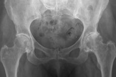
Among the typical X-ray manifestations:
- Marginal bone growths;
- Narrowed joint space;
- Signs of subchondral osteosclerosis of the acetabulum and femoral head;
- Acetabular floor protrusion.
The development of osteonecrosis of the head is indicated by these points:
- The osteonecrosis focus is surrounded by an area of osteosclerosis;
- Bone tissue is discharged under the loaded pole of the head in the form of a "crescent";
- There is an impression fracture in the loaded section of the head above the osteonecrotic focus;
- The articular surface is deformed;
- The cartilage has been destroyed.
In addition to radiography, other instrumental diagnostics are used:
- Magnetic resonance imaging and computed tomography are used to clarify the structural and other features of the pathological focus, assess the degree of lesion and localization.
- Radionuclide scanning helps to determine the focus of osteonecrosis of the head (the study is especially relevant for patients with deforming osteoarthritis on the background of sickle cell anemia).
Laboratory tests are prescribed as auxiliary diagnostic measures to differentiate or confirm the secondary pathology. Particular attention is paid to the exclusion of such diseases as gout, systemic lupus erythematosus, sickle cell anemia, seropositive and seronegative rheumatoid arthritis.
Differential diagnosis
Radiologic and tomographic examination usually provides comprehensive information about the pathology, which allows to establish the correct diagnosis. In general, deforming osteoarthritis of the hip joint should be distinguished from diseases such as:
- Lumbar osteochondrosis;
- Spinal canal stenosis;
- Paresthetic meralgia, or Berngardt-Roth disease (lateral cutaneous femoral nerve syndrome);
- Trochanteritis (acetabular bursitis);
- Metastases to the femur and pelvis;
- Coxitis;
- Pelvic fracture, femoral neck fracture;
- Fibromyalgia.
In some situations, intra-articular blockades with anesthetic agent are performed to determine the source of pain syndrome (in the absence of X-ray pathology). Hip puncture is performed with further bacteriological analysis of intra-articular fluid. If indicated, trepanobiopsy and histologic examination of the biomaterial, computer or magnetic resonance imaging of the lumbar spine may be recommended.
Treatment of the osteoarthritis of the hip joint.
Treatment measures include conservative therapy and surgical intervention. The choice of therapeutic tactics depends on the intensity of symptoms, the age of the patient, the prevalence of the pathologic focus, the severity of biomechanical intra-articular disorders, and the volume of osteonecrotic lesions.
Therapeutic procedures are aimed at reducing pain, restoring motor amplitude and functionality of the hip joint, normalizing limb length, and preserving the articulation damaged by osteonecrosis.
Non-medicamentous influences include steps such as these:
- Normalization of body weight;
- Physical therapy;
- Reducing the load on the affected limb with crutches, orthopedic devices, etc.
Drug therapy usually consists of taking analgesics (non-steroidal anti-inflammatory drugs), chondroprotectors, antispasmodics. If necessary, the doctor adjusts the basic therapy - for example, patients with rheumatoid arthritis or gout. [7]
Medications
Medications are prescribed to reduce symptoms, to repair damaged tissues and to inhibit subsequent degenerative processes. The following groups of drugs are most in demand:
- Nonsteroidal anti-inflammatory drugs that relieve pain and inflammatory reaction (Ibuprofen, Ketorol, Diclofenac, Indomethacin - in the form of tablets, injections, external preparations, suppositories);
- Corticosteroid hormonal agents that control pain syndrome (corticosteroids are more often injected directly into the joint cavity);
- Analgesics and antispasmodics (particularly Midocalm);
- Chondroprotectors (glucosamine, chondroitin, etc.).
Common drugs that require long-term and stable use are chondroprotectors, which saturate cartilage tissue with nutrients, inhibit degenerative processes, and stimulate the growth of new cells. Chondroprotectors are more effective if taken at the initial or moderate stage of pathology. The course of intake should be regular and prolonged (two months or more).
If deforming osteoarthritis is complicated by osteonecrosis of the femoral head, treatment is supplemented with hypolipidemic agents - for example:
|
Lovastatin |
The maximum dosage is 40 mg per day, and the starting dosage is 10 mg per day. Prolonged use may be accompanied by gastrointestinal disorders, headache, insomnia, dizziness. If this happens, it is necessary to consult a doctor for correction of prescriptions. |
Many experts point out the effectiveness of taking Stanozolol in the amount of 6 mg/day.
Favorable clinical and radiological dynamics is noted with the administration of vasodilators - for example, Prostacyclin derivatives.
In the early stages of osteoarthritis and osteonecrosis are effective:
|
Enoxaparin |
Low-molecular-weight heparin, an anticoagulant, is prescribed in individual dosage, after assessing the risk of thromboembolic complications and hemorrhagic consequences. The most commonly used dose is 1.5 mg/kg once daily by subcutaneous injection, on average for 10 days, under the supervision of a physician. |
|
Alendronate |
Alendronic acid preparation, taken in the morning, orally, 2 hours before breakfast. It is recommended to combine with vitamin D and calcium preparations. Treatment is usually prolonged. Possible side effects: hypersensitive reactions, abdominal pain, abdominal bloating, digestive disorders. |
|
Naropin |
Prolonged injection of the drug through a catheter into the epidural space in anesthetic concentrations (determined individually) for a week is practiced. This procedure helps prevent collapse of the femoral head. |
The above drugs should be combined with symptomatic treatment, taking nonsteroidal anti-inflammatory drugs, chondroprotectors, antispasmodics.
Physiotherapy treatment
The main recommended method of treatment of deforming osteoarthritis of the hip joint is shockwave therapy. In the first or second degree of pathology, the procedure allows you to quickly relieve pain syndrome, restore movement, slow down the destruction of articular tissues and activate recovery processes.
The impact of acoustic oscillations of infrasound frequency penetrates the affected hip joint unhindered and acts directly on the focus of the inflammatory, degenerative and dystrophic process, improving blood circulation and trophics. The treatment works in a similar way to intensive manual therapy: the blood supply to the tissues is improved, stagnation disappears and recovery is initiated.
According to experts, shockwave therapy quickly improves local metabolic processes and not only eliminates the symptoms of osteoarthritis, but also partially eliminates the cause of its development. The resulting effect is long-lasting and sustainable.
It is possible to practice physiotherapeutic treatment and at the third degree of pathology against the background of the main therapeutic measures. However, in this case, shockwave therapy is more appropriate at the stage of rehabilitation after hip arthroplasty. [8]
Surgical treatment
If deforming osteoarthritis is accompanied by severe wear and tear of the hip joint, is not amenable to medication, and severe pain occurs not only during loading, but also in a calm state, the doctor may recommend surgical replacement of the joint with a prosthesis. The surgery helps to reduce painful symptoms and restore function.
Preparation for surgery is carried out on an outpatient basis. The course of manipulation is approximately as follows: under epidural or general anesthesia, the hip joint is exposed and the head is removed along with the surface of the joint socket. An analog of the acetabular cup and a prosthesis with a synthetic head is implanted inside, which is fixed using bone cement or another method. After the intervention, the patient stays under inpatient observation for at least two weeks. The final rehabilitation is carried out in a special clinic or department. In the beginning, the patient is offered to perform appropriate exercises on crutches, and by the second month, the full allowable load on the hip joint is achieved.
Prevention
Preventive measures will not be superfluous, both for people with healthy hip joints, and for patients with deforming osteoarthritis. To prevent the development, as well as to inhibit the progression of pathology, doctors recommend:
- Adhere to the rules of proper (complete, balanced) nutrition;
- To control your own body weight;
- Keep physically fit, be active, do regular morning exercises and take long walks;
- Avoid trauma, hypothermia.
It is important to avoid overloading the hip joints, timely and qualitatively treat any injuries of the extremities (bruises, fractures, sprains) and pathologies of the bone system (flat feet, curvature of the spine, dysplasias), be physically active.
Forecast
Deforming osteoarthritis of the hip joint is better treated in the early stages of progression. The advanced form is difficult to treat, often requiring endoprosthesis. Among other possible complications:
- Infectious inflammatory pathologies;
- Pinched sciatic or femoral nerve;
- Bursitis;
- Subluxation;
- Tendovaginitis.
Osteoarthritis exacerbations are related to the periodicity of the inflammatory process. In most cases, relapses are aseptic inflammation occurring after injuries or transferred diseases. During these periods, pain increases, fever, periarticular edema may be bothersome.
To improve the prognosis, doctors advise timely referral to doctors, fulfill all their appointments, and in the presence of obesity - follow a diet. Reducing body weight helps to relieve the damaged articulation and alleviate symptoms. However, a balanced diet is shown not only to overweight people, but also to all other patients, as proper nutrition helps to improve the nutrition of cartilage and bone tissue, stabilize the water-electrolyte balance. The diet should be free from an abundance of animal and emulsified fats, sweets, preservatives, smoked meats, pickles. For the regeneration of cartilage in the body should be introduced a sufficient amount of protein - for example, in the form of white meat, dairy products, eggs. The presence of collagen in dishes is mandatory: experts advise regular consumption of all kinds of jellies, jellies, kisel, marmalade, etc.
All patients, regardless of the stage of the disease, should relieve the affected limb as much as possible - for example, use crutches, canes and other orthopedic devices. Degenerative pathology, such as deforming osteoarthritis of the hip joint, is irreversible, but early treatment offers a better chance of preserving mobility.