Medical expert of the article

New publications
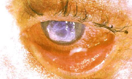
Conjunctivitis caused by physical and chemical irritants
Last reviewed: 07.07.2025

All iLive content is medically reviewed or fact checked to ensure as much factual accuracy as possible.
We have strict sourcing guidelines and only link to reputable media sites, academic research institutions and, whenever possible, medically peer reviewed studies. Note that the numbers in parentheses ([1], [2], etc.) are clickable links to these studies.
If you feel that any of our content is inaccurate, out-of-date, or otherwise questionable, please select it and press Ctrl + Enter.
Industrial and other chemicals can cause follicular conjunctivitis. Treatment of acute conjunctivitis in patients using contact lenses requires special attention. These patients are prone to the development of corneal ulcers caused by prolonged hypoxia. In some cases, the presence of pathogenic bacterial flora leads to the appearance of rapidly progressing bacterial ulcers. Negative effects can be caused by defects in the selection of contact lenses, as well as an individual reaction to wearing them.
"Artificial" conjunctivitis
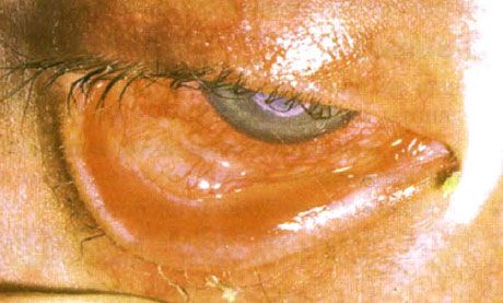
"Artificial" conjunctivitis develops due to the patient's own deliberate actions (for example, as a result of a burn or exposure to chemical irritants). The process is usually localized in the lower third of the eyeball and on the conjunctiva of the lower eyelid, accompanied by irritation of the eyelid and cheek.
Phlyctenular conjunctivitis
Phlyctenular conjunctivitis in some cases accompanies tuberculosis or staphylococcal eyelid infection, although it is usually idiopathic in origin:
- a single, limited inflammatory focus with a white center, usually located in the limbus area;
- transient course;
- duration of existence is about two weeks;
- tendency to exacerbations;
- scanty clinical symptoms.
Dendritic conjunctivitis
- Thickened nodular "woody" conglomerates in the conjunctiva.
- The cause of the disease is unknown; in some cases it occurs after surgery or an infection.
- Sometimes has an autosomal recessive type of inheritance.
- When the lesions are surgically removed, it tends to relapse. Sometimes spontaneous resorption is observed.
Biotinidase deficiency
- Conjunctivitis.
- Optic nerve atrophy.
- Hypotension.
- Cramps.
- Alopecia.
- The use of biotin is indicated.
 [ 9 ], [ 10 ], [ 11 ], [ 12 ], [ 13 ], [ 14 ], [ 15 ]
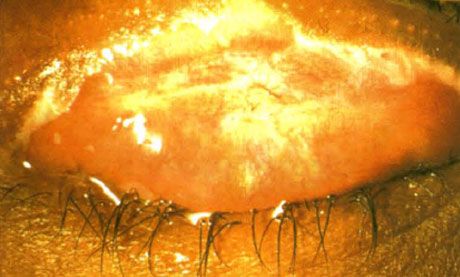
Episcleritis
- Moderate local conjunctival and episcleral injection (Fig. 6.3).
- A nodular form is also found.
- Irritation of the eyeball.
- Local and general non-steroidal anti-inflammatory therapy is indicated.
- Steroid drugs are recommended in cases that are resistant to current treatment.
Fig. 6.3. Episcleritis. Local deep injection and swelling of the episcleral tissue
Erythema multiforme - Stevens-Johnson syndrome
Cause
Apparently, the disease is a consequence of an acute allergic reaction.
Early manifestations
It occurs as a consequence of infectious diseases, most often herpes simplex, or individual intolerance to drugs, especially sulfonamides.
- Common skin rashes are “signal” lesions (prominent coin-shaped lesions of varying colors - from red to blue, painful to palpation).
- Mucous false films of red color, creating the impression of swelling and slowly resolving.
- Conjunctival pathology:
- conjunctivitis;
- mucous discharge;
- a reaction in the form of follicle formation is possible;
- conjunctival defects (Fig. 6.4);
- formation of false films;
- symblepharon;
- secondary bacterial infection.
Stevens-Johnson syndrome. Bilateral desquamative conjunctivitis with areas of necrosis. Severe keratitis, which caused scarring of the cornea. The situation was complicated by the addition of dry eye syndrome.
Late manifestations
- Scarring.
- Clogged tear ducts.
- Dry eye syndrome.
- Keratitis.
- Corneal vascularization and scarring.
- Scarring and keratinization of the eyelids.
Treatment
Acute phase
- Hospitalization.
- General use of steroid agents.
- Intensive topical application of preservative-free steroid preparations.
- Local application of preservative-free antibiotics.
- Cycloplegic drugs.
- Separation of intertissue adhesions with a glass rod.
- Treatment of skin.
Chronic phase
- For dry eye syndrome, emollients are used
- For xerosis, drugs from the retinoid group are prescribed.
- When trichiasis appears, epilation and cryotherapy are performed.
- Entropion is an indication for surgical intervention.
Xerophthalmos. Bitot's plaques appear as raised, scaly patches of conjunctiva located in the area not covered by the eyelids. As in this case, the lesions are often pigmented. (By courtesy of Mr. Michael Eckstein)
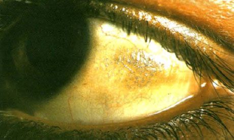
Avitaminosis A
- One of the most common causes of blindness worldwide.
- Associated with protein-calorie malnutrition.
- Accompanied by night blindness.
- Dry, wrinkled, dull conjunctiva.
- Bitot's plaques in the area of the eye slit not covered by the eyelids.
- Dry eye syndrome.
- Acute keratitis with keratomalacia and rapidly progressing corneal perforation.
What do need to examine?
How to examine?