Medical expert of the article

New publications
Confusion and diplopia
Last reviewed: 04.07.2025

All iLive content is medically reviewed or fact checked to ensure as much factual accuracy as possible.
We have strict sourcing guidelines and only link to reputable media sites, academic research institutions and, whenever possible, medically peer reviewed studies. Note that the numbers in parentheses ([1], [2], etc.) are clickable links to these studies.
If you feel that any of our content is inaccurate, out-of-date, or otherwise questionable, please select it and press Ctrl + Enter.
Binocular vision requires simultaneous bifoveal fixation by both eyes, i.e. each eye separately perceives the object of fixation, taking part in the formation of the image. The conditions necessary for binocular vision are:
- Overlapping fields of view.
- Correct neuromuscular development and coordination with the direction of the visual axes to the object.
- Normal visual pathways.
- Approximately the same image clarity and size in both eyes.
- Corresponding points of the retina, "cyclopean" eye.
- Confusion is the simultaneous perception of two superimposed but different images caused by stimulation of corresponding points (usually in the fovea) by different objects.
- Diplopia is the simultaneous perception of two images of one object. It occurs when visual images of one object are projected onto non-corresponding points of the retina. Simultaneous vision is the ability to perceive an object with both eyes at the same time.
- Visual direction is the projection of a given element of the retina in a special direction of subjective space.
- principal visual direction - a direction in external space interpreted as the line of sight. Usually it is the visual axis of the fovea;
- secondary visual directions - projected directions of extrafoveal points relative to the primary direction of the fovea.
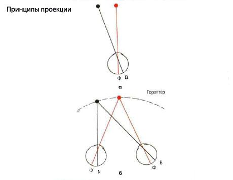
- Projection is the interpretation of the position of an object in space based on the stimulated elements of the retina.
If a red object stimulates the right foveola, and a black object located in the nasal half of the field stimulates elements of the temporal half of the retina, then the red object is interpreted by the brain as being directly projected with the head in a straight position, and the black object is interpreted as having arisen in the nasal half of the visual field. Similarly, nasal elements of the retina are projected onto the temporal half of the visual field, the upper ones onto the lower half, and vice versa.
- With both eyes open, a red object stimulates both foveae, the corresponding points of the retina. A black object stimulates not only the temporal retinal elements of the right eye, but also the nasally located retinal elements of the left eye. Thus, objects are projected into the nasal half of the visual field of the right eye and the temporal half of the visual field of the left eye. However, most of these retinal elements are corresponding points, so the object is projected into the same position in space (left).
- Retino-motor values. The image of an object in the peripheral visual field falls on the extrafoveal element. To establish fixation of the object, a saccade of a certain amplitude is required. Each extrafoveal element of the retina thus has a retino-motor value proportional to the distance from the fovea, which determines the required saccade amplitude for precise focusing of the object. The retino-motor value in the center of the foveola corresponds to zero and increases towards the periphery.
- Corresponding points are areas of the retina with the same subjective visual direction (e.g., direct projection to the fovea). Points on the nasal retina of one eye correspond to corresponding points on the temporal half of the retina of the other eye. This is the basis for normal retinal correspondence. For example, an object whose images are projected on the nasal half of the retina of the right eye and the temporal half of the retina of the left eye is projected on the right half of the visual space.
- The horopter is an imaginary plane in external space, all points of which stimulate only the corresponding elements of the retina and are therefore perceived by both eyes as one point. This plane passes through the intersection of the visual axes and thus includes the fixation point in binocular vision.
- The Panum fusion zone of binocular vision is the zone in front of and behind the horopter, within which an object is seen as single, although there is no precise stimulation of the corresponding elements. Objects outside the Panum zone are perceived as double. This is the basis of physiological diplopia. The Panum zone is narrow in the fixation zone (6 arc seconds) and widens toward the periphery, so objects within the horopter are seen as single. Objects within the Panum fusion zone are perceived as single and stereoscopic. Objects outside the Panum fusion zone are perceived as double.
- Sensory fusion is the combining of two sensory images from each eye in the visual cortex into a single visual image. Central sensory fusion combines images projected to the fovea, and peripheral sensory fusion combines images projected beyond the fovea.
- Motor fusion is the function of maintaining the correct position of the eyes to achieve bifocal fixation. The stimulus for motor fusion is the lisparity of the retinal image, which stimulates fusional vergence.
- Fusion vergence involves disjugate eye movements to overcome disparity in the retinal image. Fusion reserves can be measured using prisms or a synoptophore. Normal reserve values are:
- Convergence: about 15 D (fixation of a distant object) and 25 D (fixation of a close object).
- Divergence: about 25 D (fixation of a distant object) and 12 D (fixation of a close object).
- Vertical: 2-3 D.
- Cyclovergence: about 2.
Fusional convergence controls exophoria, while fusional divergence helps control esophoria. Fusional vergence mechanisms can be weakened by fatigue or illness, transforming phoria into tropia. The width of fusional vergence mechanisms can be increased by orthoptic exercises, such as fusional convergence when fixating a near object when convergence is weak.
- Stereopsis is the perception of depth (the third dimension, the first two being height and width). It occurs when horizontally disparate points are stimulated simultaneously by objects anterior and posterior to the fixation point, but within the Panum fusion zone. The fusion of such disparate images results in the perception of a single image in depth. The object is perceived stereoscopically (3D), since each eye sees different aspects of the object.
 [
[