Medical expert of the article

New publications
Color blindness and color perception test
Last reviewed: 29.06.2025

All iLive content is medically reviewed or fact checked to ensure as much factual accuracy as possible.
We have strict sourcing guidelines and only link to reputable media sites, academic research institutions and, whenever possible, medically peer reviewed studies. Note that the numbers in parentheses ([1], [2], etc.) are clickable links to these studies.
If you feel that any of our content is inaccurate, out-of-date, or otherwise questionable, please select it and press Ctrl + Enter.
Daltonism is a disorder of color perception. To determine it, special tests and tables are used. Let's consider the main methods of diagnosing this problem.
According to medical statistics, color blindness occurs in 0.4% of women and 8% of men. Violation of the ability to correctly perceive certain colors was officially recorded at the end of the 18th century. At the same time Dalton described the problem as not distinguishing only red color. To date, colors are a component of the symbolic system necessary for communication and control in the conditions of modern life. That is color perception has become an actively used component of vision.
Most often color blindness is hereditary, but in some cases acquired color blindness occurs. Genetic deviation is caused by the transmission of a damaged gene from mother to son with an X chromosome. Acquired form can be caused by eye injuries or diseases, chemical or drug exposure.
Depending on what shades a person distinguishes, there are several types of color blindness:
- Monochromia is the ability to distinguish only one of the three primary colors (red, green, blue). That is, a person's ability to see colors is practically impaired. With monochromia, the surrounding world looks in one color with indistinct transitions. Often this form of color blindness is accompanied by myopia and other eye diseases.
- Dichromia is an impairment in the recognition of one of the three primary colors. The most common problem is the perception of red color, which is confused with blue or green. At the same time color blind person normally perceives blue and green. In rare cases, problems with the recognition of green and blue colors are diagnosed.
- Trichromia is the most common type of color blindness. A person perceives all colors in a slightly different shade than people with normal color perception. Most often it is difficult to recognize close shades.
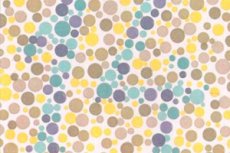
A person can independently replace problems with color perception. To diagnose color blindness, special tests are used to determine the type of the disorder. This can be the Ishihara color test and Rabkin's polychromatic tables (pictures of colored spots or dots that form a pattern recognizable by people with full vision). Regardless of what caused color blindness, the condition is incurable and irreversible.
Tests for color blindness and color perception
Daltonism is not a disease, as it refers to visual defects. Color perception is a person's ability to distinguish colors. There are several theories of color perception, the main one is Helmholtz's three-component theory. It is based on the fact that in the retina there are three types of cones, each of which is sensitive to light of a certain wavelength. That is, to the red, green and blue light spectra, which correspond to the primary colors.
Rabkin's polychromatic tables are used to identify color blindness and its manifestations. They allow to determine the degree of color perception:
- Trichromats are normal color perception.
- Protoanomalies are a perceptual disorder in the red spectrum.
- Deuteranomals - problems with the perception of the color green.
The test is performed by an ophthalmologist. For this purpose, images on the monitor or printed tables are used. The distance from the eyes to the picture should be 50-70 cm. At the same time, the monitor and the subject's eyes should be at the same level, it is not recommended to squint or tilt the head.
If the color vision disorder is acquired, it is subject to correction, but full restoration of color vision is impossible. Hereditary forms of pathology are incurable. Dantonism is not dangerous to health, but it can significantly worsen the quality of life.
Indications for the procedure
Total color blindness is a hereditary pathology. According to medical statistics, this problem is diagnosed in one in a million inhabitants of the planet. Color perception disorder is associated with gene defects at the molecular level. Color-sensitive nerve cells are located in the very center of the retina. They contain pigments, each of which is sensitive to blue, green or red. The stacking of the three primary colors in the brain's visual apparatus provides full color perception.
Due to the absence of one of the pigments, humans are unable to distinguish all colors.
- If two primary colors are recognized, the person is dichromate.
- People with a red pigment defect are more common, of which about 75% are abnormal trichromats.
- The less common blue pigment defect is tritanopia.
- People who do not distinguish between all three primary colors have a complete lack of color vision.
Indications for a color blindness test are based on various color perception disorders. The patient undergoes testing and further diagnosis, based on the results of which the doctor gives recommendations for color correction. Special contact lenses and glasses are used for this purpose.
A test for color blindness is recommended when planning a pregnancy, when one of the parents in the family had color blindness. In this case, the doctor studies the family history, conducts a set of instrumental and other diagnostic procedures to identify the carrier of the pathological gene.
Color blindness test for drivers
When obtaining a driver's license, a color blindness test is mandatory. For drivers, the inability to distinguish the color of traffic signals is not a serious problem.
According to studies, the ability to distinguish color has virtually no effect on road safety. That is, despite the fact that color blind people do not distinguish the colors of traffic lights, they can see a burning upper, middle or lower light.
The headlights of vehicles ahead are a major hazard. A driver with color blindness will not be able to tell if it is a reverse or brake light. Therefore, depending on the type of color vision impairment, a person may be denied a driver's license.
Color blindness test for children
The retina of the eye contains color-sensitive receptors - cones. Normally, there are three kinds of them, each of which is sensitive to one of the basic colors: green, blue, red. If any of the pigments is missing, the child does not distinguish one or more colors.
The color blindness test for children can detect color perception disorder.
- Most often the pathology is hereditary and is transmitted only through the mother's line. About 8% of boys and up to 0.4% of girls are color blind.
- In rare cases, the disorder develops as a result of damage to the retina or optic nerve. The acquired form has a progressive character. In this case, color blindness develops in the affected eye. The causes of the disorder can be: cataract, brain injury, prolonged use of medications.
The acquired form of color blindness is much more severe than the hereditary form. This is due to various complications for vision and the need for constant monitoring by an ophthalmologist.
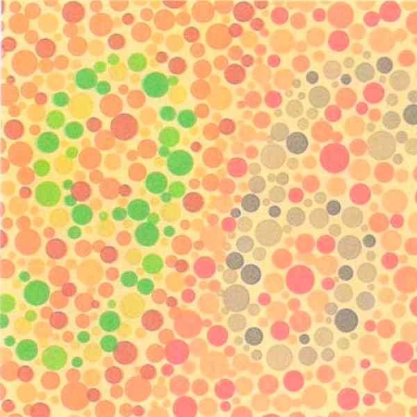
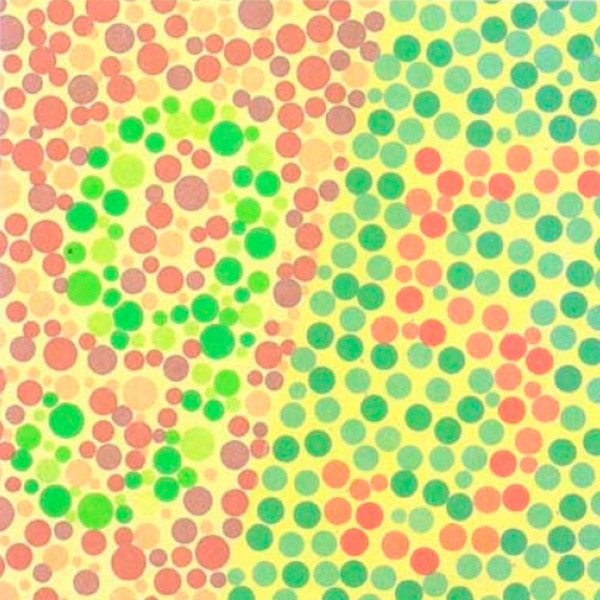
There are several methods to detect color recognition abnormalities in children. As a rule, testing is carried out for children over 3 years of age and before school. By this time, the child has already formed a color gamut and pathology can be identified. To determine the features of color perception, special polychromatic Rabkin tables are used. The drawings are circles and dots with different diameters and colors, but the same brightness.
If the child has color blindness, he will not see what is hidden in the scheme, because for him the whole picture will be homogeneous. If there are no vision problems, the child will see geometric figures and numbers made of circles of different colors.
If it is determined that the child has color blindness, the type of color blindness should be determined. This is necessary for further social adaptation. The following types of the disease are distinguished:
- Achromasia - color vision is completely absent. A person sees the surrounding world in shades of gray. This form is very rare and develops due to the absence of colored pigment in all the cones in the retina.
- Monochromasia is the perception of a single color. It is often accompanied by involuntary movements of the eyeballs (nystagmus) and photophobia.
- Dichromasia - the person distinguishes between two out of three colors.
- Protanopia is color blindness in the area of red. Children perceive red as a darker color than all other colors. They mix it with darker shades of other colors. And they see green as light gray or light yellow.
- Deuteranopia - lack of perception of green color. Green is mixed with light orange, light pink.
- Tritanopia is a perception disorder of the blue-violet spectrum. The person distinguishes shades of green and red. This type of color blindness is rare and is complicated by the absence of twilight vision.
The color blindness test allows for the timely identification of the type of congenital/acquired anomaly. In this way, parents can prepare their child appropriately not only for school, but also for later life.
Technique of the color blindness test
To test for color blindness, special tables and pictures with different colors and dots depicting numbers and figures are used. As a rule, these are the world-famous Rabkin's tables.
Abnormalities can vary. Some people see two colors because they are missing one of the pigments in the retina. There is also such a thing as complete color blindness, when a person perceives the surrounding world in gray tones.
The technique of testing is important. If the diagnostic procedure has been violated, the results of the test will be distorted.
The following rules should be followed during testing:
- The study is performed only under natural light.
- The person should be in good health (get plenty of sleep and be relaxed).
- The subject sits with his back to the window and the ophthalmologist sits across from him.
- If Rabkin's tables are used, they are shown vertically, at eye level and at a distance of 1 meter.
- The viewing time for each picture should be no more than 7 seconds.
The first two tables are seen the same way by all people, so their purpose is to visualize the testing. The remaining pictures allow you to recognize the problem. The color perception test cannot be performed online, because the monitor significantly distorts the color reality of the images.
The results are not counted, as any number of incorrect answers is a signal of visual pathology. The tests reliably establish the degree and type of impairment. Thus, one person will make a mistake already on the first problematic image, because he is unable to distinguish the red color, and another only on the last one because of problems with the recognition of green. To diagnose the type of abnormality, an additional control test is performed.
Test for color blindness type
Violation of color perception has a fairly wide classification. Test for the type of color blindness allows you to diagnose the features of pathology, causes and factors of its appearance, methods of correction. First of all, congenital and acquired color blindness are distinguished. The latter most often develops against the background of cataracts, body intoxication, CNS diseases, long-term medication.
- If a person has all three pigments present in his cones, he is trichromatic, that is, he has normal vision.
- In the absence of one pigment, a person is able to distinguish two primary colors - dichromacy. Complete absence of color perception is monochromacy.
- Monochromats are only able to detect the brightness of colors, which in turn comes in the form of cone and rod monochromats. The cone monochromats distinguish all colors as one color background. In the rod form of the pathology, the cones of the retina are completely absent. A person does not perceive more than one color and sees the surrounding world as gray.
- If the activity of pigment in the cones is reduced, this is abnormal trichromacy. It has several types, which differ depending on which color perception is impaired (protoanomaly, deuteroanomaly and tritanomaly). Color perception in such people is slightly distorted, so without special testing they may not even suspect the problem.
Various methods are used to diagnose visual features. The most popular ones include tests and such methods:
- Anomaloscopy is an examination of color vision that reveals abnormalities and their nature. The study is based on the eye's ability to perceive a given combination of red and green as yellow. Diagnosis is performed using an anomaloscope. The patient changes the proportions of red and green until the color of their mixture is identical to yellow for him.
- FALANT is a test used in the United States for new recruits to the Navy. The study consists of placing a lighthouse at a certain distance from the person on which two of the main colors (red, white, green) light up simultaneously. The test subject must name the color. To detect color blindness, the color is muted. Dichromats and many trichromats do not pass this test.
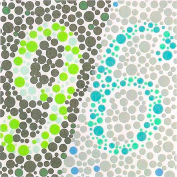
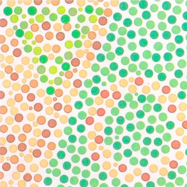
- Ishihara test - this test is widely used by Western ophthalmologists to detect color perception disorders. The test is similar to Rabkin's tables. The patient is shown cards with a background of multicolored spots on which the image is encrypted. In this case, some hidden patterns may be visible only in pathologies.
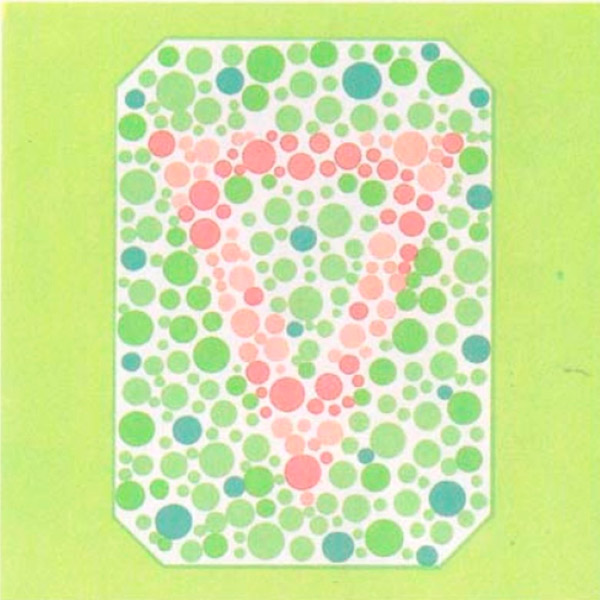
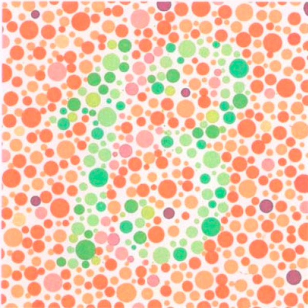
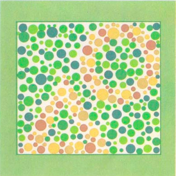
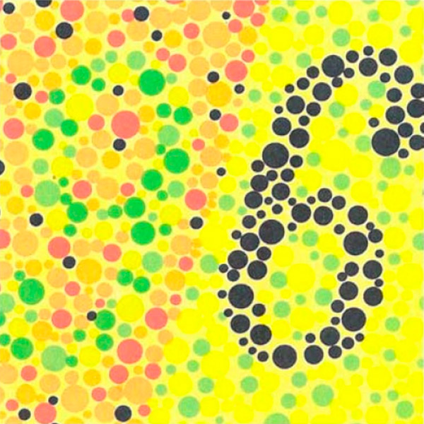
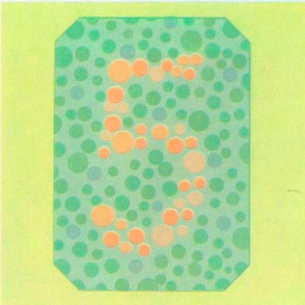
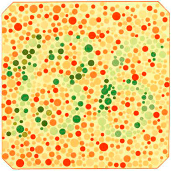
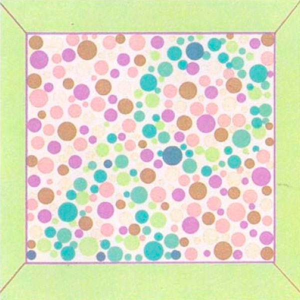
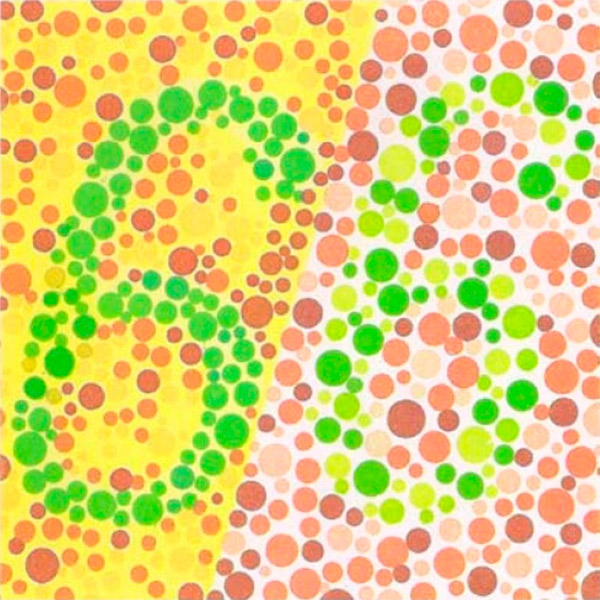
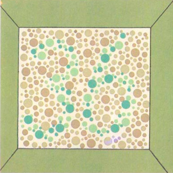
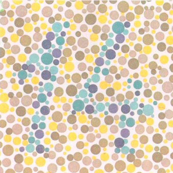
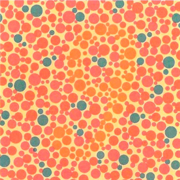
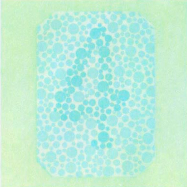
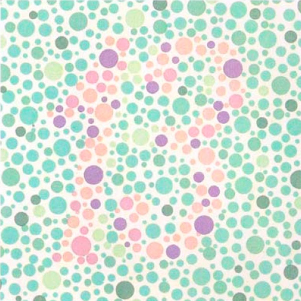
- Rabkin's Tables is a method for detecting color vision abnormalities. It most accurately determines the type and degree of color blindness, which makes it the most popular worldwide. For diagnosis, polychromatic tables are used - these are pictures of multicolored dots on which numbers, geometric figures are depicted. With problems with color recognition, a person is unable to see the hidden pattern. The test consists of 48 tables, which are divided into main 1-27 and control 28-48 groups.
Problems with color recognition can be subtle or pronounced. The color blindness test allows to detect any deviations in color perception. The test is mandatory for the military, when obtaining a driver's license and other professions in which it is important to recognize colors correctly.
The American colorblindness test
In the U.S., to assess color vision in future military personnel, the FALANT test is conducted. The essence of the American color blindness test is that at a certain distance from a person there is a beacon that emits a certain color (one of the three basic colors). The test subject's task is to determine the color of the light beam.
The light beam itself combines three colors and is passed through a special attenuating filter. Because of this, people with color blindness are unable to detect the color of the beam. The error of the American test is 30%, so people with a mild form of color blindness pass this test.
Rabkin table
Various methods and studies are used to determine color blindness and its manifestations. Rabkin's tables deserve special attention. They are recognized as one of the widely used diagnostic methods for suspected deviations in the perception of color gamut. This test most accurately determines the type and degree of color blindness.
According to the degree of color perception, people are divided into three types:
- Trichromats are the norm.
- Protoanopes are a pathology of recognition in the red spectrum.
- Deuteranopes is a disturbance in the perception of the color green.
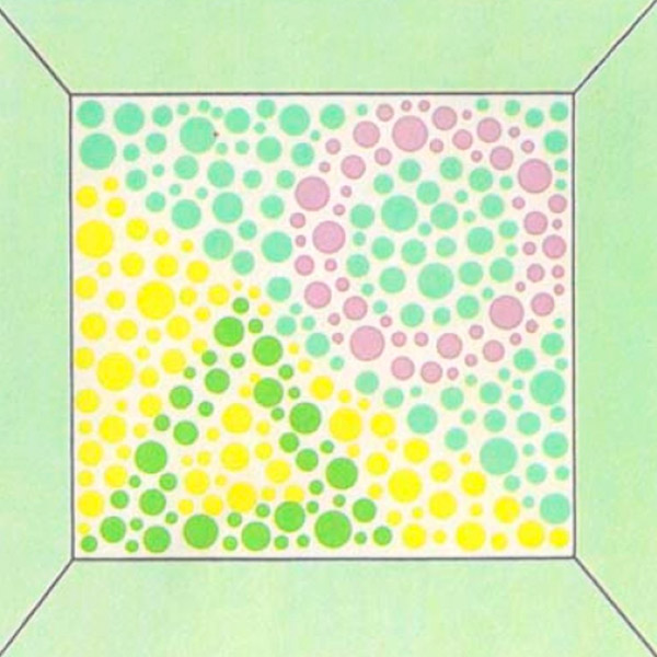
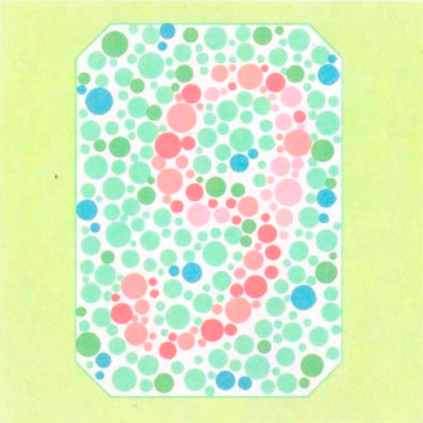
Rabkin's polychromatic tables are peculiar images with geometric figures and numbers. The drawings consist of circles of different sizes and colors, but identical in color saturation.
This is due to the fact that with pathology a person is unable to identify color, but can distinguish brightness. Also, the subject cannot discern a number or figure in a picture seen by trichromats, but identifies images indistinguishable to the healthy eye.
The test kit consists of 48 paper and ophthalmologist-calibrated tables that are divided into two groups:
- Basic - Tables 1 through 27 are used to diagnose color blindness and determine its degree.
- Control - 28 to 48 table, confirms the presence of the anomaly and allows to clarify the diagnosis.
In order for the test results to be as reliable as possible, a number of conditions are very important:
- The room where the study is taking place should have natural light.
- The test subject should sit with his/her back to the window.
- Tables are placed vertically and at eye level of the person.
- The distance from your eyes to the table should be 1 meter.
- The duration of viewing each picture is 5-7 seconds.
- The probationer must be in good health.
Main Table Features:
- The figure shows the numbers 9 and 6, which are seen by both healthy and abnormal people. This image shows how the test works and allows you to identify simulation when taking the test.
- The picture shows a square and a triangle that is visible to absolutely everyone.
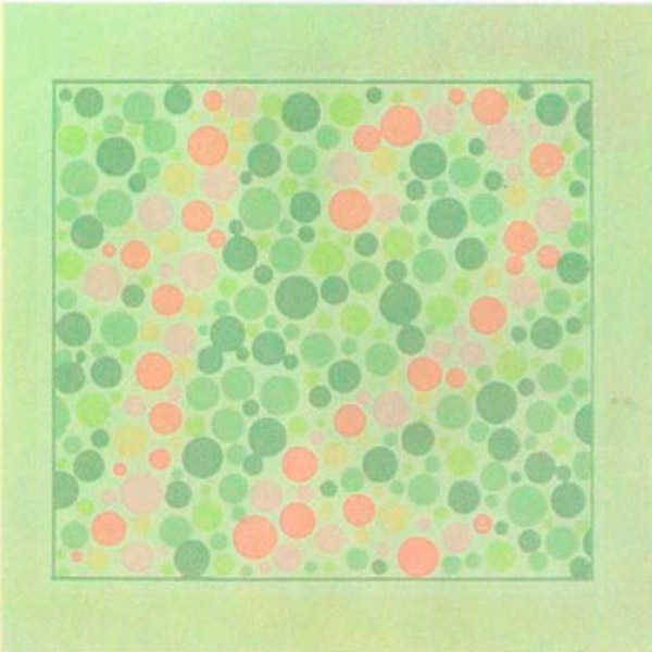
- With normal vision, a person sees 9. If there is blindness in the red or green spectrum, the person sees 5.
- In normal - triangle, in pathology - circle.
- The normal number is 13, and when disturbed, the number is 6.
- Normal is triangle and circle, colorblind people can't distinguish anything.
- Everyone sees the number 9. If a person has an acquired form of pathology, there are problems with image recognition.
- Trichromats see 5, with blindness in the red or green spectrum - the digit is difficult to discern or not visible at all.
- Normally and with problems in recognizing the green spectrum, the number 9 is seen. Subjects with red spectrum blindness can see 9,8,6.
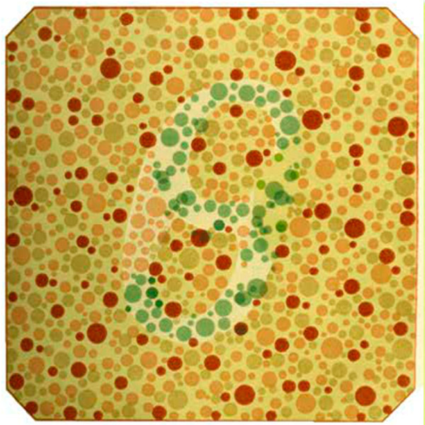
- Trichromats see 136. If there are problems with red or green spectrum - 66, 68, 69.
- Everyone sees the number 14.
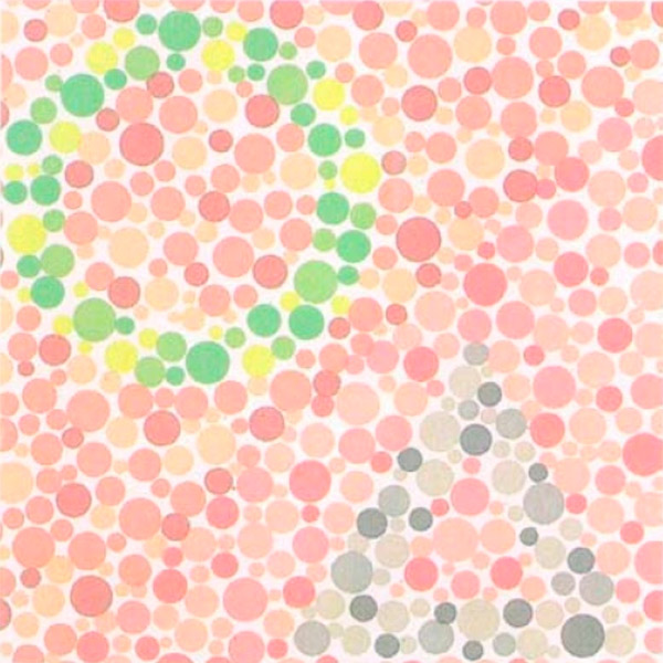
- If there are disturbances in the perception of green color, people see nothing. Normal is 12.
- Normal is a circle and a triangle.
- Trichromats are 3 and 0, protoanopes are 1 and 0, and deuteranopes are 1 and 6.
- Normal is a circle and a triangle.
- Normal is 96.
- With normal color vision, a circle and a triangle.
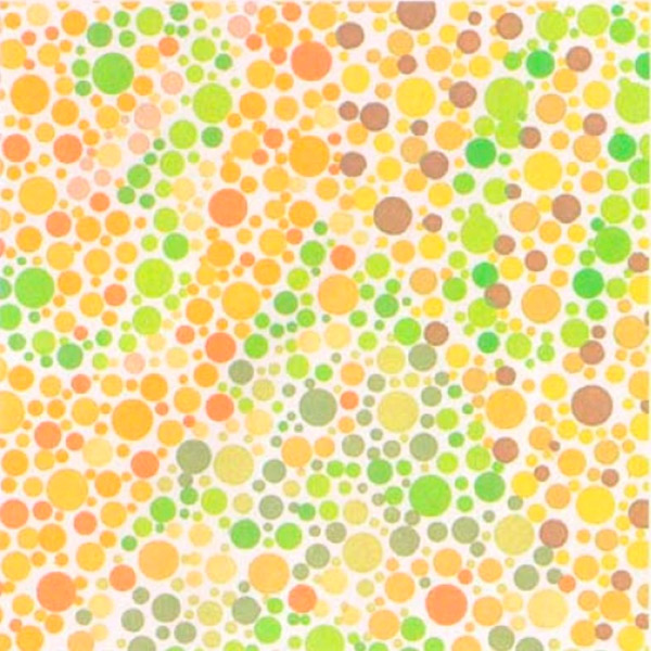
- In the absence of pathology, vertical rows are multicolored and horizontal rows (1, 3, 5, 6) are unicolored.
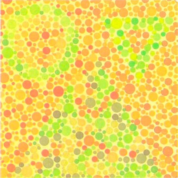
- Trichromats distinguish the number 25.
- Normally a triangle and a circle.
- Subjects with healthy color perception distinguish the number 96.
- Normal is 5, with abnormalities the image is undetectable.
- For trichromats, horizontal rows are multicolored and vertical rows are monochromatic.
- With healthy color perception, the number 2.
- Normal is 2.
- Trichromats distinguish between a triangle and a square.
- Normal trichromats, protanopes and deuteranopes distinguish the figure 4 in the table. Persons with pronounced acquired pathology of color vision do not distinguish this figure.
- Subjects with normal color perception see a triangle, while color blind people see a circle.
To evaluate the results of the Rabkin Tables test, the ophthalmologist counts the number of correct answers. If all 27 tables are passed, the person has normal vision. If there is a violation in tables 1-12 - abnormal trichromasia. If there are more than 12 incorrect answers - dichromasia. To clarify the diagnosis and identify abnormalities, the patient is shown 20 more tables.
Normal performance
The ability of the eye to perceive colors based on sensitivity to different radiation ranges of the visible spectrum is color vision. It is the main function of the cone apparatus of the retina. Depending on the length of radiation, three groups of colors are distinguished:
- Longwave (red, orange).
- Mid-wave (yellow, green).
- Shortwave (cyan, blue, violet).
Color shades are obtained by mixing primary colors (red, blue, green). If a person recognizes three primary colors, he or she is trichromatic, i.e. Has normal vision.
According to the classification of congenital color vision disorders, red is the first color (protos), green is the second (deuteros) and blue is the third (tritos). Abnormal perception of one of the three colors is designated as protomaly, deuteronomaly and tritanomaly. In this case, the pathology of recognition of one of the primary colors changes the perception of the others.
Monochromasia is diagnosed quite rarely, when a person perceives only one of the three colors. Another type of pathology of the cone apparatus is achromasia, i.e. Perception of the world in black and white.
To assess the color-discriminating ability of the eye, tests on special polychromatic tables and studies with special devices (anomaloscope) are carried out. The presence of errors in the recognition of numbers and objects in the tables, allows you to identify a disorder of color perception. Rabkin's test is considered the most reliable and recognized all over the world. If the subject has color blindness, this test determines the type of disorder, i.e. Which spectrum is not recognized by the visual organs.
How do you cheat a color blindness test?
Genetically determined color anomaly or color blindness is a peculiarity of vision that is manifested by the inability to distinguish one or more colors. According to medical statistics, color blindness affects one out of 20 people. The patient is not always aware of the diagnosis.
- Special testing is performed to detect abnormalities in the recognition of the color spectrum.
- A color blindness test is necessary for drivers, military, medical professionals and people in other professions in which it is important to perceive color correctly.
- Polychromatic tables are used in the diagnosis of pathology. With the help of multicolored circles, they depict numbers and figures.
The tables are constructed in such a way that it is practically impossible to cheat the color blindness test. The only way to cheat is to memorize the images from both the main and the control group of the tables. If a person has normal vision, he or she can immediately see the hidden images. If there is a color anomaly, the subject is unable to discern the picture.
Treatment of hereditary color blindness is impossible. If the disorder is acquired, its correction and even surgical intervention are performed, but full restoration of color vision is unlikely. For correction, special contract lenses and glasses are used. As for the prevention of color blindness, it is aimed at preserving the health of the visual organs.