Medical expert of the article

New publications
Coccidia are the causative agents of coccidioidosis
Last reviewed: 04.07.2025

All iLive content is medically reviewed or fact checked to ensure as much factual accuracy as possible.
We have strict sourcing guidelines and only link to reputable media sites, academic research institutions and, whenever possible, medically peer reviewed studies. Note that the numbers in parentheses ([1], [2], etc.) are clickable links to these studies.
If you feel that any of our content is inaccurate, out-of-date, or otherwise questionable, please select it and press Ctrl + Enter.
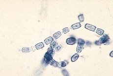

Morphology of coccidia
Coccidioides immitis is a dimorphic fungus. At room temperature (20-22 X) and in natural conditions it grows to the mycelial form. The mycelium is septate, 2-4 μm wide, without microconidia. As it grows, the cytoplasm of the fungus concentrates, the mycelial tube in the septa area becomes empty, then the cell wall of the mycelium ruptures and the mycelium disintegrates into arthrospores 1.5-2.3 μm wide and 1.5-15 μm long. Fragmentation is observed on the 10-L2-C day of cultivation.
Cultural properties of coccidia
Undemanding to nutrient media. In Sabour's medium at room temperature it forms various colonies of white, gray or brown color. Biochemical activity is low.
Antigenic structure of coccidia
When grown in liquid medium for 3 days, the mycelial form produces exoantigens HS, F (chitinase), HL, which can be determined using immunodiffusion in gel.
Pathogenicity factors of coccidia
The decrease in arthrosporogenesis in museum strains is accompanied by a decrease in their virulence.
Ecological niche - soil of endemic zones. Endemic zones are located in the Western Hemisphere between 40° north and south latitude, and 65° and 120° west longitude in the United States (western and southwestern states), as well as Central (Mexico, Guatemala, Honduras) and South (Venezuela, Paraguay, Argentina) America. The fungus is mainly found in desert and semi-desert zones, sometimes found in tropical zones and coastal forests (Northern California). Soil is the natural habitat of the fungus.
Environmental stability. Arthrospores are highly resistant to desiccation.
Sensitivity to antibiotics. Sensitive to amphotericin B, ketoconazole, miconazole, fluconazole, intraconazole. Sensitivity to antiseptics and disinfectants. Sensitive to the action of commonly used antiseptics and disinfectants, especially to heavy metal salts.
 [ 8 ], [ 9 ], [ 10 ], [ 11 ], [ 12 ]
Pathogenesis of coccidioidomycosis
After infection, arthrospores in the host organism are transformed into a tissue form - a spherule. Spherules are round formations measuring 20-90 µm, less often 2(H) µm with a thick double-contour cell wall up to 5 µm wide. When the cell wall of the spherules ruptures, the spores contained in them spread throughout the organism, which ensures the dissemination of the pathogen and the formation of secondary foci.
Secondary coccidioidomycosis develops in individuals with impaired cellular immunity. T-cell immunodeficiency causes severe pneumonia with subsequent spread of the fungus throughout the body from the primary site of inflammation.
Cellular immunity
The main role is played by T-effectors, including T-effectors of DTH, which accumulate on the 2nd-3rd week of the disease. Phagocytosis is incomplete, phagocytes are not able to protect the body at the stage of pathogen penetration. Antibodies and complement do not provide protection of the body against the pathogen. The presence of antibodies in patients with negative DTH to fungal antigens is a poor prognostic sign.
Epidemiology of coccidioidosis
Coccidioidomycosis - sapronosis. The source of the infectious agent is the soil of endemic zones, in which during the wet season of the year there is an intensive growth of fiba, and with the onset of the dry season, the mycelium disintegrates into arthrospores, which are the only infectious element. A sick person is not contagious to others.
The transmission mechanism is airborne and contact, the transmission route is airborne and dusty. Any contact with contaminated soil in endemic areas can lead to infection.
Susceptibility is high. Aspiration of 10 arthrospores is enough for infection. People with various immunodeficiencies are at the greatest risk of infection.
Symptoms of coccidioidomycosis
Symptoms of coccidioidomycosis are non-specific and are determined by the nature of the organs affected by the fungi. Secondary generalized coccidioidomycosis is characterized by a chronic course - remissions are replaced by exacerbations over decades; the presence of fistulous passages opening on the surface of the body, often remote from the focus of purulent inflammation; the presence of spherules in the pathological material.
Laboratory diagnostics of coccidioidomycosis
The materials examined include urine, sputum, blood, cerebrospinal fluid, and biopsy material.
Microscopic examination of native and Manus- or Gram-Welsch-stained preparations reveals spherules (spherical formations with a double-contour shell filled with small rounded endospores). Despite the characteristic morphology of the spherule, artifacts are possible: macrophages containing phagocytized mineral particles (dust cells), as well as accumulations of granulocyte detritus, can imitate spherical structures that are difficult to distinguish from the tissue phase of the pathogen. Diagnostics based only on the search for spherules leads to false-positive results. A simple method that allows you to exclude artifacts is to germinate the spherules: pathological material is mixed in equal volumes with distilled water, a preparation is prepared using the "distinguished drop" method, the cover glass is sealed with paraffin and incubated at 37 °C. After 4-6 hours, the true spherule germinates with mycelial threads emanating from the endospores.
Mycological examination is carried out in compliance with a special regime. On dense nutrient media, coccidiococci form colonies of leathery consistency at 37 °C, growing into the substrate, at 25 °C the mycelial form of the fungus develops. The mycelium is septate, the chlamydospores are large, located at the ends and on the sides of the mycelium. Typical arthrospores are formed on the 10th-12th day of incubation.
Biological research is conducted on hamsters and guinea pigs (males). Infection of experimental animals intratesticularly and intraperitoneally leads to the development of tissue forms of the fungus - spherules.
For serological diagnostics, RA, RP, RSK, RNGA, RIF are used. RP becomes positive in 53% of patients on the 1st week and in 91% on the 2nd-3rd week of the disease. There are no clear diagnostic titers of RSK, therefore, for diagnostic purposes, a 4-fold seroconversion is determined. An increase in the RSK titer indicates generalization of the process.
The intradermal allergy test with coccidioidin has diagnostic value only in individuals in whom it was negative at the onset of the disease; in other cases, this test can serve as an indicator of infection and is used to determine the boundaries of the endemic zone.
How to prevent coccidioidomycosis?
Specific prevention of coccidioidomycosis has not been developed. To prevent the disease, people who lack cellular immunity to the pathogen's antigens, as well as patients with a deficiency of T-lymphocytes, should avoid endemic zones. To prevent intralaboratory infections, all manipulations with suspicious cultures are performed after they are filled with sterile saline, which eliminates the spraying of arthrospores.