Medical expert of the article
New publications
Choanal atresia
Last reviewed: 29.06.2025

All iLive content is medically reviewed or fact checked to ensure as much factual accuracy as possible.
We have strict sourcing guidelines and only link to reputable media sites, academic research institutions and, whenever possible, medically peer reviewed studies. Note that the numbers in parentheses ([1], [2], etc.) are clickable links to these studies.
If you feel that any of our content is inaccurate, out-of-date, or otherwise questionable, please select it and press Ctrl + Enter.

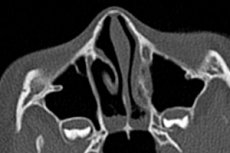
The complete absence of natural openings in various anatomical structures of the body is called atresia (from Greek a - denial of something, tresis - opening). Choanal atresia means the absence of paired openings in the posterior part of the nasal passage - posterior nasal passages, which connect the nasal cavity with the nasopharynx. [1]
Epidemiology
The frequency of this malformation is one case per 5-8 thousand live births (according to other data, three cases per 10 thousand), and in 65% of cases, children are born with unilateral choanal atresia.
At the same time, in 60-75% of cases of bilateral atresia, newborns have concomitant malformations - other craniofacial anomalies. In addition, statistics notes almost 8% of familial cases.
According to some reports, up to 30% of patients have choanal atresia in CHARGE syndrome. [2]
Causes of the choanal atresia
As choa atresia in a newborn child is a congenital pathology, its causes are related to the disruption of nasal structures during the embryonic period of intrauterine development. As a result of these disorders, a bony/cartilaginous septum or, more rarely, a fibrous (connective tissue) membrane remains between the nasal cavity (cavum nasi) and the upper part of the nasopharynx (pars nasalis pharyngis).
The genetic factor should be taken into account, especially in the presence of a complex of developmental defects, such as congenital CHARGE syndrome or cHARGE-association - with anomalies of the eye membranes, auricles, esophagus, genitalia, etc. The congenital syndromes of craniofacial (craniofacial) dysostosis or craniosyntosis (premature fusion of one or more cranial sutures) caused by gene mutations, in which there is an anomaly of the nasopharynx and posterior nasal passages, also include treacher Collins syndrome; Alfie, DiGeorgi, Apert syndromes; edwards syndrome; Crouzon, Antley-Bixler, Pfeiffer, Tessier, Beer-Stevenson, Jackson-Weiss syndromes; fetal alcohol syndrome (fetal alcohol syndrome).
When deforming nasal polyposis develops in children and young adults there is choanal stenosis, that is, their abnormal narrowing, which can be defined as narrowing of the nasal airways in the posterior choanal region, nasopharyngeal stenosis, or partial choanal atresia.
Thus, in otolaryngology, acquired choanal atresia - secondary anterior and posterior stenosis of the nasal cavity with the formation of fibrous septa - is often recognized as well. This condition can be the result of syphilis, systemic lupus erythematosus, trauma to the paranasal sinuses, surgical intervention, as well as a consequence of radiation therapy for malignant tumors of the nasopharynx.
However, choanal atresia is classified by medical experts as a congenital pathology, and practicing otolaryngologists should distinguish it from stenosis of the posterior nasal passages, which does not result in complete obstruction.
Unilateral atresia is twice as common: right-sided choanal atresia or left-sided choanal atresia, respectively. [3]
Risk factors
In addition to genetic abnormalities, various embryotoxic exposures and environmental factors are recognized as risk factors for atresia of the foramen distal nasalis.
Thus, a higher risk of this anomaly in the fetus may be exposed to expectant mothers who have taken drugs of the thioamide group in hyperthyroidism during pregnancy (to reduce the level of thyroid hormones). In such cases, the embryo may lack thyroid hormones, which negatively affects the morphogenesis of the upper respiratory organs.
In addition, studies have found a potential association of neonatal choanal atresia with high doses of vitamin B12, B3 (PP), D and zinc during pregnancy. Alcohol, tobacco smoke and caffeine have an extremely negative impact on the development of the craniofacial structures of the fetus. [4]
In 2010-12, an increase in births with choanal atresia was reported in the United States due to pregnant women's exposure to chemicals used to treat crops.
Read more:
Pathogenesis
Choans (Latin. Choane (Latin: funnel) are openings leading from the nasal cavity into the nasopharynx, bounded in the middle by the socket (edge of the bone plate); cuneiform bone - from above and behind; wing plates of this bone - from the sides, and from below - palatine bone (its horizontal plate). More information in the material - development of respiratory system organs
The formation of the choanae, originating from the gill arches of the embryo, begins in the fourth week of gestation (and continues until the eighth week) with the migration of neural crest cells into the dorsal neural folds. Next, the vertically arranged epithelial fold (oronasal membrane) between the roof of the primary oral cavity and the nasal processes (placoda nasalis) on the lateral surface of the head ruptures. The nasal processes deepen into the mesoderm, which leads to the formation of nasal fossae and then to the primary (primitive) choanae.
Theoretically, the pathogenesis of the congenital anomaly of choanal atresia may be due to the preservation of the cheek-pharyngeal (buccopharyngeal) membrane, a thin layer of ectoderm and entoderm cells covering the "oral opening" of the embryo above the cranial end of the chorda. This membrane should perforate in the sixth week of gestation, but for unknown reasons it may not, resulting in orofacial defects such as cleft palate and choanal atresia.
Also possible: preservation of bucconasal membrane (a thin layer of epithelial tissue, which should resorb at the seventh week of gestation); abnormal adherence of mesodermal tissue in the area of choanas; local disorder of mesenchymal cell migration along the neural crest, leading to defects in the formation of the embryo frontonasal protrusion of the head part and its ramifications.
But none of the assumptions of the mechanism of development of posterior nasal atresia has no evidence to date.
Symptoms of the choanal atresia
Newborns breathe through the nose because their epiglottis is higher (compared to adults), and the larynx rises during swallowing, touching the nasopharynx, and closes between the soft palate and the sides of the nasopharynx. And the ability to breathe through the mouth appears at 4-6 weeks after birth - after the lowering of the larynx.
Therefore, the classic symptoms exhibited by bilateral choanal atresia in neonates are due to complete impairment of respiratory function.
For example, the infant has cyclic cyanosis indicative of episodes of asphyxia: lividity of the skin, which decreases when crying (when the child opens his mouth wide and breathes in and out) and repeats as soon as crying stops and the infant closes his mouth. In such cases, emergency medical attention is required - endotracheal intubation or tracheotomy.
Unilateral atresia (i.e. Absence of only one posterior nasal passage) is often detected later in life (at 5-10 months of age or much later) and its first signs are unilateral nasal congestion. In addition, there is persistent discharge from one nostril - rhinorrhea, snoring and stridor (noisy breathing), as well as chronic sinusitis. [5]
Complications and consequences
Bilateral choanal atresia leads to acute neonatal respiratory distress syndrome due to complete obstruction of the nasal airways.
Consequences and complications of unilateral atresia: distortion of facial proportions, impaired growth of the upper and lower jaws and formation of a pathological bite - due to improper craniofacial development; the appearance of obstructive night apnea and other respiratory problems associated with impaired functioning of the upper respiratory tract. [6]
Diagnostics of the choanal atresia
If bilateral neonatal choanal atresia is suspected, preliminary clinical diagnosis is made by a neonatologist in an emergency by inserting a nasogastric tube through the infant's nasal cavity. Suspicion of this congenital anomaly is confirmed if the catheter cannot be inserted.
To confirm the diagnosis, imaging is necessary: endoscopy (examination) of the nasal cavity, CT scan of the nose, paranasal sinuses and paranasal bone structures.
Unilateral choanal atresia is the most common form and may have no associated birth defects, so it may not be diagnosed immediately after birth.
In unilateral atresia, instrumental diagnostics are also performed: anterior and posterior rhinoscopy; endoscopy of the nasal cavity and nasal computed tomography; rhinomanometry - study of nasal respiratory function.
Differential diagnosis
The differential diagnosis includes nasal breathing problems that may be due to: deviated nasal septum or cartilage dislocation; nasal cavity stenosis and congenital hypertrophy of the lower nasal bones; isolated stenosis of the nasal foramen pear-shaped (anterior bony restriction of the nasal skeleton); an anthrochoanal polyp, dermoid cyst of the nasal cavity, or nasolacrimal duct cyst; hemangioma or nasal garmatoma.
Who to contact?
Treatment of the choanal atresia
In case of choanal atresia, only surgical treatment by transnasal endoscopic resection and choanoplasty with prior CT or MRI of the nasal cavity is performed to restore their patency.
Surgical intervention for bilateral choanal atresia is usually performed within the first three months of life, and in cases of unilateral atresia, after the child is two years old. [7]
All details in the publication - restoration of choanal atresia
Prevention
Given the known risk factors for this birth defect, preventive measures can be considered proper management of pregnancy and extreme caution in prescribing any medications to expectant mothers.
And responsible couples have a way to prevent having a child with a genetically determined syndrome, such as medical-genetic counseling.
Forecast
Bilateral choanal atresia is life-threatening for the newborn, but with timely and effective treatment and no association with congenital syndromes, the prognosis is usually considered good.
Books on Atresia of the choanae
- Pediatric Otolaryngology: Principles and Practice Pathways" - by Christopher J. Hartnick et al. (Year of release: 2015)
- "Scott-Brown's Otorhinolaryngology, Head and Neck Surgery" - Author: John C Watkinson et al. (Year of publication: 2020)
- "Cummings Otolaryngology: Head and Neck Surgery" - Author: Paul W. Flint et al. (Year of release: 2020)
- "ENT: An Introduction and Practical Guide" - Author: Sharan K. Naidoo (Year of release: 2018)
Literature used
- Palchun, Magomedov, Alexeeva: Otorhinolaryngology. National manual. GEOTAR-Media, 2022.
- Congenital choanal atresia in children. Textbook for medical students. Kotova E.N., Radtsig E.Yu. 2021

