Medical expert of the article

New publications
Chemosis: Causes, Symptoms, Diagnosis, and Treatment
Last updated: 30.03.2026

All iLive content is medically reviewed or fact checked to ensure as much factual accuracy as possible.
We have strict sourcing guidelines and only link to reputable media sites, academic research institutions and, whenever possible, medically peer reviewed studies. Note that the numbers in parentheses ([1], [2], etc.) are clickable links to these studies.
If you feel that any of our content is inaccurate, out-of-date, or otherwise questionable, please select it and press Ctrl + Enter.
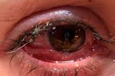
Chemosis is a pronounced swelling of the conjunctiva, the transparent mucous membrane covering the sclera and inner surface of the eyelids. Clinically, it appears as a translucent, sometimes bluish-pale or yellowish, "bulge" of the conjunctiva, which may even bulge between the eyelids. The Merck Manual describes chemosis as a severe swelling of the conjunctiva with ballooning and possible prolapse. [1]
It's important to understand that chemosis is not a disease in itself, but a symptom. It occurs when the conjunctiva responds to inflammation, allergy, infection, trauma, venous congestion, lymphatic dysfunction, burns, surgery, or orbital pathology by increasing vascular permeability and fluid accumulation. Therefore, treatment for chemosis always depends not on the swelling itself, but on its cause. [2]
In many cases, chemosis is benign and reversible, such as with seasonal allergies or mild viral conjunctivitis. However, it can sometimes be a significant warning sign of a more serious condition: orbital cellulitis, thyroid ophthalmopathy, severe chemical trauma, carotid-cavernous fistula, or severe postoperative lymphatic drainage disorder. This is why severe chemosis should not be considered "ordinary redness of the eye." [3]
The clinical significance of chemosis is determined not only by the risk of pain and discomfort. Severe swelling can make it difficult for a person to completely close the eye, which creates a risk of exposure keratopathy, or corneal drying and damage. In severe orbital conditions, chemosis can be associated with decreased vision, proptosis, limited eye movement, and even the threat of irreversible vision loss. [4]
Code according to ICD-10 and ICD-11
In the international version of ICD-10, chemosis is not identified as a separate line item with its own name, but is included in the category H11.4 Other vascular disorders and cysts of the conjunctiva, where conjunctival hyperemia and conjunctival edema are explicitly listed among the inclusions. This is important because, in clinical practice, chemosis is essentially a form of conjunctival edema. [5]
The clinical revision of ICD-10 uses a more detailed code, H11.42 Conjunctival edema, and then specifies the side: right eye, left eye, bilateral process, or unspecified eye. This detailing is convenient for documentation, but essentially describes the same phenomenon—conjunctival edema, or chemosis. [6]
In ICD-11, chemosis is included in the category 9A61.4Y Other specified vascular diseases of the conjunctiva. The synonyms and inclusions for this category explicitly list conjunctival oedema and conjunctival chemosis, and also provide post-coordination by laterality and anatomical zone of the conjunctiva. This means that in ICD-11, chemosis is coded more precisely and transparently than in the international ICD-10. [7]
In practice, the code should always be contextualized. If chemosis is part of allergic conjunctivitis, orbital cellulitis, thyroid ophthalmopathy, or a postoperative complication, the underlying disease should also be reflected in the diagnosis. This is especially important for a medical article, because chemosis itself describes a morphological feature, not a definitive causal diagnosis. [8]
| Clinical situation | ICD-10 | ICD-11 | Comment |
|---|---|---|---|
| Chemosis as conjunctival edema | H11.4 | 9A61.4Y | In ICD-10 it is included in the category of vascular disorders of the conjunctiva. |
| Conjunctival edema with indication of the side in clinical modification | H11.42 and the party's qualifying code | 9A61.4Y with post-coordination | A more detailed entry for documentation |
| Chemosis as part of allergic conjunctivitis | Both the underlying disease and the ocular manifestation are coded depending on the situation. | The main nosology and local symptom are coded. | A causal diagnosis is more important than an isolated symptom. |
| Chemosis in orbital cellulitis or thyroid ophthalmopathy | They primarily encode the ground state | Same rule | Chemosis is part of a broader pathology |
Source for the table: ICD-10 of the World Health Organization, ICD-11 and clinical modification of ICD-10. [9]
Epidemiology
There are virtually no specific population statistics for chemosis, as it is a symptom rather than a distinct disease. Therefore, epidemiology must usually be described in terms of the diseases and clinical situations in which chemosis is particularly common. This approach is more honest and clinically useful than attempting to quantify the "prevalence of chemosis" across all causes. [10]
The most common everyday context is allergic conjunctival inflammation. According to the ICON international consensus, ocular allergies, including allergic conjunctivitis, can affect up to 40% of the population, while the StatPearls review cites a range of approximately 10%-30% of the general population for the simple allergic form. Typical symptoms include itching, lacrimation, hyperemia, and chemosis. [11]
Infectious forms also have epidemiological significance. Conjunctivitis remains one of the most common reasons for visits to primary care physicians, and in the United States, approximately 1% of visits to primary care physicians are related to conjunctivitis. In acute hemorrhagic viral conjunctivitis, chemosis is a characteristic symptom complex, along with hemorrhage and eyelid edema. [12]
A separate area is surgical ophthalmology and aesthetic eyelid surgery. In a 2024 study, intraoperative chemosis occurred in 9.17% of cases of phacoemulsification. In postoperative aesthetic eyelid surgery, the incidence of persistent chemosis in the literature ranged from approximately 1% to 34.5%, and the risk depended on the surgical technique and lateral canthoplasty. These are not general ophthalmological statistics, but they are very important figures for clinical practice. [13]
Another important context is thyroid ophthalmopathy. Current consensus does not provide precise percentages for chemosis in this disease, but it emphasizes that chemosis is a common clinical sign, along with proptosis, periorbital edema, and diplopia, and that severe, visually threatening forms of the disease occur in approximately 2%-8% of patients. This makes chemosis an important marker of severity in orbital practice. [14]
| Epidemiological context | What is known |
|---|---|
| Isolated prevalence of chemosis | Not securely installed |
| Allergic conjunctivitis | Approximately 10%-30% of the population, according to some consensus estimates, eye allergies are up to 40% |
| Conjunctivitis as a reason for visits | About 1% of primary care visits in the US |
| Intraoperative chemosis during phacoemulsification | 9.17% in one modern study |
| Postoperative chemosis after lower eyelid surgery | Ranges from 1% to 34.5% have been reported in the literature. |
| Severe thyroid ophthalmopathy with vision risk | Approximately 2%-8% of cases of thyroid ophthalmopathy |
Source for table: ICON, StatPearls, BMC Ophthalmology, Thyroid Eye Disease Consensus. [15]
Reasons
The most common causes of chemosis are allergies, infections, and trauma. The Cleveland Clinic specifically lists bacterial and viral conjunctival infections, allergens, and conjunctival damage from foreign bodies or mechanical impact as the most common triggers. In this group, chemosis typically develops rapidly and is often accompanied by redness, itching, lacrimation, or discharge. [16]
The second major cause is chemical and physical irritation. Smoke, dust, household chemicals, burns, medicinal drops, contact lenses, and mechanical eye rubbing can all trigger a local inflammatory reaction with increased vascular permeability. When caused by toxic or medicinal substances, chemosis is often asymmetrical and associated with severe ocular surface discomfort. [17]
The third group of causes involves the orbit, not just the ocular surface. In orbital cellulitis, chemosis occurs due to orbital tissue inflammation and venous congestion; in thyroid ophthalmopathy, it is associated with autoimmune inflammation of the orbital tissues, edema, proptosis, and impaired venous and lymphatic drainage. In these cases, chemosis is no longer considered "simple conjunctivitis" but requires an evaluation of the orbital process. [18]
Systemic causes are less common but clinically very important. In review and clinical literature, chemosis has been described in heart failure, hypoproteinemia, nephritis, systemic capillary syndrome, venous congestion, and carotid-cavernous vascular disorders. In these situations, the conjunctiva becomes a kind of "window" into systemic fluid or venous imbalance. [19]
Finally, the postoperative cause is very important. Chemosis often occurs after eyelid and periorbital surgery, especially after lower blepharoplasty. Modern reviews attribute it to a combination of inflammation, dryness of the ocular surface, conjunctival manipulation, impaired lymphatic and venous drainage, and periorbital edema. [20]
| Group of reasons | Typical examples |
|---|---|
| Allergic | Seasonal and year-round allergic conjunctivitis, atopic forms |
| Infectious | Viral and bacterial conjunctivitis, orbital cellulitis |
| Traumatic and toxic | Foreign body, chemical burn, irritants, drug reaction |
| Orbital | Thyroid ophthalmopathy, orbital cellulitis, orbital vascular disorders |
| Systemic | Heart failure, hypoproteinemia, systemic capillary syndrome |
| Postoperative | Blepharoplasty, canthoplasty, and other surgeries on the eyelids and around the eye |
Source for table: Cleveland Clinic, Merck Manual, Thyroid Eye Disease Consensus, and Current Postoperative Reviews.[21]
Risk factors
Risk factors depend directly on the cause. Allergic chemosis is associated with atopy, seasonal pollen exposure, exposure to house dust mites, animal dander, fungal allergens, and other allergic diseases, including allergic rhinitis, asthma, and eczema. However, more severe allergic forms, such as vernal and atopic keratoconjunctivitis, have a higher risk of corneal complications. [22]
Risk factors for infectious chemosis include contact with contaminated hands and objects, wearing contact lenses, ocular surface trauma, sinusitis, and adjacent infections associated with orbital cellulitis. Orbital cellulitis often develops as a spread of infection from the paranasal sinuses rather than as an isolated ocular infection. This is one reason why the combination of chemosis with fever and pain upon eye movement requires particular caution. [23]
Autoimmune thyroid disease and smoking are significant for thyroid eye disease. The American Thyroid Association and the European Thyroid Association consensus emphasize that smoking is associated with the development and progression of thyroid eye disease, and smoking cessation should be discussed with all patients. In this group, chemosis is often associated with eyelid retraction, proptosis, and dry eye. [24]
Postoperative chemosis has its own set of established risk factors. Recent publications mention advanced age, male gender, hormonal therapy, eyelid laxity, lagophthalmos, dry eye, ocular allergies, aggressive coagulation, prolonged scleral exposure, and trauma to the conjunctiva and its lymphatic drainage. This is important because some of these factors can be assessed and corrected preoperatively. [25]
Finally, there are also general behavioral risk factors: eye rubbing, poor hand hygiene, improper use of eye drops, and working with chemicals without protection. These don't explain all cases, but they increase the likelihood of superficial trauma, irritation, or infection, and therefore the risk of chemosis. [26]
| Risk factor | What are the most common causes of this? |
|---|---|
| Atopy and seasonal allergens | Allergic chemosis |
| Contact lenses | Infectious and toxic chemosis |
| Sinusitis | Orbital cellulitis |
| Smoking | Thyroid ophthalmopathy |
| Dry eye and lagophthalmos | Postoperative and chronic chemosis |
| Aggressive lower eyelid surgery | Postoperative chemosis |
| Rubbing eyes and poor hygiene | Irritable and infectious chemosis |
Source for table: MSD Manual, Thyroid Ophthalmopathy Consensus Statement, Current Reviews of Postoperative Chemosis. [27]
Pathogenesis
The pathogenesis of chemosis is simple: fluid accumulates in the conjunctiva. However, the causes of this accumulation can vary. The Cleveland Clinic describes the general mechanism as follows: after damage or irritation of the conjunctiva, the body directs additional fluid, blood cells, and immune cells to the affected area, leading to swelling. [28]
In allergies, hypersensitivity mediators play a leading role, primarily histamine and other inflammatory signals that increase vascular permeability. Therefore, allergic chemosis often develops rapidly, is accompanied by itching, and subsides after removal of the allergen, cold compresses, and topical antiallergic therapy. In severe cases, the inflammation becomes more persistent and can affect not only the conjunctiva but also the cornea. [29]
In infections, the mechanism is somewhat different. Here, inflammatory vasodilation, vascular permeability, and exudation predominate, driven by a viral or bacterial process. If the infection is limited to the conjunctiva, chemosis usually remains a superficial manifestation. If the orbit is involved, as in orbital cellulitis, the swelling intensifies due to deep inflammation and venous congestion. [30]
In thyroid ophthalmopathy, chemosis is associated with orbital autoimmune inflammation. Current reviews and consensus documents describe inflammation of the orbital tissue and extraocular muscles, tissue expansion, and impaired venous outflow as the basis for proptosis, periorbital edema, and chemosis. In such cases, conjunctival edema is part of a broader orbital syndrome. [31]
Following eyelid and periorbital surgery, mechanical trauma, ocular surface dehydration, and impaired lymphatic drainage play a significant role. Reviews of postoperative chemosis emphasize that it is the combination of inflammation, conjunctival manipulation, and lymphatic dysfunction that makes some cases protracted and prone to chronicity. [32]
Symptoms
The most characteristic symptom is visible swelling of the conjunctiva. Mild chemosis may appear as a localized "bubble" on the sclera, while more severe chemosis may appear as a translucent pad of tissue extending between the eyelids. The Cleveland Clinic notes that severe chemosis may make it difficult for a person to fully close the eye. [33]
Subjectively, patients most often describe a foreign body sensation, irritation, lacrimation, burning, and a sensation that the eye "interferes with blinking." If the cause is an allergy, itching, eyelid swelling, and a watery or stringy discharge are particularly typical. Infections often cause redness and mucopurulent discharge. [34]
Pain is uncommon in simple allergic chemosis. If severe pain, decreased vision, photophobia, limited ocular motility, or proptosis occur alongside chemosis, these are indications of a more serious cause—an orbital process, severe trauma, corneal damage, or another acute pathology. These symptoms should prompt a more urgent approach. [35]
In thyroid ophthalmopathy, chemosis is rarely the only symptom. It is usually associated with dryness, a gritty sensation, periorbital edema, eyelid retraction, proptosis, and diplopia. In orbital cellulitis, chemosis is accompanied by pain, fever, decreased ocular motility, and sometimes visual impairment. [36]
In chronic or postoperative chemosis, complaints may be less pronounced but more persistent: constant irritation, cosmetic discomfort, a sensation of protruding tissue, deterioration of the tear film, and the need for frequent rewetting drops. These forms are often underestimated and persist for weeks or months. [37]
| Symptom | What more often involves |
|---|---|
| Itching and watery eyes | Allergic chemosis |
| Purulent discharge | Bacterial process |
| It's hard to close your eyes | Severe swelling and risk of corneal damage |
| Pain when moving the eye | Orbital cellulitis or other orbital pathology |
| Proptosis | Orbital process |
| Diplopia | Thyroid ophthalmopathy or orbital pathology |
| Photophobia and decreased vision | More severe involvement of the anterior segment or orbit |
Source for table: Cleveland Clinic, MSD Manual, Thyroid Eye Disease Consensus. [38]
Classification, forms and stages
There is no single, universally accepted classification of chemosis for all clinical practice. This is because chemosis is a symptom that occurs in a wide variety of conditions, from allergies to orbital infections and postoperative lymphatic dysfunction. Therefore, in practice, working rather than dogmatic descriptive schemes are used. [39]
The most convenient clinical classification is based on the duration of the disease. A distinction is made between acute chemosis, which develops rapidly due to allergy, infection, trauma, or chemical irritation; subacute chemosis; and chronic or persistent chemosis, which persists for weeks or months. Postoperative literature even describes a protracted "type 3" chemosis, lasting up to 6 months or more, often due to lymphatic dysfunction. [40]
The second practical principle is to classify the causes: allergic, infectious, traumatic, toxic, orbital, postoperative, and systemic. This classification is more convenient for the physician because it immediately suggests a diagnostic and treatment algorithm. For example, itching and bilaterality point toward allergy, while pain with eye movement and proptosis point toward the orbit. [41]
The third approach is to classify chemosis by severity. Mild chemosis barely interferes with eyelid closure or vision. Moderate chemosis causes significant discomfort and is sometimes cosmetically noticeable. Severe chemosis interferes with eyelid closure, causes conjunctival prolapse, impairs tear film, and increases the risk of exposure keratopathy. For thyroid ophthalmopathy, specialized scales for orbital disease activity and severity are also used. [42]
It's useful to describe the process as unilateral or bilateral. Bilateral chemosis is more common with allergies and systemic conditions, while severely unilateral chemosis prompts a more careful search for local infection, trauma, foreign body, postoperative problem, vascular anomaly, or orbital pathology. This is not an absolute rule, but it is a very useful clinical clue. [43]
| Classification principle | Options |
|---|---|
| By duration | Acute, subacute, chronic, persistent |
| Because of | Allergic, infectious, traumatic, toxic, orbital, postoperative, systemic |
| On the side | One-sided, two-sided |
| By severity | Light, moderate, heavy |
| According to clinical context | Isolated superficial, orbital-associated, postoperative |
Source for table: practice charts from contemporary reviews and consensus statements on conjunctival and orbital pathology. [44]
Complications and consequences
The most common complication of severe chemosis is incomplete closure of the eyelids. When the conjunctiva becomes severely swollen and bulges, the eyelids no longer fully cover the cornea. This leads to dryness of the ocular surface, disruption of the tear film, and the risk of exposure keratopathy. This is especially dangerous in cases of thyroid ophthalmopathy and postoperative chemosis. [45]
Chronic chemosis itself rarely leads to blindness, but it can perpetuate a vicious cycle of irritation, friction, dryness, and recurrent swelling. This is especially true in long-term postoperative cases, where persistent lymphatic dysfunction and mechanical stress make the process persistent and unpleasant for the patient. In these situations, chemosis becomes not so much dangerous as chronically annoying. [46]
Complications are much more serious when chemosis is part of orbital cellulitis. The MSD Manual notes that orbital cellulitis can cause vision loss due to ischemic retinopathy and optic nerve compression, limited eye movement, and intracranial complications, including cavernous sinus thrombosis, meningitis, and brain abscess. In this context, chemosis is an important, but far from the only, symptom of a severe disease. [47]
In thyroid ophthalmopathy, complications are also associated not so much with chemosis as with the orbital disease itself. Modern reviews highlight the risk of exposure keratopathy and compressive optic neuropathy. Therefore, chemosis combined with proptosis and eyelid retraction should be considered as a possible component of a visually threatening picture. [48]
This is why the prognosis for chemosis is determined by the cause. Allergic and mild infectious chemosis usually resolve without sequelae. Orbital, severe postoperative, and thyroid-related chemosis require more active monitoring, as they are critical to preserving the ocular surface, eyelid function, and sometimes vision itself. [49]
| Complication | When does it occur more often? |
|---|---|
| Exposure keratopathy | With severe swelling and incomplete closure of the eyelids |
| Conjunctival prolapse | In severe chemosis |
| Chronic irritation and dryness | In case of protracted postoperative and orbital process |
| Limitation of eye movements | For orbital cellulitis and thyroid ophthalmopathy |
| Loss of vision | In severe orbital causes |
| Intracranial complications | For orbital cellulite |
Source for table: MSD Manual, Thyroid Eye Disease Consensus Statement, Cleveland Clinic. [50]
When to see a doctor
Almost any new, pronounced chemosis should be seen by a doctor, especially if the cause is not obvious. Even when the swelling appears "allergic," it is difficult to confidently rule out a foreign body, toxic reaction, keratitis, or the onset of orbital pathology without an examination. In ophthalmology, it is the combination of symptoms, not the specific type of conjunctiva, that determines the urgency of the situation. [51]
The Cleveland Clinic recommends seeking immediate medical attention for chemosis if you experience difficulty closing the eye completely, blurred or decreased vision, photophobia, severe pain, a foreign body sensation, excessive discharge, especially yellow or green, or blood in the white of the eye. This is a practical list of warning signs that is useful for initial patient management. [52]
A separate emergency situation is suspected orbital cellulitis. If chemosis is accompanied by pain with eye movement, fever, severe eyelid swelling, decreased ocular motility, proptosis, and visual impairment, urgent assistance is needed, as this may be a visually and life-threatening process. In such cases, urgent visualization of the orbit and sinuses is often required. [53]
In thyroid ophthalmopathy, urgent referral is required if signs of a visually threatening course appear: decreased vision, deterioration of color perception, rapidly increasing proptosis, and severe corneal exposure. The consensus of the American Thyroid Association and the European Thyroid Association explicitly emphasizes the need for urgent referral if a visually threatening course is suspected. [54]
If chemosis develops after surgery and does not improve, but rather persists or worsens over the course of weeks, a follow-up examination is also necessary. For early postoperative swelling, waiting is often acceptable, but persistent chemosis may indicate lymphatic dysfunction, lagophthalmos, dry eye, or the need for more active intervention. [55]
Diagnostics
Diagnosis begins with a simple but crucial sequence: the doctor evaluates vision, pain, ocular motility, corneal condition, the presence of discharge, the affected side, and the context of the swelling. For "red eye," a physical examination with visual acuity, external appearance, and slit-lamp examination is considered the general standard. In ophthalmology, this basic set of tests helps quickly determine whether the problem is superficial or a deeper orbital process. [56]
Slit-lamp examination is particularly important because it allows for the assessment of the nature of chemosis, corneal transparency, fluorescein staining, tear film condition, and the presence of concomitant keratitis or epithelial defects. Clinical reviews emphasize that detailed slit-lamp biomicroscopy is critical for the diagnosis and management of conjunctival edema, and it is advisable to evert the eyelids to detect mechanical causes, foreign bodies, trichiasis, or tarsal conjunctival pathology. [57]
The next step is to determine the cause. If itching, bilaterality, lacrimation, and seasonality predominate, an allergy is more likely. If there is purulent discharge, viral symptoms, or a pronounced conjunctival reaction, an infectious process is more likely. If pain with eye movement, proptosis, limited mobility, or fever are present, the priority shifts to orbital cellulitis. [58]
Instrumental diagnostics are not necessary for everyone, but rather based on indications. If orbital cellulitis is suspected, computed tomography or magnetic resonance imaging is typically performed. In thyroid ophthalmopathy, imaging is recommended in atypical and severe cases, as well as during surgical planning. To assess activity and rule out facial expressions, consensus recommends contrast-enhanced computed tomography or magnetic resonance imaging, and for surgical planning, native computed tomography. [59]
Laboratory tests are also prescribed based on the context. If infection is suspected, a complete blood count and inflammatory markers may be needed; in the case of thyroid ophthalmopathy, a thyroid profile and autoantibodies; in the case of systemic edema, biochemistry, protein, and cardiovascular assessment. For common allergic chemosis, a comprehensive laboratory program is usually unnecessary, as the diagnosis is usually clinical. [60]
| Diagnostic step | What does it give? |
|---|---|
| Visual acuity test | Allows you to immediately identify a threat to your vision |
| Slit lamp examination | Confirms chemosis and evaluates the cornea and ocular surface |
| Eversion of the eyelids | Helps to find a mechanical cause or hidden foreign body |
| Eye movement testing | Important for excluding the orbital process |
| Computed tomography or magnetic resonance imaging | Needed if orbital cellulitis or severe thyroid ophthalmopathy is suspected |
| Laboratory tests | Assigned based on probable cause |
Source for table: Ophthalmology Reviews, MSD Manual, Thyroid Eye Disease Consensus. [61]
Differential diagnosis
Chemosis must be distinguished not only from other causes of red eye but also from other forms of localized conjunctival protrusion. For example, with subconjunctival hemorrhage, the patient also notices a noticeable change in the white of the eye, but this is a collection of blood, not watery edema. With conjunctival lymphangiectasia, the protrusion is chronic, localized, and associated more with lymphatic changes than acute inflammation. [62]
Differentiating between superficial conjunctivitis and orbital pathology is crucial. Preseptal cellulitis typically causes marked eyelid swelling without affecting vision or eye movement, whereas orbital cellulitis is accompanied by chemosis, proptosis, pain with eye movement, limited mobility, and sometimes decreased vision. These features help distinguish the relatively more superficial process from a deep orbital infection. [63]
Chemosis in thyroid ophthalmopathy must be differentiated separately from a common allergic or infectious process. In thyroid ophthalmopathy, proptosis, eyelid retraction, diplopia, soft tissue swelling of the orbit, and a chronic course, rather than just redness and discharge, are usually prominent. Therefore, thyroid history and orbital signs are crucial. [64]
Postoperative chemosis should be distinguished from lagophthalmos, severe dry eye, conjunctivochalasis, and mechanical postoperative problems. In some cases, it is these conditions that contribute to the swelling, not vice versa. Therefore, persistent postoperative chemosis always requires an assessment of not only the conjunctiva itself, but also the position of the eyelid, the completeness of closure, and the condition of the tear film. [65]
For practice, it's helpful to remember a simple rule. Itching and seasonality suggest allergies. Discharge and contagiousness suggest conjunctivitis. Pain with eye movement, proptosis, and fever suggest orbital cellulitis. Chronic proptosis, eyelid retraction, and a history of systemic thyroiditis suggest thyroid ophthalmopathy. Asymmetry and persistence suggest a localized or orbital process. [66]

Treatment
Treatment of chemosis always begins with answering one question: why the swelling occurred. Chemosis itself is not treated with a single, universal regimen, as antihistamine drops, antibiotics, corticosteroids, decongestants, and surgery all address different issues. If the cause is incorrectly identified, temporarily reducing swelling may mask the problem but not resolve it. Therefore, the first principle of treatment is a causal approach. [67]
For mild superficial chemosis, especially allergic or irritant, basic measures often include cold compresses, artificial tears, reducing contact with the allergen or irritant, and avoiding eye rubbing. The Cleveland Clinic specifically recommends cold compresses and lubricating drops as home remedies for mild cases, while the MSD Manual for allergic conjunctivitis emphasizes the importance of avoiding allergens, cold compresses, and artificial tears. [68]
If allergic inflammation is the primary cause, the mainstay of drug therapy is topical antihistamines, mast cell stabilizers, or a combination of these. Current guidelines and reviews also recommend topical nonsteroidal anti-inflammatory drugs, short courses of topical corticosteroids, and sometimes cyclosporine for more persistent cases. Oral antihistamines may also be necessary for severe concomitant respiratory allergies. [69]
In bacterial conjunctivitis, treatment depends on the severity and clinical presentation. If typical bacterial conjunctivitis is present, topical antibacterial therapy and hygienic measures are used. However, if chemosis develops not as part of superficial conjunctivitis, but as a result of orbital cellulitis, topical drops are no longer sufficient. In this situation, urgent systemic antibacterial therapy, ophthalmologist observation, and often visualization of the orbit and sinuses are required. [70]
For viral conjunctivitis, treatment is usually symptomatic. In most cases, this includes cold compresses, artificial tears, and infection control. However, for severe adenoviral cases with a pronounced inflammatory reaction and corneal damage, modern reviews discuss iodine-containing regimens, and for long-term corneal infiltrates, immunomodulatory drops such as cyclosporine or tacrolimus. However, these decisions are made by ophthalmologists, not the patient. [71]
If chemosis is associated with thyroid eye disease, treatment follows the guidelines for the underlying orbital disease. The consensus of the American Thyroid Association and the European Thyroid Association recommends topical ocular measures and lifestyle modifications for all patients, and systemic immunomodulatory therapy for active moderate to severe cases. In recent years, targeted approaches have emerged in this area, and new methods, such as teprotumumab, are being discussed as an important addition to systemic therapy for active thyroid eye disease. Chemosis is reduced not by suppressing it with drops, but by decreasing orbital inflammation. [72]
In severe chemosis with incomplete eyelid closure, corneal protection becomes a priority. This requires frequent rewetting drops, thick ointments at night, sometimes protective dressings, night masks, and other measures against exposure keratopathy. In thyroid ophthalmopathy, consensus specifically recommends topical protective measures to prevent corneal exposure. This is an important step, as the cornea is the first to suffer if chemosis and eyelid edema interfere with eye closure. [73]
Postoperative chemosis is most often initially treated conservatively. Modern reviews describe ocular surface lubrication, decongestants, anti-inflammatory agents, compression techniques, head elevation, ice, and manual eyelid compression as standard first steps. In many cases, early postoperative chemosis resolves spontaneously within 1-2 months, but not always. [74]
If postoperative chemosis becomes persistent, the approach changes. Recent literature discusses surgical techniques, including snip-conjunctivoplasty, sometimes with methylene blue and fibrin glue, as well as more targeted conjunctival interventions for long-standing prolapse and lymphatic dysfunction. This is no longer a routine first-line approach, but a solution for protracted and functionally significant cases that do not respond to conservative therapy. [75]
Finally, when systemic causes arise, it's not the eye that's treated, but the entire body. If chemosis is associated with heart failure, hypoproteinemia, systemic capillary syndrome, or severe venous congestion, topical drops may only slightly alleviate discomfort. The real effect occurs when the fluid balance, protein status, venous outflow, or underlying systemic disease are corrected. Therefore, chemosis is a good example of a symptom that requires not only ophthalmological but sometimes also general therapeutic considerations. [76]
| Cause of chemosis | Main treatment approaches |
|---|---|
| Allergic | Cold compresses, artificial tears, topical antihistamines, mast cell stabilizers, topical corticosteroids if needed |
| Bacterial conjunctivitis | Local antibacterial therapy and hygiene |
| Viral conjunctivitis | Symptomatic therapy, sometimes special regimens for severe cases |
| Orbital cellulitis | Urgent systemic antibiotics, imaging, observation, sometimes surgery |
| Thyroid ophthalmopathy | Local eye protection, smoking cessation, systemic anti-inflammatory and targeted therapy as indicated |
| Postoperative | Lubrication of the eye, cold, decongestants, anti-inflammatory therapy, compression, if persistent, surgical correction is possible |
| Systemic edema | Treatment of the underlying general disease |
Source for table: Cleveland Clinic, MSD Manual, Thyroid Eye Disease Consensus, Current Reviews of Postoperative Chemosis. [77]
Prevention
Prevention of chemosis is always tied to its most likely cause. For superficial forms, this primarily involves protecting the conjunctiva from irritants, maintaining hand hygiene, handling contact lenses correctly, and avoiding eye rubbing. These measures are simple, but they reduce the risk of both infectious and mechanical inflammation of the ocular surface. [78]
If a person has a known eye allergy, it is important to prevent exposure to the allergen and initiate topical antiallergic therapy early during the acute season. The MSD Manual and recent reviews emphasize that avoiding allergens and using topical antiallergic agents can significantly reduce the severity of symptoms, including chemosis. Prevention is particularly effective in this group. [79]
In postoperative ophthalmological and plastic surgery practice, chemosis prevention revolves around careful surgical technique and protection of the ocular surface. Current publications recommend minimizing conjunctival dehydration, carefully managing lymphatic and venous pathways, using humidification, cooling, and proper postoperative care. Preoperative assessment for dry eye, allergies, and lagophthalmos is particularly important. [80]
In thyroid orbitopathy, one of the most important preventive measures is smoking cessation. The consensus of the American Thyroid Association and the European Thyroid Association clearly indicates the need to explain to patients the importance of smoking cessation to reduce the risk of orbital disease progression. This is one of the most convincing modifiable measures for at-risk groups. [81]
In patients with chronic systemic diseases, prevention boils down to good control of the underlying condition. If heart failure, thyroid disease, or chronic ocular surface inflammation can be controlled, the risk of recurrent chemosis is reduced. Therefore, prevention is always twofold: ophthalmological and systemic. [82]
Forecast
The prognosis for chemosis is usually favorable if the cause is superficial and recognized promptly. If caused by an allergic or mild infection, swelling typically subsides within hours, days, or several weeks, especially if the trigger is removed and appropriate topical therapy is initiated promptly. The Cleveland Clinic clearly states that some causes resolve within a few days, although some forms can last for weeks or longer. [83]
Postoperative chemosis also regresses in most cases, but more slowly. Modern reviews emphasize that in the early postoperative phase, it often resolves spontaneously, but in protracted cases, it can persist for weeks or months. Therefore, a good prognosis is possible here as well, but only if the process is not accompanied by lagophthalmos, dry eye, or lymphatic dysfunction. [84]
The prognosis becomes guarded if chemosis is part of orbital cellulitis, severe thyroid ophthalmopathy, or another deep orbital process. In these cases, conjunctival edema itself is not the most dangerous component, but it can be an early marker of a visually threatening condition. In such cases, the outcome is determined by the speed of diagnosis and treatment of the underlying process. [85]
In practice, it's helpful to remember a simple principle. The more the chemosis resembles a localized superficial response, the better the prognosis. The more closely it is associated with the orbit, abnormal ocular closure, or systemic disease, the higher the risk of complications and the more important prompt evaluation. [86]
| Clinical variant | Normal forecast |
|---|---|
| Allergic | Usually good |
| Mild viral or irritant | Usually good |
| Postoperative early | Often favorable, but a protracted form is possible |
| Chronic postoperative | Depends on eliminating the cause |
| Thyroid | Determined by the severity of orbital disease |
| Orbital cellulitis | Potentially serious, depending on the urgency of treatment |
Source for table: Cleveland Clinic, MSD Manual, Thyroid Eye Disease Consensus. [87]
FAQ
Is chemosis always conjunctivitis?
No. Chemosis is often associated with conjunctivitis, but can also occur with allergies, trauma, chemical irritation, orbital cellulitis, thyroid ophthalmopathy, systemic edema, and after eyelid surgery.[88]
Can chemosis resolve on its own?
Yes, mild allergic or post-surgical chemosis often resolves on its own or with simple local therapy. However, if pain, decreased vision, difficulty closing the eye, or orbital symptoms occur, don't wait. [89]
Is it dangerous to rub your eye if you have chemosis?
Yes, it can increase irritation and swelling. The Cleveland Clinic specifically advises against rubbing your eyes, as this worsens the condition and can further injure the ocular surface. [90]
Are antibiotics necessary for all cases of chemosis?
No. If the cause is allergic or non-infectious, antibiotics will not solve the problem. They are only necessary when a bacterial process is confirmed or strongly suspected. [91]
Why is it difficult to close the eye with chemosis?
Because the swollen conjunctiva begins to bulge between the eyelids, mechanically interfering with full closure. This is especially characteristic of severe chemosis and is important due to the risk of corneal desiccation. [92]
When is computed tomography or magnetic resonance imaging (MRI) needed for chemosis?
Typically, when an orbital process is suspected: pain with eye movement, proptosis, limited mobility, decreased vision, fever, or an atypical presentation. In thyroid ophthalmopathy, imaging is also needed in severe and atypical cases. [93]
Does chemosis have its own staging system?
There is no universal staging system. In practice, chemosis is described by duration, severity, affected side, and cause. In certain postoperative situations, specific classifications of persistent chemosis are used. [94]
Key points from experts
Below are practical conclusions based on the experts' areas of work and publication activity. These are not verbatim quotes.
| Expert | Regalia | Practical thesis |
|---|---|---|
| Leonard Biellori | MD, Professor of Medicine, Associate Professor of Ophthalmology and Visual Sciences, Co-Director of the Immunophthalmology Service, Rutgers New Jersey Medical School | In case of chemosis with severe itching, bilaterality and seasonality, first of all, it is necessary to think about the allergic mechanism and start with allergen control and local antiallergic therapy. [95] |
| James A. Garrity | MD, Whitney and Betty MacMillan Professor of Ophthalmology, Emeritus, Mayo Clinic College of Medicine and Science, specialist in general ophthalmology, neuro-ophthalmology, and orbital pathology | Chemosis becomes truly dangerous not when it is simply noticeable, but when it is combined with orbital signs: proptosis, impaired eye mobility, pain and decreased vision. [96] |
| Henry B. Birch | MD, Chief of the Division of Diabetes, Endocrinology, and Metabolic Diseases, National Institute of Diabetes and Digestive and Kidney Diseases, USA, Co-author of the consensus statement on thyroid eye disease | In chemosis due to thyroid ophthalmopathy, it is necessary to treat not only the ocular surface, but also the orbital autoimmune inflammation, and early referral to a specialized team and smoking cessation are especially important. [97] |