Medical expert of the article

New publications
The causative agent of pneumocystosis (Pneumocystis jiroveci)
Last reviewed: 06.07.2025

All iLive content is medically reviewed or fact checked to ensure as much factual accuracy as possible.
We have strict sourcing guidelines and only link to reputable media sites, academic research institutions and, whenever possible, medically peer reviewed studies. Note that the numbers in parentheses ([1], [2], etc.) are clickable links to these studies.
If you feel that any of our content is inaccurate, out-of-date, or otherwise questionable, please select it and press Ctrl + Enter.
Pneumocystis is a disease caused by opportunistic fungi; it is characterized by the development of pneumonia in individuals with weakened immunity (prematurity, congenital or acquired immunodeficiency, HIV infection). Pneumocystis jiroveci is classified as an opportunistic yeast-like fungi. However, according to their morphological and other properties, sensitivity to antimicrobial drugs, they are typical protozoa.
")
Morphology and physiology of Pneumocystis jiroveci
The life cycle of Pneumocystis includes the formation of trophozoites, precysts, cysts and intracystic gels. The trophozoite is oval or amoeboid, 1.5~5 μm in size. It is covered with a pellicle and a capsule. Trophozoites attach to first-order pneumocytes using pellicle outgrowths (unlike the endogenous stages of Cryptosporidium, which live in second-order pneumocytes in the lungs). Rounding, grophozoites form a thickened cell wall, turning into a precyst and a cyst. The cyst, 4-8 μm in size, has a thick three-layer wall that stains intensely for polysaccharides. A rosette of 8 daughter bodies (sporozoites) forms inside the cyst. These intracystic bodies are 1-2 μm in diameter, have a small nucleus and are surrounded by a two-layer membrane. After leaving the cyst, they transform into extracellular trophozoites.
Epidemiology and clinical picture of pneumocystosis
The source of infection is people. The route of transmission is airborne dust. The incubation period is from 1 to 5 weeks. Pneumocystis is an opportunistic infection with lung damage, the leading AIDS marker infection. Pneumocystis pneumonia occurs with shortness of breath, fever and dry cough. Death occurs with respiratory failure. But usually it is an asymptomatic infection; over 70% of healthy people have antibodies to pneumocystis. Most healthy children become infected with the fungus at the age of 3-4 years.
 [
[ Microbiological diagnostics of pneumocystosis
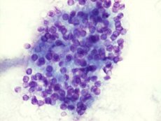
The microscopic method includes microscopy of a smear from a biopsy, lung tissue, sputum, stained according to Romanovsky-Giemsa: the cytoplasm of the parasite is blue, and the nucleus is red-violet. Special staining methods that reveal the cell wall of pneumocysts include toluidine blue staining and Gomori-Grocott silvering. RIF, ELISA and PCR are also used for diagnostics. Detection of IgM or an increase in the level of IgG antibodies in paired sera indicates acute pneumocystis infection.