Medical expert of the article

New publications
Pneumocystis
Last reviewed: 06.07.2025

All iLive content is medically reviewed or fact checked to ensure as much factual accuracy as possible.
We have strict sourcing guidelines and only link to reputable media sites, academic research institutions and, whenever possible, medically peer reviewed studies. Note that the numbers in parentheses ([1], [2], etc.) are clickable links to these studies.
If you feel that any of our content is inaccurate, out-of-date, or otherwise questionable, please select it and press Ctrl + Enter.
Pneumocystis is a causative agent of a respiratory lung disease that occurs in people from a risk group. This disease is not typical for healthy people, since the causative agent is opportunistic. The prevalence of pneumocystis pneumonia among the population is low, but among people with primary immunodeficiency states, this pathology is very common: in patients with leukemia, lymphogranulomatosis, other oncopathologies, with congenital immunodeficiencies, as well as with HIV infection. In patients with AIDS, pneumocystosis is a "marker" of the disease and occurs in more than half of those infected.
 [
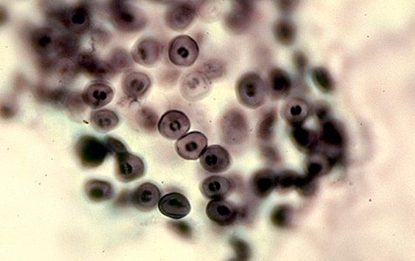
[ Structure of Pneumocystis
Pneumocystis carinii is a microorganism that was isolated from a sick person from the respiratory tract in the bronchi at the bifurcation site (carina), which is where the name of this species comes from. This pathogen naturally lives in the lungs of many animals, as well as in some people, from whom the infection occurs. The route of infection is airborne. However, people with a normal immune status may not get sick, but only be carriers, since pneumocystis is opportunistic. In immunodeficiency states, clinical symptoms of the disease develop.
When studying the structure of this microorganism, there were many discussions about which Kingdom to classify this species. The structural features of RNA, mitochondria, and protein membrane structures allowed it to be classified as Fungi, but the absence of ergosterol and the life cycle features confirm that Pneumocystis is a Protozoan.
The structure of pneumocystis is not so simple. This is due to the inconsistency of the structure of cellular elements due to the complex cell cycle. The sizes of the simplest vary from 1 to 10 micrometers, depending on the stage of the cycle. Therefore, according to the microscope, there can be various forms - from small forms with a thin cell wall to large ones with a thicker wall.
Pneumocystis is an extracellular parasite and is localized mainly in the alveolocytes of the first and second order. The microorganism can exist in four main forms: trophozoite, precyst, cyst, and sporozoite.
Trophozoite is a form of existence characterized by a significant cell diameter and irregular shape. The membrane is thick and has pseudopod-shaped outgrowths, so the shape of the trophozoite is not constant. These structures are designed for close contact of the pathogen with the alveolocyte cell. Inside the cell is cytoplasm with organelles characteristic of many microorganisms: mitochondria, fish-somes, vacuoles with cell juice and lipid and carbon components. The nucleus takes up enough space and is surrounded by two membranes to protect the genetic material.
Precysts are oval in shape, without outgrowths, small in size and have a thin cell membrane. In the middle of these structures, the nuclei divide to form cysts.
Cysts are also round in shape, but their size is larger, as they contain special bodies - precursors of sporozoite. Cysts have a shell and a thick three-layer membrane, which after rupture has an irregular shape and thus the cycle repeats.
Pneumocystis reproduces by simply dividing its genetic material in two, and then dividing the contents of the cytoplasm with the membrane.
Life cycle of Pneumocystis
Pneumocystis is a percellular parasite, but at the same time, various forms of existence allow it to penetrate into the cell. Infection occurs through airborne droplets. A cyst enters the human body, which, with a sufficient immune response, is neutralized by immunocompetent cells. If not, the cyst grows and continues its development cycle further with the formation of mature forms. The entire cycle can be divided into two phases - sexual and asexual.
The life cycle of Pneumocystis is quite complex and goes through several life forms in stages: trophozoite, precyst, cyst, sporozoite. The trophozoite is a vegetative form that attaches to the alveolocyte with its pseudopodia and closely interacts with the cell membrane. Then, by cell division, two mature cells are formed, and thus reproduction occurs. This is the so-called asexual phase of Pneumocystis development.
The trophozoite forms a precyst, which has a huge nucleus and nutrients necessary for the future cyst concentrated around it. As time passes, the nucleus divides and a cyst is formed, which usually has 8 nuclei inside. Microorganisms - sporozoites - emerge from the cyst. They have a single set of genetic information, and when they merge with each other, they again form a trophozoite, and the development cycle repeats itself. This is the sexual phase of development.
Cysts, when they enter the human body, are localized in the alveoli. With intensive reproduction of trophozoites and cysts, there are more and more and alveolocytes are desquamated, then clinical manifestations occur. The first immune reactions to this process develop due to cellular immunity. Macrophages and T-helpers react to foreign agents and try to phagocytize them, but cysts have the ability to be inside the macrophage and not be affected by its lysosomal enzymes. Therefore, the cellular immune reaction is not enough for a comprehensive immune response and elimination of pneumocystis. When the humoral link of immunity is launched with the help of cascade mechanisms under the influence of T-helpers, immunoglobulins affect trophozoites and infected macrophages. That is why, in people with pathology of the immune response, this disease develops very quickly, because for adequate protection, a good level of both local cellular and humoral immunity is necessary.
Symptoms of Pneumocystis pneumonia
The incubation period of the disease is from one to five weeks. It depends on the age and the degree of immunosuppression of the body. Often the disease can proceed as a common acute respiratory disease, then the clinical signs are weakly expressed and a person can die against the background of a mild course.
Taking into account the morphological changes in the lungs, several clinical stages are distinguished:
- edema stage – occurs during the onset of infiltration changes and is characterized by symptoms of intoxication and increasing respiratory distress.
- stage of atelectasis – the disruption of the secretion outflow from the alveoli contributes to their sticking together and the development of pulmonary atelectasis. Clinically, cough appears, respiratory failure increases.
- emphysema stage – lasts an indefinite period of time, depending on the effectiveness of treatment. Symptoms are reduced, but residual effects in the lungs in the form of emphysematous bullae produce a box-like sound when percussed.
Symptoms of Pneumocystis pneumonia differ in adults and children. Children can get sick in case of prematurity, pathology of the central nervous system, perinatal injuries, intrauterine infections. In this case, the disease develops in the 3-4 month of the child's life. Then the child loses weight, refuses to breastfeed, his sleep is disturbed, symptoms of shortness of breath and perioral cyanosis appear. The child coughs like whooping cough, sometimes with the release of foamy sputum. On the radiograph, there may be changes like interstitial infiltrates or like "cloudy" lungs.
In adults, clinical signs develop a week after infection in patients treated with immunosuppressants, and after 2-3 months in patients with AIDS. The disease begins with a rise in temperature to subfebrile numbers, moderate cough, shortness of breath during physical exertion and pain in the chest area. In the absence of treatment, a week later, the symptoms intensify, cyanosis and high temperature appear. Severe course of the disease is due to rapid diffuse spread of inflammation to both lungs. This increases respiratory failure and, against the background of general immunosuppression, is dangerous due to pulmonary edema.
In HIV-infected patients, the peculiarities of pneumocystosis are the sluggish development of symptoms of the disease, which often contributes to a fulminant course with a fatal outcome. Therefore, in patients with AIDS, there are certain indications for the beginning of preventive treatment of pneumocystis pneumonia, even if there are no special clinical manifestations.
Diagnosis of Pneumocystis carinii infection
Considering the fact that the symptoms of Pneumocystis pneumonia are not specific and the disease often proceeds without pronounced clinical manifestations, but with a fulminant course, etiological verification in this case is very important for timely treatment.
Clinical manifestations are not pathognomonic, therefore, based on the anamnesis and objective examination, the doctor can only determine the presence of pneumonia, and its nature is difficult to suspect.
An important fact of the anamnesis is the presence of oncopathology, treatment with cytostatics, HIV infection in the patient. This allows us to suspect this type of pneumonia against the background of a significant decrease in immune reactivity. Therefore, it is important to examine such a contingent of patients very carefully and carry out preventive measures.
Therefore, laboratory and instrumental diagnostic methods are the leading ones in verifying the diagnosis.
Chest X-ray is a mandatory method for diagnosing and confirming pneumonia. Characteristic changes are the phenomenon of "white lung" or "cloudy lung", but these symptoms are not so common and at the initial stages these changes do not yet develop. In children, pneumocystosis can be expressed on the X-ray as interstitial pneumonia.
Bronchoscopy is recommended to obtain bronchial lavage and further examination of the secretion.
Pneumocystis in sputum can be detected if there is a significant amount of them in the alveoli. Sputum examination is one of the reliable methods of verifying the diagnosis. In addition to sputum, bronchoalveolar lavage can be used as material for examination. A microscopic method is used with Romanovsky-Giemsa staining of the material, and purple cells with a red nucleus are detected. But this method does not always give a result, since a sufficient amount of the pathogen may not have gotten under the microscope lens. A more accurate method is parasitological. The material obtained from the patient is sown on a nutritious medium and the pathogen grows in a few days, which confirms the diagnosis.
These methods are rarely used in modern conditions, since it takes a long time to obtain the result, and a laboratory with equipment is also required, which is not available in every medical institution. Therefore, serological diagnostic methods are currently widespread.
An analysis for the qualitative determination of pneumocysts can be carried out with the study of not only sputum, but also blood. The polymerase chain reaction method is used - a molecular genetic method based on the detection of DNA in the patient's material.
A simpler serological method of research (blood serum research) is the detection of antibodies to pneumocystis. Since immunoglobulins are produced against the pathogen, their level or presence indicates the activity of the process. The level of immunoglobulins of class G and M is determined by the enzyme immunoassay or immunofluorescence method. An increased level of immunoglobulins of class M indicates an acute infection, and with an increase in immunoglobulins G, a long-term chronic infection is possible.
Treatment and prevention of pneumocystosis
Treatment of this disease is a complicated task, since antibiotics do not act on the pathogen. In addition, treatment should be started as early as possible and only specific. Before starting therapy, the severity of the disease should be determined, which is characterized by the degree of respiratory failure by the level of partial pressure of oxygen in the blood.
The etiological treatment of pneumocystosis is the use of sulfamethoxazole/trimethoprim – biseptol. In mild cases, oral administration of the drug or intravenous infusions at a dose of 100 mg/kg and 20 mg/kg, respectively, are prescribed. However, given the presence of concomitant immunodeficiency in patients, these drugs cause many side effects: skin rash, anemia, leukopenia, agranulocytosis, nausea, dyspeptic manifestations. Therefore, the optimal course of treatment is 2 weeks.
In severe cases, Pentamidine is added to this drug - a drug that has a specific effect, since it damages the reproductive systems of pneumocysts. It is used in a dose of 4 mg / kg when diluted in 5% glucose. The course of treatment is 2-3 weeks.
This is only etiotropic therapy, but symptomatic antipyretic agents, detoxification therapy, rehydration, antifungal drugs and antibiotics for HIV-infected patients are also used.
Prevention of pneumocystosis is necessary due to the complexity of the disease and its complicated course in the contingent of patients. Prevention methods can be non-specific and specific - medicinal. Non-specific prevention methods are characterized by examination of patients from the risk group in case of epidemiological indications, as well as correct and proper antiretroviral therapy in patients with AIDS. For such people, the correct daily routine, adequate nutrition, and elimination of bad habits are of great importance.
Specific methods of prevention are the use of etiotropic medications. The same drugs are used for prevention as for treatment. The indication for such primary prevention is the level of CD4 cells below 300, as this is considered the level of risk of pneumocystis infection.
Pneumocystis is the causative agent of a very complex disease, which, without specific clinical signs, must be diagnosed at an early stage and prescribed the correct treatment, since the consequences can be very serious. Pneumocystis develops in people with primary or secondary immunodeficiencies and these conditions are mutually aggravating. Therefore, in certain groups of patients, it is necessary to prevent this disease by both specific and non-specific methods.