Medical expert of the article

New publications
The causative agent of melioidosis
Last reviewed: 04.07.2025

All iLive content is medically reviewed or fact checked to ensure as much factual accuracy as possible.
We have strict sourcing guidelines and only link to reputable media sites, academic research institutions and, whenever possible, medically peer reviewed studies. Note that the numbers in parentheses ([1], [2], etc.) are clickable links to these studies.
If you feel that any of our content is inaccurate, out-of-date, or otherwise questionable, please select it and press Ctrl + Enter.
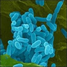
Melioidosis - like glanders, it occurs as severe septicopyemia in acute or chronic form with the formation of abscesses in various organs and tissues. The causative agent of melioidosis was isolated and described by A. Whitmore and K. Krishnaswamy in 1912.
The causative agent of melioidosis is Burkholderia pseudomallei (according to the old classification - Pseudomonas pseudomallei) - a gram-negative rod with rounded ends, 0.3-0.6 x 3-6 μm in size, located singly or in short chains. In old cultures, filiform, short and thick rods, coccobacteria, etc. are found. It does not form spores, freshly isolated bacteria often have a pseudocapsule. The microbe is motile; lophotrichus, in young cultures - monotrichus. Like the causative agent of glanders, it often gives bipolar staining, since there are inclusions of polyhydroxybutyric acid located at the poles. The content of G + C in DNA is 69 mol %. Strict or facultative aerobe, grows on a medium in which the only source of nitrogen is ammonium sulfate, and carbon is glucose. The optimum temperature for growth is 37 ° C, the pH of the medium is neutral. On MPA with 3-5% glycerol, shiny, smooth S-colonies grow after 24 hours; dissociation is possible later, the colonies acquire a yellowish-brown color and become folded. In MPB with glycerol, uniform turbidity appears after 24 hours, subsequently a sediment forms without clearing the medium, and on the 2nd-3rd day a delicate film appears on the surface, adhering to the wall of the test tube. Then the film thickens and becomes folded. Many strains of the causative agent of melioidosis, when growing on media, initially emit an unpleasant putrid odor, which is then replaced by a pleasant truffle aroma. On blood agar, it sometimes produces hemolysis. Ferments glucose, lactose and other carbohydrates with the formation of acid. As the culture ages, the enzymatic activity decreases. Liquefies gelatin and coagulated whey. Peptonizes milk, but does not coagulate. Does not form indole. Has denitrifying properties and lecithinase activity.
In terms of antigens, the causative agent of melioidosis is quite homogeneous. It has somatic (O), membrane (K), mucous (M) and flagellar (H) antigens, and the somatic O-antigen is related to the O-antigen of the glanders causative agent.
The causative agent of melioidosis produces two heat-labile toxins. One of them causes hemorrhagic and necrotic lesions, the second causes death of laboratory animals (lethal toxin) without damaging tissues at the injection site.
 [
[ Epidemiology of melioidosis
The source of melioidosis are rodents (rats, mice), cats, dogs, goats, sheep, pigs, cows, horses, among which epizootics can occur. In endemic areas, the pathogen is found in soil, water of open reservoirs contaminated with excrements of sick animals. The possibility of human infection not only by contact, but also by alimentary means is not excluded. A sick person is not contagious to others. In Russia, for many decades, cases of melioidosis among people have not been observed. The disease occurs in a number of countries in Southeast Asia, Europe, Africa, North and South America, Australia.
The causative agent of melioilosis dies at a temperature of 56 °C within 30 minutes, a 1% phenol solution or a 0.5% formalin solution kills it within 10 minutes. It survives in water and soil for up to 1.5 months, in animal corpses - up to 12 days.
Symptoms of melioidosis
Human infection occurs mainly through damaged skin or mucous membranes when in contact with water or soil. which contain the causative agent of melioidosis. The incubation period of melioidosis is from 4 days to several months. The causative agent of melioidosis multiplies in the blood, spreads throughout the body, which leads to the formation of abscesses in various organs and tissues.
The course of melioilosis can be acute and chronic. The prognosis is always serious, the disease can last for months and even years.
Laboratory diagnostics of meliolosis
Bacteriological, serological and biological methods are used. To isolate a pure culture, blood, sputum, pus from abscesses, nasal discharge and urine, as well as cadaveric material are taken. The blood of patients is inoculated on glycerin MPB, any other material - on glycerin agar. The pathogen, unlike other pseudomonads, is resistant to polymyxin at a concentration of 400 μg/ml.
Along with sowing the material on the media, guinea pigs or hamsters are infected: the blood of the sick is injected intraperitoneally, other material - subcutaneously or by rubbing into the scarified skin. If the result is positive, edema, necrosis, ulceration develops at the injection site, and abscesses appear in the lymph nodes. When opening a dead animal, multiple abscesses are found in the internal organs; a pure culture can be easily isolated from them.
To detect specific antibodies in the blood of patients or those who have recovered from the disease, the RSC, RPGA, and agglutination reaction are used. The increase in antibody titers in these reactions is an important diagnostic sign, but even in this case it is not always possible to differentiate melioidosis from glanders.
Specific prevention of meliolosis
Specific prevention of melioidosis has not been developed. General prevention comes down to deratization measures in areas unfavorable for melioidosis, preventing rodents from accessing water sources, housing and food. Swimming in stagnant water bodies and drinking undisinfected water are prohibited. Sick domestic animals are isolated, treated (or destroyed).