Medical expert of the article
New publications
Bunyaviruses
Last reviewed: 04.07.2025

All iLive content is medically reviewed or fact checked to ensure as much factual accuracy as possible.
We have strict sourcing guidelines and only link to reputable media sites, academic research institutions and, whenever possible, medically peer reviewed studies. Note that the numbers in parentheses ([1], [2], etc.) are clickable links to these studies.
If you feel that any of our content is inaccurate, out-of-date, or otherwise questionable, please select it and press Ctrl + Enter.

The Bunyaviridae family (from the name of the Bunyamwera area in Africa) is the largest in terms of the number of viruses included in it (over 250). This is a typical ecological group of arboviruses. It is divided into five genera:
- Bunyavirus (over 140 viruses, grouped into 16 antigen groups, and several ungrouped) - transmitted mainly by mosquitoes, less often by midges and ticks;
- Phlebovirus (about 60 representatives) - transmitted mainly by mosquitoes;
- Nairobivirus (about 35 viruses) - transmitted by ticks;
- Uukuvirus (22 antigenically related viruses) - also transmitted by ixodid ticks;
- Hantavirus (more than 25 serovariants). In addition, there are several dozen bunyaviruses that are not assigned to any of the genera.
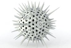
The viruses contain single-stranded negative-weight fragmented (3 fragments) RNA with a molecular weight of 6.8 MDa. The nucleocapsid has helical symmetry. Mature virions are spherical and 90-100 nm in diameter. The envelope consists of a 5-nm-thick membrane covered with 8-10-nm-long surface projections. The surface projections consist of two glycopeptides that combine to form cylindrical morphological units 10-12 nm in diameter with a 5-nm-diameter central cavity. They are arranged to form a surface lattice. The membrane to which the surface subunits are fixed consists of a lipid bilayer. The cord-like nucleoprotein is located directly beneath the membrane. Bunyaviruses have three major proteins: one nucleocapsid-associated protein (N) and two membrane-associated glycoproteins (G1 and G2). They reproduce in the cell cytoplasm, similar to flaviviruses; maturation occurs by budding into intracellular vesicles, then the viruses are transported to the cell surface. They have hemagglutinating properties.
Bunyaviruses are sensitive to elevated temperatures, fat solvents and temperature fluctuations. They are very well preserved at low temperatures.
Bunyaviruses are grown in chicken embryos and cell cultures. They form plaques in cell monolayers under agar. They can be isolated by infecting 1-2-day-old white suckling mice.
Of the diseases caused by bunyaviruses, the most common are mosquito fever (pappataci fever), Californian encephalitis, and Crimean (Congo) hemorrhagic fever (CCHF-Congo).
Pathogenesis and symptoms of bunyavirus infections
The pathogenesis of many human bunyavirus infections has been studied relatively little, and the clinical picture has no characteristic symptoms. Even in diseases that occur with symptoms of CNS damage and hemorrhagic syndrome, the clinical picture varies from extremely rare severe cases with a fatal outcome to latent forms, which predominate.
The carrier of mosquito fever is the mosquito Phlebotomus papatasi. The incubation period is 3-6 days, the onset of the disease is acute (fever, headache, nausea, conjunctivitis, photophobia, abdominal pain, leukopenia). 24 hours before and 24 hours after the onset of the disease, the virus circulates in the blood. All patients recover. There is no specific treatment. Prevention is non-specific (mosquito nets, use of repellents and insecticides).
California encephalitis (carrier - mosquito of the genus Aedes) begins suddenly with a severe headache in the frontal region, an increase in temperature to 38-40 "C, sometimes vomiting, lethargy and convulsions. Less often, signs of aseptic meningitis are observed. Fatal cases and residual neurological effects are rare.
Crimean (Congo) hemorrhagic fever occurs in the south of our country and in many other countries. Infection occurs through tick bites of the genera Hyalomma, Rhipicephalus, Dermacentor, and by contact. The virus was isolated by M. P. Chumakov in 1944 in Crimea. The incubation period is 3-5 days. The onset is acute (chills, fever). The disease is based on increased permeability of the vascular wall. Growing viremia causes the development of hemorrhages, severe toxicosis, up to infectious toxic shock with disseminated intravascular coagulation. Mortality is 8-12%.
Immunity
As a result of a bunyavirus infection, long-term immunity is formed due to the accumulation of virus-neutralizing antibodies.
Laboratory diagnostics of bunyavirus infections
Bunyaviruses can be isolated from pathological material (blood, autopsy material) during intracerebral infection of suckling mice, which causes paralysis and death. Viruses are typed in the neutralization reaction, RSK, RPGA and RTGA. In the serological method, paired sera are examined in RN, RSK or RTGA (it should be taken into account that the Crimean hemorrhagic fever virus does not have hemagglutinin).


 [
[