Medical expert of the article

New publications
Bronchography
Last reviewed: 29.06.2025

All iLive content is medically reviewed or fact checked to ensure as much factual accuracy as possible.
We have strict sourcing guidelines and only link to reputable media sites, academic research institutions and, whenever possible, medically peer reviewed studies. Note that the numbers in parentheses ([1], [2], etc.) are clickable links to these studies.
If you feel that any of our content is inaccurate, out-of-date, or otherwise questionable, please select it and press Ctrl + Enter.
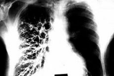
Bronchography is a medical procedure or diagnostic test that is used to visualize the bronchi (branches of the airways) and lungs by injecting a contrast agent and x-rays or other imaging techniques. Bronchography allows doctors to evaluate the condition of the bronchi, identify possible changes such as tumors, structural abnormalities or obstructions, and helps in establishing diagnoses and planning treatment.
The bronchography process may include the following steps:
- PatientPreparation: The patient may require some preparation, such as an overnight fast before the procedure to reduce the risk of vomiting. Consent for the procedure and discussion of medical history may also be required.
- Contrastagent administration: Contrast agent (usually iodine-containing) is injected into the bronchi through a tracheostomy (a catheter inserted through a hole in the throat) or with a bronchoscope (a flexible tubular instrument that is inserted through the nose or mouth and delivered to the bronchi).
- Bronchial Imaging: When the contrast agent is delivered to the bronchi, x-rays or other imaging techniques are taken to obtain detailed images of the bronchi and lungs.
- Analyzing the results: The resulting images are evaluated by a radiologist or other specialist to identify changes and make a diagnosis.
Bronchography can be performed for a variety of purposes, including diagnosing tumors, determining the cause of bronchial obstruction, evaluating bronchial abnormalities, and assessing the effectiveness of treatment for respiratory diseases. It is usually performed in specialized medical centers and under the supervision of experienced specialists.
Indications for the procedure
Bronchography may be ordered in the following cases:
- Evaluation of tumors and neoplasms: Bronchography can be used to detect and evaluate tumors, cysts, polyps, and other neoplasms in the bronchi and lungs. It can help diagnose lung cancer and other diseases.
- Determining the cause of airway obstruction: If a patient has signs of airway obstruction, such as coughing, shortness of breath, or severe chest pain, bronchography can help doctors determine the cause of the obstruction, such as bronchoconstriction, foreign bodies, or other abnormalities.
- Evaluation of bronchial anomalies: Bronchography can be useful for evaluating bronchial anomalies in children and adults.
- Investigation of bronchial infections and inflammation: In some cases, bronchography can help diagnose bronchial infections and inflammation in the bronchi.
- Surgical planning: Prior to lung or bronchial surgery, bronchography may be used to better plan the surgical procedure and locate neoplasms.
- Monitoring treatment efficacy: In patients with chronic lung disease, bronchography may be performed to assess treatment efficacy and monitor bronchial and lung health.
The indications for bronchography may vary depending on the specific clinical situation and the physician's recommendations. The decision about the need for the study is usually based on the patient's medical history, symptoms, and the results of other diagnostic tests.
Preparation
Preparation for bronchography can vary depending on the individual patient and the specific requirements of the healthcare provider, but usually includes the following general steps:
- Consultation with your doctor: Before having bronchography, it is important to consult with the doctor who will perform the procedure. Your doctor will tell you about the procedure, explain why it is necessary, and discuss your medical and allergy histories with you.
- Rascal: You will usually be advised not to eat or drink anything for a few hours before your bronchography. This is to prevent the risk of vomiting during the procedure.
- Allergy test for contrast agent: In some cases, bronchography may require the use of a contrast agent that is injected into the bronchi for better visualization. If you are allergic to the contrast agent, your doctor will take appropriate steps to prevent a reaction.
- Medications: If you are taking any medications, discuss this with your doctor. You may need to be advised to take or stop your medication before the procedure.
- Pregnancy Awareness: If you are pregnant or suspect you may be pregnant, tell your doctor. Bronchography may pose a risk to the fetus and the decision to have the procedure should be considered.
- Consent: You will need to give consent for bronchography after your doctor has explained the procedure and risks to you in detail.
Be sure to follow your doctor's recommendations and ask for any additional instructions that may be related to your specific case. Bronchography is a procedure that is performed by specialists and requires careful preparation to ensure safe and accurate diagnosis.
The device for carrying out the procedure
The bronchography procedure is a method of imaging and diagnosing the bronchi (airways) and lungs using X-rays and a contrast agent. Specialized medical devices and equipment are used to perform bronchography. The main apparatus for bronchography is an x-ray machine or x-ray machine, which is used to produce images of the bronchi and lungs.
The bronchography procedure can be performed either in an inpatient setting or in clinics and hospitals that have the necessary equipment and experienced medical staff. The following equipment and supplies may be needed to perform bronchography:
- X-ray machine: It is used to take x-rays of the bronchi and lungs.
- Contrast agent: A contrast agent is injected into the bronchi to make them easier to see on X-rays. It may be liquid or foam.
- Bronchoscope: A bronchoscope is a flexible, tubular instrument that is inserted into the bronchi through the patient's mouth or nose. It may contain a camera for visual inspection of the bronchi and allows the injection of a contrast agent.
- Monitor and computer: Bronchography images are displayed on a monitor for real-time observation and for subsequent analysis.
- Sterile instruments and materials for the procedure: This includes instruments for injecting contrast agent and performing bronchography.
Bronchography is an invasive procedure and is usually performed under local or general anesthesia to ensure patient comfort and minimize discomfort. [1]
Technique of the bronchograms
Here is the general technique for performing bronchography:
- Patient preparation: The procedure and preparation for the procedure are explained to the patient. A history is usually taken prior to bronchography, including information about allergies to the contrast agent and possible pregnancies.
- Contrast agent administration: Before the procedure, the patient may be asked to take a medication or intravenous contrast agent that helps to highlight the bronchial tubes on x-rays. [2], [3]
- Positioning: The patient is usually in the supine position on the x-ray table or the physician may perform bronchography in a special procedure room. The physician or radiologist assists the patient into a specific position to allow better access to the bronchial tubes.
- Injection of contrast agent into the bronchi: Contrast agent can be injected into the bronchial tubes in one of the following ways:
- Through a tracheostomy: If the patient has a tracheostomy (a cannula in the trachea), contrast agent can be injected through this cannula.
- Through a gastroesophageal tube (GET): The GET can be inserted through the nose or mouth and down into the stomach and then through the trachea into the bronchi.
- Bronchoscopic: A bronchoscope, which is a flexible tube with a camera at the end, can be inserted into the bronchi through the mouth or nose and contrast material is injected through it.
- X-rays: After injecting a contrast agent, a doctor or radiologist takes X-rays to visualize the bronchi and assess the condition of their structures.
- Evaluation and interpretation of results: The x-rays obtained are analyzed and interpreted to identify any abnormalities, pathologies, or other changes in the bronchial tubes.
Contraindications to the procedure
Bronchography can be a useful diagnostic procedure in many cases, but it also has certain contraindications and limitations. Contraindications to bronchography may include the following:
- Allergy to contrast agent: If the patient is known to be allergic to contrast agents that may be used in bronchography (e.g. Iodine), this may be a contraindication. Doctors can take steps to reduce the risk of allergic reactions, but in some cases the study may not be desirable.
- Severe patient conditions: If the patient is in a severe condition, such as acute heart failure, respiratory failure or shock, bronchography may be contraindicated due to the risk of worsening the general condition.
- Absolute contraindications: In some cases there are absolute contraindications, e.g. If the patient does not consent to the procedure or if patient safety conditions are not met (e.g. If respiratory support cannot be provided during the procedure).
- Need for other diagnostic methods: If there is another safe and effective diagnostic method that can provide the necessary information about the condition of the bronchi and lungs, bronchography may be delayed or avoided.
Normal performance
By "normal values" in the case of bronchography, the following aspects are generally meant:
- Bronchial clearance: Normally, bronchography allows visualization of the bronchi and confirmation of bronchial clearance without any significant narrowing, blockage, or other abnormality. The lungs and bronchi should normally be free of tumors, foreign objects, or other pathologic changes.
- Assessment of bronchial structure: Bronchography can be used to assess the structure of the bronchi, including their size and shape. This can help identify abnormalities or changes such as bronchial dilatation (dilation) or narrowing.
- Respiratory dynamics: During bronchography, respiratory dynamics and air movement in the bronchi can be assessed. This can be useful in determining the degree of respiratory obstruction or other functional abnormalities.
- No complications: It is important that bronchography is performed without complications such as allergic reactions to the contrast agent or infections.
Understanding bronchography results and their interpretation should be performed by a qualified physician or radiologist who is experienced in interpreting data from this procedure. Normal values may vary depending on age, gender, and other factors, and only a physician can make a final judgment about the condition of a patient's bronchi and lungs based on bronchography results.
Complications after the procedure
Bronchography is an invasive procedure that may be associated with some complications and risks. Complications after bronchography may include the following:
- Allergic reactions to contrast agent: Sometimes the contrast agent used during bronchography may cause an allergic reaction in the patient. This can manifest as skin rashes, itching, redness of the skin, breathing difficulties and even anaphylactic shock (very rare). Medical personnel are always prepared to handle such reactions.
- Infections: Insertion of the bronchoscope into the respiratory tract may increase the risk of infection. It is therefore important to ensure aseptic conditions during the procedure and to ensure that sterility is maintained.
- Bleeding: Some bleeding from the bronchi or lungs may occur after bronchography, especially if biopsies or tumors were biopsied or removed during the procedure. This is usually easily controlled by medical staff.
- Pain and discomfort: Patients may experience pain and discomfort in the throat, chest, or back after the procedure, especially if a bronchoscopy has been performed.
- Pneumothorax: It is rare but still possible to develop a pneumothorax (airborne severe pneumothorax) after bronchography, especially if a lung biopsy was performed during the procedure.
- Risk for patients with comorbidities: Patients with certain conditions, such as bronchial asthma or chronic obstructive pulmonary disease (COPD), may experience an increase in symptoms after bronchography.
After bronchography, the medical staff monitors the patient's condition and provides necessary medical care in case of complications.
Care after the procedure
Care after bronchography may include the following recommendations:
- Condition monitoring: After the procedure, the patient is usually under medical supervision to check on their recovery from the procedure. Medical staff will monitor your condition and provide medical care as needed.
- Stay in Observation: Depending on the nature and results of the procedure, you may be asked to stay in observation or hospitalized for a short period of time. This may be necessary for additional observation and monitoring for possible complications.
- Eating: You may be asked to abstain from food and drink for a period of time after the procedure to avoid the risk of choking or vomiting. You will gradually be able to start eating according to your doctor's recommendations.
- Mouth and throat examination: If bronchography was performed by mouth, it is important to take good care of the mouth and throat after the procedure. This may include rinsing the mouth with warm saline water and avoiding food and drink for a period of time.
- Postoperative instructions: Your doctor or medical staff will provide you with detailed instructions for care after the procedure, including taking medications, exercise regimen, no driving, and other recommendations.
- Relief from discomfort: After the procedure, you may feel a slight irritation in your throat, dryness or slight pain. This should be reported to the medical staff so that they can offer appropriate relief, for example through gargling or pain medication.
- Contact your doctor in case of complications: If you experience any serious symptoms or complications after bronchography, such as severe pain, bleeding, difficulty breathing, or fever, contact your doctor immediately.
After a bronchography procedure, it is important to strictly follow medical advice and instructions to ensure a good recovery and minimize the risks of complications.
List of authoritative books and studies related to the study of bronchography
- "Flexible Bronchoscopy" (Author: Ko-Pen Wang, 2012) - This book describes the principles and techniques of flexible bronchoscopy and may contain information about bronchography.
- "Bronchoscopy and Central Airway Disorders: A Patient-Centered Approach" (authors: Momen M. Wahidi et al., 2012) - A book that addresses various aspects of bronchoscopy, including bronchography.
- "Diagnostic Bronchoscopy: Past, Present, and Future" (by George E. Zavoyski, 2007) - A review of the development of diagnostic bronchoscopy and its prospects.
- "Flexible Bronchoscopy" (Authors: Authors Collective, 2020) - An article discussing current flexible bronchoscopy techniques and their applications.
- "Endobronchial Ultrasound-Guided Transbronchial Needle Aspiration: A State-of-the-Art Review" (Authors: Authors' Collective, 2017) - A review of current methods of endobronchial ultrasound navigation and needle aspiration through the bronchoscope for the diagnosis of lung disease.
Literature
Fundamentals of Radiation Diagnosis and Therapy. National Manual on Radiation Diagnostics and Therapy. Edited by S.K. Ternovoy, GEOTAR-Media, 2013.