Medical expert of the article
New publications
Biopsy in gynecology
Last reviewed: 04.07.2025

All iLive content is medically reviewed or fact checked to ensure as much factual accuracy as possible.
We have strict sourcing guidelines and only link to reputable media sites, academic research institutions and, whenever possible, medically peer reviewed studies. Note that the numbers in parentheses ([1], [2], etc.) are clickable links to these studies.
If you feel that any of our content is inaccurate, out-of-date, or otherwise questionable, please select it and press Ctrl + Enter.

Diagnostics and differential diagnostics of diseases of the external genitalia, vagina, cervix, endometrium. This diagnostic method plays a decisive role in identifying background, precancerous conditions and malignant neoplasms.
In gynecological practice, incisional biopsy (excision of a piece of tissue), targeted (under the control of extended colposcopy or hysteroscopy) and aspiration (material for examination is obtained by aspiration) are used.
It is possible to excise a piece of the pathological formation or perform a total biopsy - excision of the entire pathologically altered area located superficially over a small area.
An incisional biopsy is performed using a scalpel.
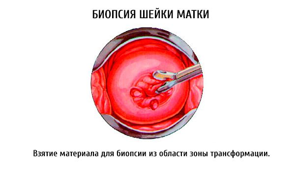
Cervical biopsy
A cervical biopsy is performed if cancer or other diseases are suspected.
Excision of cervical tissue is performed after a colposcopic examination, as this allows for precise determination of the area of the cervix for biopsy.
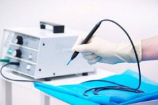
In a knife biopsy, a wedge-shaped section of tissue is excised with a scalpel. To do this, the cervix is exposed with mirrors, fixed with bullet forceps and pulled to the area of the vaginal entrance. A section of the cervix with the underlying tissue is excised with a scalpel. If necessary, 1-2 catgut sutures are applied to the wound. A biopsy can also be performed with a conchotome or a loop electrode. The excised piece of tissue is sent for histological examination.
 [ 9 ]
[ 9 ]
Technique of knife biopsy of the cervix
After disinfecting the vulva, perineal skin and vagina with iodine solution, the cervix is exposed using speculums, treated with alcohol, grasped with bullet forceps and brought down. A wedge-shaped excision of tissue is made with a scalpel with the base outward (more than 1 cm in size) and the apex in the thickness of the tissue so that it includes pathologically altered (erosion, leukoplakia, etc.) and healthy tissue. Do not grasp the epithelial cover of the excised piece with tweezers, so as not to damage it. Bleeding from the wound is stopped by tamponade of the vagina or by applying 1-2 catgut sutures to the wound. The site for collecting material is best selected using a colposcope. If this is not possible, the technique of lubricating the cervix with Lugol's solution can be used. A biopsy is taken from an area that has not absorbed the dye.

For aspiration biopsy, aspirate is taken from the uterine cavity on the 25th-26th day of the menstrual cycle in menstruating women, in the absence of a regular cycle, in the perimenopausal period - 25-30 days after bloody discharge. Aspiration can be performed using a Braun syringe with an intrauterine cannula. The aspirated contents are applied to a glass slide and a thin smear is prepared. The method can be used as a screening method.
To perform it, the vagina is exposed using speculums. The cervix (anterior lip) is grasped with bullet forceps. After probing the uterus, the tip of the syringe is brought to the bottom of the uterus. Then, while simultaneously pulling the plunger of the syringe towards you, the tip is moved alternately to the sides, thus sucking out the contents from different parts of the endometrium. Often, pieces of tissue sufficient for histological examination are obtained.
Endometrial biopsy
It is performed on an outpatient basis using a special instrument (a curette from the company "Pipel"), which allows a section of the endometrium to be obtained through aspiration.

