Medical expert of the article
New publications
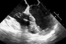
Anechogenic mass in the pericardium
Last reviewed: 29.06.2025

All iLive content is medically reviewed or fact checked to ensure as much factual accuracy as possible.
We have strict sourcing guidelines and only link to reputable media sites, academic research institutions and, whenever possible, medically peer reviewed studies. Note that the numbers in parentheses ([1], [2], etc.) are clickable links to these studies.
If you feel that any of our content is inaccurate, out-of-date, or otherwise questionable, please select it and press Ctrl + Enter.

Anechogenic masses are sometimes visualized on ultrasound. This is often a tumor. However, it can also be a sign of thrombosis, embolism, or even a parasite. Most often, however, it is still a tumor. In this case, anechogenic areas indicate an unfavorable character of the course of the tumor process. Such a tumor may be inoperable, and often ends in death. In general, anechogenic formation is any formation in the human body that does not reflect ultrasound. It is not a diagnosis, it is one of the diagnostic signs that allows the doctor to make a diagnosis. Echogenicity depends on the ability of the structure to absorb ultrasound, which is due to the morphological features of the organ, the structure itself. To a large extent, echogenicity depends on the amount of fluid in the structure. The less fluid contains the object, the higher its echogenicity, and the more it will be visible on the screen as a bright spot. The less fluid, the lower the echogenicity. Such a structure will be visible as a dark spot on the screen.
The presence of any anechogenic mass requires further differential diagnosis to determine its exact localization, its characteristics. Often an anechogenic mass in the pericardial cavity indicates the presence of a cyst. If the diameter of such a cyst does not exceed 5 cm, they can regress. However, if such a formation is quite large, and exceeds 5 cm, this indicates its tolerance to the effects of drugs, various types of therapy. Accompanying signs of the tumor process, is the presence of arterial hypertension, violation of excretory processes, the development of stasis, impaired blood and lymph circulation. When anechogenic areas are detected in patients over 50 years of age, it is often a malignant neoplasm, which in most cases is untreatable, inoperable. In some cases, it is possible to remove the anechogenic area using laparoscopy. In this case, surgical methods of treatment are necessarily combined with drug treatment. Often selected appropriate hormonal therapy, treatment with iodine preparations. In any case, for the selection of treatment, requires additional diagnostics. For diagnosis, such methods as Dopplerography, X-ray examination, laparoscopy, biopsy, MRI, CT can be used. Laboratory methods of research can also be used, in particular, tests for hormones, biochemical screenings. As a rule, if such a formation is isolated for the first time, a wait-and-see tactic is used. The patient is monitored. Further tests and repeated detection of the mass indicates the need to search for treatment methods.
This is particularly important when a tumor process is suspected. Thus, if it is suspected that an anechogenic mass is a tumor, it is necessary to resort to differential diagnosis. In particular, cytologic, histologic methods of research are widely used. Often, not single, but multiple tumors are formed in the heart cavity. In this case, the blood circulation, outflow of lymph and tissue fluid is sharply disturbed. Characteristic symptoms are the appearance of dyspnea, severe edema, cyanosis.
Tumors are difficult to diagnose. They can be asymptomatic, however, they are mostly detected by accidental diagnosis, e.g. Fluoroscopy.
Anechogenic areas in some cases may develop against the background of parasitic infection that has penetrated into the pericardial cavity. In parasitic lesions of the pericardium, parasitic cysts may form, which are cavities filled with mucus with the products of parasite activity, or with eggs. It is they during ultrasound and are detected as anechogenic areas. Parasitic cysts differ from ordinary cysts in that daughter vesicles and scolexes can form in the cyst cavity. After the death of the parasites that are contained in the cavity, it undergoes calcification. Abruptly, the process of calcification occurs. Sometimes histoplasmosis, a process of calcification of the surrounding tissue, develops. These areas are also often anechogenic.
An anechoic area may also represent a normal cyst. For example, a connective tissue cyst, which is a benign tumor, develops over a long period of time and forms areas that do not reflect ultrasound. Often in the heart cavity are formed not single, but multiple cysts. In this case, the blood circulation, lymph and tissue fluid outflow are sharply disturbed.
Pericardial tumors can be visualized on ultrasound as anechogenic areas. Conventionally, all pericardial tumors can be divided into primary and secondary tumors. At the same time, secondary tumors are more often observed. Of benign tumors, the most common are such as fibroma, or fibromatosis, fibrolipoma, hemangioma, lymphagioma, dermoid cyst, teratoma, neurofibroma. All of these tumors have some common features. First of all, they are all visualized as anechogenic structures. Therefore, differential diagnosis is necessary to make a definitive diagnosis.
It is also not uncommon to see pseudotumors (thrombotic masses) as anechogenic areas. Such tumors are also called fibrinous polyps.

