Hepatic lobule, as a morphofunctional unit of the liver
Last reviewed: 23.04.2024

All iLive content is medically reviewed or fact checked to ensure as much factual accuracy as possible.
We have strict sourcing guidelines and only link to reputable media sites, academic research institutions and, whenever possible, medically peer reviewed studies. Note that the numbers in parentheses ([1], [2], etc.) are clickable links to these studies.
If you feel that any of our content is inaccurate, out-of-date, or otherwise questionable, please select it and press Ctrl + Enter.
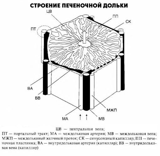
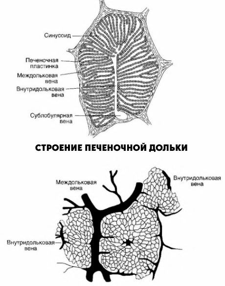
Hepatic lobule is a morphofunctional unit of the liver. In the center of the lobule is the central vein. Central veins, connecting with each other, eventually fall into the hepatic veins, the latter, in turn, flow into the lower vena cava. The wedge has the form of a prism 1-2 mm. It consists of radially arranged double rows of cells (hepatic plates, or beams). Between the rows of hepatocytes are intra-lobular bile ducts, their ends facing the central vein are closed. The resulting bile is sent to the periphery of the lobules. Between the hepatic plates are the sinusoidal capillaries, where blood is mixed, entering the liver via the portal vein and its own hepatic artery. On the periphery of the hepatic lobe are triads: interlobular veins (to which the portal vein branches), interlobular arteries (to which the own artery of the liver is branched) and interlobular bile ducts (which, merging with each other, eventually form the right and left hepatic ducts).

Thus, inside the hepatic lobule, bile moves from the center to the periphery and is then removed from the liver through the common bile duct. Blood from the portal vein and the liver's own artery, mixing the intrahepatic lobule, moves from its periphery to the center and is withdrawn through the central veins into the system of the inferior vena cava.

The hepatic lobe is delimited from the others by a connective tissue envelope containing collagen and elastin fibers. The total number of hepatic lobules is about 0.5 million. In 1 minute, 1.2 liters of blood flows through the liver of the adult, almost 70% of which passes through the portal vein.
The functional unit includes a sinusoid with the surrounding space between its endothelium and hepatocytes (Disse space), adjacent hepatocytes and the bile canaliculus. Some authors believe that the structure of the liver should be considered proceeding from the structure of the leading and withdrawing blood vessels, their interlacing,
For clinical evaluation, the state of sinusoids is important. They have three departments: peripheral, intermediate and central. Intermediate department is 90% of their length. It, in contrast to the peripheral and central department, does not have a basal membrane. Between the endothelium of the sinusoid and hepatocytes there are spaces communicating with the peri- portal spaces; together with intercellular slits they serve as the beginning of the lymphatic system. It is in these spaces that the various substances come into contact with the cytoplasmic membrane of the hepatic cell.
Endothelium of sinusoids contains pores that provide the transition to hepatocytes of various molecules. Some of the endothelial cells provide a sinusoidal structure, while others, such as stellate reticuloendotheliocytes (Kupffer cells), have phagocytic function or are involved in the renewal and neoplasm of connective tissue. These cells account for 40% of all endothelial cells. At the same time, 48% of endothelial cells perform structural function and 12% - fibroplastic.
The peripheral parts of the hepatic lobe are formed by small hepatocytes, they participate in the regeneration process and act as a border plate, separating the parenchyma of the lobule from the connective tissue of the portal field. Interlobular veins of the v. Portae and arterioles of the hepatic artery, the cholangiols emerge that flow into the interlobular hepatic ducts. Between the hepatocytes and the connective tissue there are spaces called the Mol spaces.
The portal tract on the periphery of the lobule looks like a triangle with the terminal branches of the portal vein, the hepatic artery and the interlobular bile duct, termed the triad. It consists of lymphatic fissures with a lined endothelium and nerves, braided blood vessels. A rich network of nerve fibers penetrates the hepatic lobules to hepatocytes and endothelial cells.
The connective tissue in the form of reticulin and collagen fibers, as well as basal membranes of sinusoids, blood vessels and bile ducts of the portal tract in children is very tender and only in elderly people it forms coarse fibrous accumulations.
Ultrastructure of hepatocyte
It was established that different parts of the hepatocyte membrane perform specialized functions. So, bilateral transport is carried out on a sinusoidal surface, where substances reaching the liver through the portal vein system enter the hepatocyte, and those that secrete by the hepatocyte leave it. The membranes of the tubules of neighboring hepatocytes form hepatic tubules, preventing the release of secreted substances back into the sinusoid. In the mitochondria of the hepatocyte, oxidation and metabolism of various substances, including fatty acids, processes of gluconeogenesis, accumulation and release of energy pass through. The nucleus and nucleoli are surrounded by a membrane that connects to the endoplasmic reticulum, which is a long network of tubules and cisterns involved in various biochemical processes, including the synthesis of protein, triglycerides, the metabolism of a number of drugs. The endoplasmic reticulum serves as a part of the microsomal fraction obtained by ultracentrifugation of liver homogenate. Apparatus Golgi (lamellar complex) carries out the "packing" of proteins and participates in the secretion of bile components. Peroxisomes contain enzymes (including oxidase and catalase) and are involved in the metabolism of proteins and bile acids. The hepatocyte has a cytoskeleton, consisting of actin filaments, distributed throughout the cell and concentrated in the plasma membrane. Lysosomes contain hydrolase enzymes and play an important role in intracellular digestion of substances.


 [
[