Medical expert of the article

New publications
X-ray signs of stroke
Last reviewed: 06.07.2025

All iLive content is medically reviewed or fact checked to ensure as much factual accuracy as possible.
We have strict sourcing guidelines and only link to reputable media sites, academic research institutions and, whenever possible, medically peer reviewed studies. Note that the numbers in parentheses ([1], [2], etc.) are clickable links to these studies.
If you feel that any of our content is inaccurate, out-of-date, or otherwise questionable, please select it and press Ctrl + Enter.
Cerebral circulatory disorders lead to a variety of clinical effects - from transient ischemic attacks to stroke, the third most common cause of death. In most cases, blood flow disorder is associated with atherosclerotic vascular lesions, which at first may manifest itself with not very expressive symptoms - headache, memory loss, sleep disorders, etc.
Ultrasound examination of the neck vessels plays an important role in the recognition of chronic cerebral circulatory disorders.
Atherosclerosis can affect intracerebral vessels, but much more often it develops in the extracranial sections of the arteries that supply blood to the brain. Most often, changes develop in the area of the bifurcation of the common carotid artery and it is here that they can be successfully eliminated by endarterectomy and reconstructive operations on the brachiocephalic vessels.
Ultrasound diagnostics is performed using one-dimensional Dopplerography and two-dimensional color Doppler mapping. Dopplerograms determine the position, shape, and condition of the lumen of the vessels. In this case, it is possible to register even small narrowing of the arteries and individual atherosclerotic plaques on their inner surface. Then, changes in blood flow in the brachiocephalic vessels, asymmetry in the blood flow velocity in both carotid or vertebral arteries, a decrease in the blood flow velocity in any of the vessels, vortex and retrograde blood movements are established.
In cases where the question of endovascular or surgical treatment is raised, angiography, or CT or MRI angiography, is performed. Angiograms provide the most accurate assessment of the condition of both the brachiocephalic and cerebral vessels.
In the diagnosis of acute cerebrovascular disorders - infarctions, intracerebral and meningeal hemorrhages - CT and MRI currently play a major role.
Infarction occurs due to blockage of a cerebral vessel. It is customary to distinguish three forms of cerebral infarction: extensive, lacunar and subcortical atherosclerotic encephalopathy. In the first hours after the development of infarction, changes are not detected on CT scans, but after 6-8 hours, a poorly defined area of low density with fuzzy edges is detected, which corresponds to the edema zone. On magnetic resonance tomograms performed in the T2-weighted image mode, edema is detected earlier than on CT scans. Within 2-5 days, the contours of the infarction become clearer and it is more noticeable that it has a wedge-shaped form and reaches the cortex of the brain in some direction. Large foci of infarction most often occur in the area of the middle cerebral artery. The edema disappears after a few weeks. Often, a hemorrhagic component may appear in the infarction zone, which is well visualized on CT.
As the infarction is organized, its area may become practically indistinguishable from the image of the surrounding brain tissue. However, the density of the affected area then decreases again, since after 1-2 months, as a rule, a post-infarction cyst is formed in it, surrounded by atrophic brain tissue. As a result of the cicatricial process, the nearest section of one of the cerebral ventricles is pulled to the infarction zone.
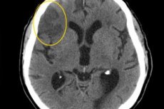
Intracerebral or meningeal hemorrhage (hematoma) is immediately detected on a CT scan as an area of increased density. This is because the absorption of X-rays by blood (52 HU) and erythrocytes (82 HU) exceeds that of the brain tissue (30-35 HU). In the area of intracerebral hemorrhage, the absorption is 40-90 HU, and this area is especially noticeable because there is an edema zone around it (18-28 HU).
If the hemorrhage is accompanied by a breakthrough of blood into the cerebrospinal fluid spaces, then areas of increased density are determined in the cerebral ventricle. Gradually, the intensity of the hemorrhage shadow decreases, and then a posthemorrhagic cyst usually forms in its place. Subdural and epidural hematomas also cause areas of increased density, but there is no edema zone around them. In addition, they are adjacent to the bones of the skull and have an oval or ribbon-like shape. Naturally, large hematomas cause displacement of brain structures, including the cerebral ventricles.
In recognizing developmental defects of cerebral vessels and their aneurysms, angiography, of course, sets the tone. However, certain data can also be obtained from non-invasive studies - CT and MRI. Angiograms determine the position, shape and size of the aneurysm and the presence of a thrombus in it. Aneurysms of the cerebral arteries are usually small - 0.3-0.7 cm in diameter. Most often, aneurysms are located in the anterior communicating and middle cerebral arteries. In 25% of patients, aneurysms are multiple.
Angiograms allow us to detect arteriovenous fistulas and arteriovenous deformations. They are characterized by the presence of a large number of dilated vessels with shunting of blood directly from the arterial bed to the venous bed (there is no capillary network). If the malformation is large enough, it can also be suspected when analyzing computer tomograms.