Medical expert of the article

New publications
Whole-body plethysmography
Last reviewed: 03.07.2025

All iLive content is medically reviewed or fact checked to ensure as much factual accuracy as possible.
We have strict sourcing guidelines and only link to reputable media sites, academic research institutions and, whenever possible, medically peer reviewed studies. Note that the numbers in parentheses ([1], [2], etc.) are clickable links to these studies.
If you feel that any of our content is inaccurate, out-of-date, or otherwise questionable, please select it and press Ctrl + Enter.
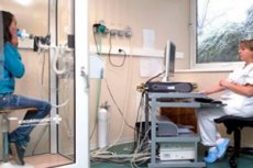
A little-known type of diagnostics – plethysmography – is a method of recording changes in the volumes of internal organs or individual parts of the body, which are accompanied by changes in blood circulation in them. This method is often used to assess the quality of vascular tone.
To implement the plethysmography method, special devices are used - plethysmographs, of various modifications - electrical, water, photographic, mechanical.
Indications for the procedure
In what situations does a doctor refer a patient for diagnostic plethysmography? Such a referral may be given to patients with multiple vascular pathologies, with blood flow disorders in different organs, with chronic heart problems. Thus, indications for plethysmography often include:
- prolonged vascular spasm of unknown etiology;
- vasospastic angiotrophoneurosis, Raynaud's disease;
- varicose veins;
- deep thrombosis;
- circulatory disorders in the brain;
- diseases of the respiratory tract - in particular, the lungs and bronchial tree.
It is possible to conduct plethysmography to record and evaluate the effectiveness of the treatment. In addition, this diagnostic method helps to clearly define the quality of conditioned reflexes of the vessels.
Referrals for plethysmography are issued by general practitioners, as well as phlebologists, neurologists, and vascular surgeons.
Preparation
Before proceeding with plethysmography, it is necessary to prepare the patient - first of all, the doctor must explain to the patient why this procedure is used and what it can clarify.
- A week before the plethysmography test, the patient is prohibited from drinking alcohol or taking any medications not prescribed by the doctor.
- Before the examination, the patient must visit the restroom and put on special clothing provided by the health worker.
- It is better to come to the plethysmography procedure early to sit, relax and calm down. The patient's breathing should be even and calm.
 [ 3 ]
[ 3 ]
Technique plethysmography
The plethysmography procedure is performed on a specific area of the body, depending on which organ or limb needs to be examined. For example, if the doctor needs to check the lower limbs, the electrodes are attached to the legs. A special device records any changes in electrical resistance, depending on the respiratory function and changes in blood flow inside the venous vessels.
Plethysmography consists of the following stages:
- First, the patient lies down straight, with the lower limbs elevated at an angle of no more than 35 degrees. The ankle joints should be above the heart level, which will improve blood flow in the legs.
- The patient bends the limb at the knee and rotates it at the hip joint, transferring the body weight to the leg being examined.
- The doctor attaches the electrodes from the plethysmograph to the skin of the ankles. The electrodes should be positioned in isolation, the distance between them should be approximately 8 cm. The cuff is fixed on the thigh, slightly above the knee joint.
- The doctor uses the device to inflate the cuff to 50-60 cm of water column. In this case, the venous vessels are compressed, and the arterial blood supply is not disrupted. The cuff is left inflated for several seconds (up to one minute), until the curve on the device stabilizes.
- The plethysmograph records information: the state of the curve displays the degree of venous filling with blood, as well as the change in this filling when the cuff is deflated. The study is usually performed on the other limb. If the doctor has doubts, the procedure can be repeated.
- To evaluate the results of plethysmography, the doctor uses a curve with the maximum filling of the vessels with blood and with the strongest blood flow.
Normally, with short-term compression of venous vessels, their filling with blood increases. And when the compression stops, a sharp outflow is observed.
In case of thrombotic complications, blood flow is disrupted: the outflow is slowed down when the cuff is deflated, and the filling of the vessels with blood is disrupted.
Impedance plethysmography
The essence of impedance plethysmography is to record the general electrical resistance of tissues to the intermittent action of high-frequency currents. Since liquid biological media have a higher coefficient of electrical conductivity, this method allows one to quickly determine the dynamics of blood flow and the type of capillary circulation in the examined areas of the body.
Conducting impedance plethysmography leads to an objective assessment of the filling of any section of the vascular venous network, both in a calm and in an excited state of blood circulation. The main difference between the method and standard rheovasography is that impedance plethysmography uses currents with higher frequencies.
Plethysmography is recognized as an absolutely safe and painless technique with a high information content coefficient.
Occlusion plethysmography
Venous occlusion plethysmography is prescribed to assess the volume and speed of blood flow in the legs and arms. During the procedure, any fluctuations in the volume of the organ are recorded against the background of compressed blood outflow through the venous vessels. Normally, the values of the volumetric blood flow in a calm state should be in the range of 2.9 ± 0.37 ml per minute per 100 cubic centimeters of tissue.
In the area where the pathology is located, the volumetric blood flow may exceed the degree of blood flow in the same areas of the healthy limb by 5 to 10 times.
The installation of special electrodes during plethysmography helps to assess not only the state of blood flow, but also to find out the degree of elasticity of the veins in the legs, as well as to determine the quality of blood outflow to the right heart chambers. The violations found during the study indicate that there is thrombosis, or dysfunction of the valves of the venous vessels, or deterioration of the venous blood flow from the abdominal organs.
Pulmonary plethysmography
The general method of plethysmography allows direct measurement of the bronchial resistance value against the background of calm, measured breathing. The essence of the study is the synchronous measurement of the air flow rate and pressure differences in the closed cabin space in which the patient is located.
The pressure readings inside the cabin are measured in relation to the fluctuations in alveolar pressure – this pressure is measured taking into account the proportional coefficient between the volumes inside the cabin and the volumes of gas in the pulmonary system.
Plethysmography is good at detecting even small areas of bronchial narrowing, unlike spirography, which can detect deterioration in bronchial patency caused by a collapse of their lumen during exhalation.
Contraindications to the procedure
Plethysmography has virtually no contraindications: the procedure is considered completely safe. However, the study is not performed in the following situations:
- if the patient suffers from claustrophobia or severe mental disorders;
- if the patient is in an excited state, his breathing is restless;
- if the room where the procedure will be performed is cold, cold extremities may distort the results of the study;
- if the patient is under the influence of alcohol, chemicals or medications;
- if the patient experiences severe pain that does not allow him to relax fully.
Reviews
It is quite difficult to give a clear assessment of the state of blood circulation in a certain area, so plethysmography is simply irreplaceable in some cases. This type of examination helps many patients establish the correct diagnosis if there is a need to differentiate vascular pathology of functional and organic etiology.
No other type of diagnostics can identify damaged and healthy vascular network in a sick person in a short time.
If a doctor needs to determine the presence of severe pathologies of regional blood flow, then plethysmography can establish the typical and pathogenetic features of the dystonic condition with high accuracy.
In narrow medical circles, plethysmography is used to study vasoactive medications, or more precisely, to track their pharmaceutical effect on blood vessels. It was plethysmography that at one time made it possible to prove that some substances, such as caffeine, have a positive effect on cerebral vascular dystonias not because they cause vasodilation, but because they are excellent tonics.
According to medical experts, plethysmography is unique and cannot be replaced by any other diagnostic method.