
Medical expert of the article

New publications
Ultrasound for prostatitis: when is it really necessary, what does it show, and how does it affect treatment?
Last updated: 11.04.2026

All iLive content is medically reviewed or fact checked to ensure as much factual accuracy as possible.
We have strict sourcing guidelines and only link to reputable media sites, academic research institutions and, whenever possible, medically peer reviewed studies. Note that the numbers in parentheses ([1], [2], etc.) are clickable links to these studies.
If you feel that any of our content is inaccurate, out-of-date, or otherwise questionable, please select it and press Ctrl + Enter.
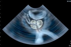
Ultrasound examination for prostatitis is often perceived as the primary way to "see inflammation," but modern urology views it differently. In acute bacterial prostatitis, the diagnosis is primarily clinical: it is made in the context of an acute urinary tract infection with systemic symptoms and signs of prostate involvement. In chronic forms and chronic pelvic pain, imaging is not considered mandatory for every patient. [1]
This doesn't make ultrasound examination secondary or unnecessary. On the contrary, it's especially valuable when the doctor needs to answer a specific question: is there an abscess, how enlarged is the gland, does urine remain in the bladder after urination, is there a dilation of the upper urinary tract, stones, or another condition masquerading as prostatitis. [2]
In practical terms, an ultrasound examination for prostatitis is not so much a "prostatitis test" as a tool for clarifying the situation. It helps distinguish between uncomplicated and complicated cases, identify the causes of a poor response to treatment, and determine whether the inflammation is associated with obstruction, residual urine, or significant prostate enlargement. [3]
Below we discuss when an ultrasound examination is really necessary, what type is chosen, what exactly it can show, where its capabilities end, and why a good examination result does not cancel out a urine test, culture, and clinical examination. [4]
When is an ultrasound examination really necessary?
In acute bacterial prostatitis, an ultrasound examination is not automatically required for every patient on the first day. Current sources emphasize that the starting point remains complaints, fever, perineal or pelvic pain, a gentle digital rectal examination, urinalysis, and urine culture. Radiological examination is usually initiated when the course appears atypical, severe, or poorly responds to treatment. [5]
The most important situation for imaging is suspected prostate abscess. European and American sources agree that if there is no clinical improvement within 48 hours despite adequate therapy, or if fever persists for more than 36 hours, an abscess should be considered and imaging should be considered. This is where ultrasound becomes not an "additional option," but a practically significant step. [6]
The situation is different in chronic bacterial prostatitis and chronic prostatitis with chronic pelvic pain. A clinical review from the United States National Library of Medicine specifically states that imaging is not routinely recommended in the evaluation of chronic prostatitis, but may be used selectively in refractory cases to exclude abscesses, calcifications, and anatomical abnormalities. [7]
Even when infection is not the issue, ultrasound remains useful for severe urinary dysfunction. The European Association of Urology's Guidelines for Male Lower Urinary Tract Symptoms recommend assessing residual urine, as well as using upper urinary tract ultrasound in men with large residual urine, blood in the urine, or signs of urolithiasis. In men with prostatitis, this can often help determine whether the complaints are masked by severe obstruction. [8]
Finally, an ultrasound examination is necessary when the clinical picture does not correspond to "pure" prostatitis. If symptoms persist, relapses persist, urine flow is poor, or evidence of concomitant prostate enlargement appears, the examination helps shift the discussion from abstract "inflammation" to a more precise urological scenario. [9]
| Clinical situation | The need for ultrasound examination | Why do they do it? |
|---|---|---|
| First typical episode of acute bacterial prostatitis without severe symptoms | It's not always necessary right away | Exclusion of complications when clinically necessary |
| No improvement after 48 hours of treatment | High | Search for an abscess |
| The fever lasts more than 36 hours | High | Search for abscess and complications |
| Chronic or recurring symptoms | Selectively | Exclusion of calcifications, anatomical causes, residual urine |
| Weak stream, feeling of incomplete emptying, urinary retention | Often useful | Measurement of residual urine, assessment of obstruction |
| Blood in urine or suspected stones | Healthy | Evaluation of the urinary tract and associated pathology |
Sources for the table. [10]
What types of ultrasound examinations are used?
In common parlance, this is often simply referred to as a "prostate ultrasound," but in reality, two main approaches are used. The first is transabdominal ultrasound, which is performed through the anterior abdominal wall. The second is transrectal ultrasound, which involves inserting the probe into the rectum and positioning it as close as possible to the prostate. These methods are not competing, but rather address different objectives. [11]
A transabdominal examination is more convenient and less invasive. It is well suited for assessing the bladder, residual urine, and sometimes prostate volume, as well as the upper urinary tract. This is why, in men with prostatitis and urinary dysfunction, a simpler abdominal examination is often started with a more detailed one rather than a more detailed one. [12]
A transrectal examination provides a more detailed picture of the gland itself and surrounding structures. RadiologyInfo and European urology guidelines emphasize that this approach is better suited for visualizing the prostate, accurately assessing its size, searching for intraparenchymal changes, and performing image-guided procedures. Therefore, if an abscess is suspected or if an accurate assessment of the gland's shape is needed, this option is more preferable. [13]
For measuring prostate volume, transrectal ultrasound is more accurate than digital prostate examination and more accurate than rough abdominal wall assessment. The European Association of Urology explicitly states that transrectal ultrasound is superior to digital prostate examination and is better for assessing prostate size, especially when treatment decisions depend on it. [14]
The choice of examination method always depends on the question being addressed. If the goal is to determine how much urine remains after urination, whether hydronephrosis is needed, or whether there is gross enlargement of the gland, a transabdominal approach is often sufficient. If a detailed examination of the prostate itself is needed, an abscess is suspected, or puncture and drainage are planned, a transrectal approach is more logical. [15]
| Type of research | What is visible better? | Pros | Restrictions |
|---|---|---|---|
| Transabdominal | Bladder, residual urine, kidneys, prostate gross volume | Less invasive, convenient, good for initial evaluation of the urinary tract | Less precise detailing of the prostate itself |
| Transrectal | The prostate, its shape, volume, focal changes, abscess | More accurate for the gland, convenient for puncture and drainage | More unpleasant, depends on the operator's experience |
| Upper urinary tract examination | Kidneys, hydronephrosis, stones, indirect signs of obstruction | Important for difficult urination | Doesn't answer all questions about the prostate itself |
Sources for the table. [16]
What can an ultrasound show about prostatitis?
The first thing an ultrasound examination can do effectively is assess the size and shape of the prostate. This is important not because volume automatically confirms prostatitis, but because many patient complaints are associated not only with inflammation but also with concomitant enlargement of the gland, a prominent middle lobe, and signs of bladder outlet obstruction. This is where the examination helps differentiate prostatitis from other urological problems. [17]
The second practical benefit is measuring residual urine. The European Association of Urology recommends measuring residual urine in men with lower urinary tract symptoms, as an increase in this level is associated with the risk of symptom progression and acute urinary retention. For men with prostatitis, this is one of the most valuable findings: it explains why symptoms persist and why standard therapy may not be as effective as expected. [18]
The third important task is examination of the upper urinary tract. If the patient has a large residual urine load, blood in the urine, or a history of urolithiasis, an ultrasound examination of the kidneys and bladder helps detect hydronephrosis, stones, and other causes of pain or obstructed urinary flow. This is not so much a diagnosis of prostatitis as an assessment of its urological consequences or associated pathology. [19]
The most clinically significant ultrasound finding in prostatitis is a prostate abscess. According to a review by the United States National Library of Medicine, transrectal ultrasound is usually the initial investigation for suspected abscesses and can detect them as hypoechoic cavities with well-defined walls and septa; furthermore, it is under this guidance that aspiration and drainage can be performed immediately. [20]
Finally, transrectal ultrasound may reveal prostatic calcifications. In chronic bacterial prostatitis, these are sometimes significant as a possible reservoir of infection and a factor in recurrence. However, their presence alone does not prove prostatitis, as such calcifications are also often found in men with benign prostatic enlargement. This is a very important limitation of interpretation. [21]
| Possible find | What does it mean? | How does it affect tactics? |
|---|---|---|
| Enlarged prostate | May explain some of the urinary symptoms | Helps differentiate inflammation from underlying obstruction |
| Middle lobe, which modifies the contour of the bladder outlet | A mechanical component of urination disorder is possible | Affects the choice of treatment for combined symptoms |
| High residual urine | The bladder is not completely emptied | Requires assessment of risk of urinary retention and obstruction |
| Hydronephrosis or kidney changes | Possible consequences of urinary outflow obstruction | Requires closer urological monitoring |
| Hypoechoic cavities with septa | Suspected abscess | Image-guided drainage is possible |
| Prostate calcifications | Possible background of a chronic process, but not proof of diagnosis | Interpret only in conjunction with clinical findings and tests. |
Sources for the table. [22]
What ultrasound can't do
The main limitation of ultrasound is that it cannot independently confirm the bacterial nature of prostatitis. Bacteria are detected not by the transducer, but by urine culture, culture after a massage test in certain clinical scenarios, and a general clinical and laboratory assessment. Therefore, even a very "inflammatory-like" image on the screen is no substitute for microbiology. [23]
In chronic prostatitis and chronic pelvic pain, the limitations are even more pronounced. A review by the United States National Library of Medicine explicitly states that imaging is not routinely recommended and is used selectively. This reflects the problem: some findings are nonspecific, and some patients have significant complaints with very modest or vague imaging findings. [24]
Even calcifications, which are often perceived by patients as "there it is, confirmation of chronic inflammation," are not a reliable diagnostic sign. The same review emphasizes that prostate calcifications are often found in benign enlargement of the gland, so they cannot be used alone to make a diagnosis. [25]
Transrectal ultrasound also has technical limitations. It depends on the specialist's experience, can be uncomfortable for the patient, does not always accurately reveal the spread of the disease beyond the prostate, and does not, by itself, resolve the issue of adjacent tissue involvement. In severe cases, where widespread infection or emphysematous abscess is suspected, computed tomography or magnetic resonance imaging may be more useful. [26]
It's important not to confuse ultrasound with ultrasound-guided biopsy. Prostate biopsy can be performed under image guidance, but if bacterial prostatitis is suspected, it is not a way to "prove inflammation" and carries a risk of complications, including infection and sepsis. In clinical practice, biopsy is needed primarily to rule out cancer, not to confirm prostatitis. [27]
| Question | Why ultrasound alone is not enough |
|---|---|
| Are there bacteria? | It's the crops that decide it, not the image. |
| Is there such a thing as chronic bacterial prostatitis? | Clinical laboratory verification is required. |
| Is it possible to confirm the diagnosis using calcifications? | No, they are non-specific. |
| Is it possible to rule out all other diseases? | No, sometimes other imaging methods and tests are needed. |
| Can the test replace a biopsy or MRI? | No, each method has its own task. |
Sources for the table. [28]
How the study is conducted and how to prepare for it
In terms of safety, prostate ultrasound is considered a favorable method: it does not use ionizing radiation, is considered non-invasive in its routine diagnostic use, and requires minimal preparation. RadiologyInfo emphasizes that the examination is safe, painless, and can be repeated as needed. [29]
If a transrectal examination is planned, a full bladder is usually not necessary. Guy's and St. Thomas' Hospital advises that a full bladder is not required for this examination; regular medications can be continued, and comfortable clothing is sufficient. This is beneficial for the patient, as many men delay the examination, anticipating complex preparation. [30]
If a transabdominal examination is performed through the anterior abdominal wall instead, the conditions are different. This option is less accurate for the prostate itself, but is convenient for assessing the bladder and residual urine; it generally requires a full bladder. The same British leaflet states that before this type of examination, patients are usually asked to drink approximately 500 ml of water approximately 30 minutes before the procedure. [31]
The transrectal examination itself is usually short. RadiologyInfo and British publications describe it as a procedure lasting approximately 15-20 minutes, in which the patient lies on their side with their legs bent, and a probe covered with a protective sheath and lubricated with gel is gently inserted into the rectum to obtain images from different angles. [32]
A routine transrectal examination is often described as mildly uncomfortable, but not painful. Guy's and St. Thomas' Hospital states that unless the patient is already experiencing significant pain due to their condition, the examination itself is usually painless, although some discomfort is possible. Following a routine examination, the patient can immediately return to normal activities. [33]
| Stage | Transrectal examination | Transabdominal examination |
|---|---|---|
| Preparing the bladder | A full bladder is usually not necessary. | A full bladder is usually required. |
| Clothing and preparation | Comfortable clothes, minimal preparation | Comfortable clothes, water before the examination |
| Position | On your side with your legs bent | Lying on your back |
| Feelings | Minor discomfort is possible | Usually easily tolerated |
| Duration | Usually about 15-20 minutes | Often even shorter |
| Best use | Detailed assessment of the prostate | Residual urine, bladder, kidneys |
Sources for the table. [34]
How the study results change treatment
If an ultrasound scan does not reveal an abscess and the clinical picture is consistent with uncomplicated bacterial prostatitis, treatment is usually continued as usual with antibiotics and observation. This is important because a "normal" scan does not negate the need for comprehensive treatment: it merely helps ensure that no significant purulent complication is currently evident. [35]
If the examination reveals signs of an abscess, the approach changes. The United States National Library of Medicine notes that transrectal ultrasound not only aids in diagnosis but also allows for immediate puncture and aspiration of the contents for therapeutic drainage and culture. In such a situation, ultrasound is no longer simply diagnostic, but therapeutic and navigational. [36]
The discovery of a large residual urine output changes the symptomatic discussion. Instead of the abstract "inflammation is interfering with urination," a specific indicator emerges, requiring an assessment of the risk of urinary retention, the degree of obstruction, and the likely response to medication. For acute bacterial prostatitis and urinary retention, urological guidelines recommend consultation with a urologist and consideration of suprapubic drainage, rather than simply waiting. [37]
If the examination reveals significant enlargement of the prostate gland or middle lobe, it can help explain why the patient experiences a prolonged weak stream, straining, and incomplete voiding. In this situation, ultrasound helps differentiate symptoms of inflammation from the associated symptoms of benign prostate enlargement and influences the choice of medications or subsequent interventions. [38]
When a test doesn't fully explain the symptoms, this is also a useful result. It encourages not endless repetition of the same test, but rather a reassessment of the diagnosis: is there chronic pelvic pain, a stricture, a neurological component, or a cause for which magnetic resonance imaging is more informative. Thus, a good ultrasound examination is sometimes important not for what it "finds," but for what it narrows down. [39]
| Research result | What does this change? |
|---|---|
| Abscess not found | Standard treatment and monitoring are usually continued. |
| An abscess was found | Need a question about drainage and seeding |
| High residual urine | The risk assessment for urinary retention and obstruction is changing |
| Marked enlargement of the prostate | The contribution of concomitant benign enlargement is clarified |
| The study is uninformative. | A different diagnostic pathway may be needed |
Sources for the table. [40]
FAQ
Should all men with prostatitis have an ultrasound?
No. In acute bacterial prostatitis, the diagnosis is often made clinically, and imaging is used if complications are suspected or there is a poor response to treatment. In chronic prostatitis and chronic pelvic pain, testing is also not considered mandatory for every patient and is used selectively. [41]
Which is better: examination through the abdomen or through the rectum?
It depends on the task. The abdomen is more convenient for assessing the bladder, residual urine, and kidneys, while the rectum provides a better view of the prostate itself, its volume, focal changes, and possible abscesses. Therefore, the question is not "which is better in general," but rather the clinical question the physician is addressing. [42]
Will an ultrasound show whether it is bacterial prostatitis or not?
In itself, no. Bacterial origin is confirmed by cultures and clinical laboratory data. Ultrasound examination helps identify complications, residual urine, calcifications, gland size, and abscess, but does not replace microbiology. [43]
Is a transrectal examination painful?
It typically causes only mild discomfort. Authoritative patient literature describes it as a generally safe procedure that typically does not cause significant pain unless the patient already experiences severe pain in the area due to a medical condition.[44]
What should I do if the report says prostate calcifications?
Don't panic and don't automatically consider this a confirmation of chronic prostatitis. Calcifications can occur both in chronic bacterial infections and in benign prostate enlargement. Their significance is assessed only in conjunction with the patient's complaints, tests, and clinical picture. [45]
Can ultrasound replace magnetic resonance imaging?
Not always. Ultrasound is excellent for many practical applications, but when the process is extensive, the anatomy is complex, other prostate lesions are suspected, or when deeper tissue characterization is needed, magnetic resonance imaging may be more useful. [46]
If everything is normal on the ultrasound, does that mean there is no prostatitis?
No. A normal or uninformative examination does not rule out either acute bacterial prostatitis or chronic pelvic pain. A diagnosis is made based on a combination of complaints, examination, tests, and sometimes additional studies. [47]
Key points from experts
The following are not verbatim quotes, but editorially formulated conclusions based on modern guidelines and reviews. [48]
Gernot Bonkat, MD, Professor, Chairman of the European Association of Urologists' Panel on Urological Infections. Current guidelines suggest that, in the case of bacterial prostatitis, ultrasound is primarily valuable for identifying complications and urological risk factors, rather than as a universal test for initial diagnosis. The primary focus remains on clinical evaluation, urine culture, and timely recognition of abscesses or urinary retention. [49]
Daniel Engeler, Associate Professor, Chair of the European Association of Urology's Panel on Chronic Pelvic Pain: In the field of chronic pelvic pain, ultrasound should not be used routinely in every patient. The phenotype of symptoms and the exclusion of specific causes of pain are more important, and imaging is used when it genuinely changes the diagnosis or management. [50]
H. Henry Lai, MD, professor of urology at the University of Iowa, is the lead author of the 2025 American Urological Association guideline for male chronic pelvic pain. The American approach to chronic pelvic pain in men emphasizes targeted diagnostic testing rather than routinely ordering every available test. For ultrasound, this means a simple rule: use it when it is necessary to exclude a specific pathology or clarify the anatomical cause of symptoms. [51]
Prathit A. Kulkarni, MD, an infectious disease specialist at Baylor College of Medicine and the Michael E. DeBakey Veterans Affairs Medical Lifeline, authored a 2026 review on bacterial prostatitis. Current infectious disease research demonstrates that imaging is no substitute for clinical judgment. Acute bacterial prostatitis is recognized by the combination of systemic infection and signs of prostate involvement, and ultrasound becomes especially important when imaging already suggests an abscess or when the patient is not progressing as expected. [52]
Conclusion
Ultrasound examination for prostatitis is a useful, safe, and very practical tool, but it is not the "ultimate test for prostate inflammation." Its strengths are the detection of abscesses, measurement of residual urine, assessment of prostate size, the upper urinary tract, and any urologic factors that make the disease more severe or complicated. [53]
The most reasonable modern approach is to not automatically screen everyone, but also not to underestimate it in cases of fever, poor response to treatment, urinary retention, severe bowel symptoms, or suspected complications. It is at these points that ultrasound examination ceases to be a formality and begins to have a real impact on treatment decisions. [54]