Medical expert of the article
New publications
Bronchoalveolar fluid processing
Last reviewed: 06.07.2025

All iLive content is medically reviewed or fact checked to ensure as much factual accuracy as possible.
We have strict sourcing guidelines and only link to reputable media sites, academic research institutions and, whenever possible, medically peer reviewed studies. Note that the numbers in parentheses ([1], [2], etc.) are clickable links to these studies.
If you feel that any of our content is inaccurate, out-of-date, or otherwise questionable, please select it and press Ctrl + Enter.

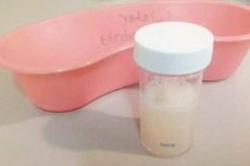
The primary objective of bronchoalveolar lavage is to obtain cells, extracellular proteins, and lipids present on the epithelial surface of the alveoli and terminal airways. The cells obtained can be evaluated cytologically as well as biochemically, immunohistochemically, microbiologically, and electron microscopically. Routine procedures include total and cell counts and, if possible, detection of lymphocytes by monoclonal antibody staining.
Normal bronchoalveolar lavage fluid from non-smokers contains 80-90% alveolar macrophages, 5-15% lymphocytes, 1-3% polymorphonuclear neutrophils, less than 1% eosinophils, and less than 1% mast cells, as well as bronchial and squamous epithelial cells. The ratio of T-lymphocyte subpopulations CD4/CD8 = 2:2.
Analysis of the bronchoalveolar lavage cytogram in interstitial lung diseases reveals the dominant cell population, which, by determining the nature of alveolitis, allows, with a certain degree of probability, to speak in favor of the diagnosis of "sarcoidosis, exogenous allergic alveolitis", etc. Quantitative assessment of the cellular composition of bronchoalveolar lavage should be based not so much on the absolute number of cells, but on determining the percentage ratios of cell populations in the patient and comparing them with similar indicators of healthy donors.
Depending on the cellular composition of bronchoalveolar lavage, alveolitis can be classified into two types: type 1 - an increase in lymphocytes (characteristic of sarcoidosis, hypersensitivity pneumonitis, tuberculosis, berylliosis, fungal infections), type 2 - an increase in neutrophils (characteristic of idiopathic pulmonary fibrosis, asbestosis, pneumoconiosis, chronic obstructive pulmonary disease).
Cytological examination of bronchoalveolar lavage plays an important role in the diagnosis of inflammatory changes in small bronchi and bronchioles. For ALS in chronic bronchitis, an increase in the cytogram of the proportion of neutrophilic leukocytes and a decrease in macrophages is characteristic. O.M. Grobova et al. (1989) studied the cytogram of bronchoalveolar lavage in chronic bronchitis and the possibility of using it to clarify the degree of inflammation activity in the bronchial tree. Three degrees of activity of the inflammatory process in the bronchoalveolar environment were identified.
- At the first stage of the inflammatory process activity, the cytogram shows a significant increase in the neutrophil content (p<0.001). The number of cylindrical, integumentary and squamous epithelial cells, which are absent in the bronchoalveolar lavage of healthy people, increases sharply.
- For the second degree of activity of the inflammatory process, a sharp increase in the relative number of neutrophils is characteristic (p<0.001), the number of columnar epithelial cells is significantly reduced.
- At the III degree of activity of the inflammatory process the number of cells in bronchoalveolar lavage increases (p< 0.01). The number of neutrophils increases significantly (p<0.01), while the number of lymphocytes does not change. The number of all types of epithelial cells and destroyed cells decreases.
In addition to determining the type of cellular elements, the material obtained using diagnostic bronchoalveolar lavage is used to study the functional activity of alveolar macrophages and other immunological, biochemical and microbiological studies.
During bronchoscopy, the tracheobronchial tree normally looks like this. The glottis is of regular shape. The vocal folds are fully mobile. The subglottic space is free. The trachea is free, the carina is sharp and mobile. The openings of the fourth-order bronchi are free, round or oval, their spurs are sharp and mobile. The mucous membrane of all visible bronchi is pale pink, with a delicate vascular pattern. The openings of the mucous glands are pinpoint. The secretion is mucous, liquid, in small quantities.

