Medical expert of the article

New publications
Sprengel's disease
Last reviewed: 04.07.2025

All iLive content is medically reviewed or fact checked to ensure as much factual accuracy as possible.
We have strict sourcing guidelines and only link to reputable media sites, academic research institutions and, whenever possible, medically peer reviewed studies. Note that the numbers in parentheses ([1], [2], etc.) are clickable links to these studies.
If you feel that any of our content is inaccurate, out-of-date, or otherwise questionable, please select it and press Ctrl + Enter.
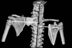
The shoulder girdle forms the support for the upper limbs. It includes the collarbones, shoulder blades and muscles. The shoulder blade connects the humerus to the collarbone. It is flat, triangular, and shaped like a shovel. Deformation of the shoulder joint, in which the shoulder blade is located higher than its normal position, is turned out and looks like a wing, is called Sprengel's disease after the German surgeon who first described it. It can be either unilateral or bilateral.
Causes Sprengel's disease
The cause of the pathology lies in the disruption of intrauterine development of the fetus. This is a congenital disease. The shoulder blades of the embryo are located high, but as it develops, the skeletal system grows, including the entire shoulder girdle. The shoulder blades lengthen, taking the place prescribed to them by nature. Disruption of full fetal growth leads to Sprengel's disease, often combined with other skeletal defects. [ 3 ]
Risk factors
Possible factors contributing to the disruption of embryonic development are:
- hereditary predisposition;
- harmful working conditions in production;
- infectious diseases;
- severe toxicosis;
- uterine pathologies.
Pathogenesis
Many scientists have tried to explain the pathogenesis of Sprengel's disease, but this issue has not yet been fully clarified; there are only assumptions. [ 4 ] The only thing they agree on is that the defect begins to develop in the early stages of pregnancy, before the appearance of the buds of the upper limbs (earlier than the 4th-5th week). Embryologically, the scapula develops together with the upper limb; it appears during the fifth week in the upper dorsal and lower cervical region together with the rudiment of the arm and descends to its final anatomical position to one of the second-eighth thoracic vertebrae by the 12th week of pregnancy. [ 5 ], [ 6 ]
The deformation is usually associated with hypoplasia or atrophy of the muscles, and the combination of these factors leads to disfigurement and functional limitation of the shoulder. There are 2 types of deformation: muscular and bone. The first case is less severe and affects the trapezius and rhomboid muscles, the second is associated directly with the scapula bone.
Symptoms Sprengel's disease
The first signs of the disease become noticeable immediately after birth: the shoulder blade (usually one) is shorter than the other, located higher and severely deformed. Upward arm movements are limited.
Sprengel's disease leaves its mark on the appearance - a short neck, low hairline, asymmetrical shoulders. Often the pathology is not limited to a cosmetic defect, but also pains caused by excessive tension of nerve fibers. Patients note a feeling of obstruction when moving the shoulder blade, in some cases clicking sounds appear.
Stages
The cosmetic aspect of the deformity was classified by Cavendish into four grades in an attempt to simplify the indications for treatment.[ 7 ]
- Grade I (very mild) - The shoulders are level; the deformity is not visible when the patient is dressed.
- Grade II (mild) - Shoulders are almost at the same level; deformity is visible as a curvature of the neck when the patient is dressed.
- Grade III (Moderate) - The shoulder joint is elevated by 2-5 centimeters; visible deformity.
- Grade IV (severe) - The shoulder joint is elevated; the upper angle of the scapula is near the back of the head.
Complications and consequences
Ignoring the disease of the shoulder girdle leads to further processes of its deformation. This worsens the mobility of the upper limbs, increases pain symptoms, and has a negative effect on other organs.
Diagnostics Sprengel's disease
Abnormal development of the scapula is visible to the naked eye. Instrumental diagnostics X-ray allows us to detect a partial or complete connection between the scapula and the cervical spine, the so-called omovertebral bone, which is observed in one third of patients. Computed tomography (CT) with three-dimensional (3-D) reconstruction and magnetic resonance imaging (MRI) are currently necessary for the diagnosis of coexisting pathologies and treatment planning. [ 8 ], [ 9 ]
Advanced conditions are characterized by changes in the back muscles, which are confirmed by electromyography.
Differential diagnosis
Differentiation of Sprengel's disease is carried out with birth trauma of the brachial plexus, Erb-Duchenne paralysis, and thoracic scoliosis.
Who to contact?
Treatment Sprengel's disease
There are 2 directions of treatment of Spregel's disease: conservative and surgical. In the early stages, with not clearly expressed changes and minor dysfunctions, they do without surgery, strengthen the muscles of the shoulder and chest, and efforts are also directed at increasing the motor activity of the upper limbs. Patients with bilateral deformities or Cavendish grade 1 deformity can be observed by an orthopedist to assess the dynamics of the disease.
For this purpose, massage, swimming, and therapeutic exercise are prescribed. Ozokerite and paraffin applications are effective.
Surgical treatment
Progression of the deformity with age, development of secondary changes in the shoulder girdle, hypotrophy of its muscles, initially severe pathology of bone and muscle tissue are indications for surgical treatment. Surgical intervention at the age of up to 2 years is technically more complex. [ 10 ], [ 11 ] Surgical intervention is best recommended for patients aged 3 to 8 years with moderate or severe cosmetic or functional deformity. The presence of concomitant congenital anomalies may be a contraindication to surgery. [ 12 ]
The goal of surgery for Sprengel's deformity is cosmetic and functional improvement, however, the disease is often associated with other anomalies such as torticollis and congenital scoliosis, which limit the amount of correction that can be performed.
There are more than 20 methods of surgical treatment of the disease, one of the most effective is to lower the scapula to the level of the healthy one and fix it to the underlying rib, in particular, partial resection of the scapula and release of the long head of the triceps for the treatment of Sprengel's deformity [ 13 ], fixation of the upper angle of the scapula to the lower thoracic spine [ 14 ], vertical scapular osteotomy [ 15 ], surgical treatment using the Mears method [ 16 ], Woodward's operation. [ 17 ]
For 3 weeks, a plaster cast fixes the upper limb in an abducted position. From the fifth day, the patient is prescribed massage sessions, UHF, and electrophoresis. In 3 cases out of 30, complications were observed after surgery in the form of brachial plexus paralysis. [ 18 ] Within six months, as a result of drug and physiotherapy treatment, such neurological disorders passed.
Prevention
The main role in preventing further deformation of the shoulder blades, as well as after surgery, belongs to therapeutic physical training, swimming, volleyball. They are designed to adapt the back to physical activity, strengthen the muscles.
Forecast
Unfortunately, the serious defect caused by Spregel's disease cannot be completely corrected. The prognosis is more favorable the sooner you contact a specialist.
Использованная литература