
Medical expert of the article

New publications
Retrograde pyelography: indications, preparation, procedure, risks, and interpretation of results
Last updated: 06.04.2026

All iLive content is medically reviewed or fact checked to ensure as much factual accuracy as possible.
We have strict sourcing guidelines and only link to reputable media sites, academic research institutions and, whenever possible, medically peer reviewed studies. Note that the numbers in parentheses ([1], [2], etc.) are clickable links to these studies.
If you feel that any of our content is inaccurate, out-of-date, or otherwise questionable, please select it and press Ctrl + Enter.
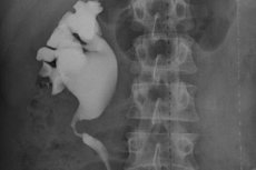
Retrograde pyelography is an X-ray examination of the upper urinary tract in which a contrast agent is injected directly into the ureter via a cystoscope and a thin catheter, rather than into a vein. This allows the physician to obtain an image of the ureter and renal pelvis and assess its patency, contours, stenosis, filling defects, and the level of blockage. The examination is usually performed under anesthesia and is often combined with cystoscopy and other urological procedures. [1]
Historically, this technique was used much more widely, but in modern practice, it is no longer the first choice for most patients. Today, it is more often prescribed as a targeted examination when it is necessary to clarify local anatomy, assess urinary obstruction, prepare for stent placement, perform ureteroscopy, or obtain an answer in a situation where conventional imaging has proven insufficiently informative. For this reason, retrograde pyelography remains an important, albeit more specialized, tool in modern urology. [2]
The main difference from intravenous pyelography is the route of contrast administration. During intravenous testing, contrast enters the bloodstream, is filtered by the kidneys, and then excreted into the urinary tract. During retrograde pyelography, contrast is advanced backward through the urethra, bladder, and ureteral orifice, allowing the physician to more accurately delineate a specific section of the ureter or renal pelvis. [3]
Another important feature of the procedure is that it is often not only diagnostic but also part of the treatment phase. During the same procedure, the urologist can place a ureteral stent, perform ureteroscopy, clarify the catheter position, determine the level of obstruction, and, in some cases, immediately change the treatment strategy. This is convenient for the patient, as diagnostic information and practical decisions are often obtained within a single episode of care. [4]
Despite the development of computed tomography and magnetic resonance urography, this method remains relevant. It is especially valuable when it is necessary to visualize the lumen and patency of the ureter, confirm a local obstruction, clarify the nature of the stenosis, or obtain an image before endourological intervention. Therefore, retrograde pyelography is no longer an outdated technique, but a specialized tool for appropriate clinical situations. [5]
| Characteristic | Retrograde pyelography | Intravenous pyelography | Computed tomography urography |
|---|---|---|---|
| Route of contrast administration | Through a cystoscope and catheter into the ureter | Through a vein | Usually through a vein |
| What is especially clearly visible | Lumen of the ureter and renal pelvis-calyceal system, block level | General excretory picture | Anatomy, stones, tumors, surrounding tissues |
| Where is it most commonly used today? | Targeted, before or during a urological procedure | Less often than before | Often as the primary visualization method |
| Is cystoscopy necessary? | Yes, as a rule | No | No |
| Is it possible to install a stent immediately? | Yes | No | No |
The table summarizes the differences between the methods according to current descriptions from Cleveland Clinic, Johns Hopkins, and RadiologyInfo. [6]
When is retrograde pyelography really necessary?
One of the most common indications is suspected ureteral or upper urinary tract obstruction. This could be due to a stone, stricture, blood clot, tumor, external compression, or other cause of obstruction. In such situations, it is important for the physician not only to see dilation of the renal pelvis but also to understand the location and severity of the obstruction. [7]
The procedure is also used for hematuria, when it is necessary to clarify the condition of the upper urinary tract. According to the American Urological Association algorithm, if hematuria persists after a negative renal ultrasound, urographic imaging or retrograde pyelograms may be performed. This emphasizes that this method does not replace the entire modern diagnostic pathway, but remains an important alternative or adjunct in complex cases. [8]
In urological oncology, retrograde pyelography remains valuable when upper urinary tract tumors are suspected. The European Association of Urology states that retrograde ureteropyelography remains an option for detecting urothelial carcinoma of the upper urinary tract, and that flushing cytology from the renal pelvis and ureter is preferable before contrast administration, as contrast can degrade the quality of cytological material. In practice, this is especially important when endoscopic evaluation, contrast, and sample collection must be combined. [9]
Another area of application is ureteral trauma. When ureteral injury is suspected, computed tomography urography is usually considered the method of choice, but if the data remain unclear, the European Association of Urology explicitly states that retrograde or antegrade urography is the best method for confirmation. This makes retrograde pyelography particularly valuable in postoperative and trauma scenarios. [10]
Finally, the method is widely used as part of an endourological procedure. It assists before stent placement, during ureteroscopy, and in planning interventions for stones, strictures, and obstructions. In real-world clinical practice, the study is often not separate from the treatment phase, but rather serves as a guide for the urologist's subsequent actions. [11]
| Clinical situation | Why is the study prescribed? |
|---|---|
| Suspected ureteral stone | Specify the level and degree of the block |
| Ureteral stricture | Assess the location and extent of the stenosis |
| Hematuria | Further evaluate the upper urinary tract |
| Suspected upper urinary tract tumor | Detect filling defect, combine with cytology and endoscopy |
| Preparing for stent placement | Obtain an accurate anatomical picture |
| Suspected ureteral injury | Confirm contrast leakage or obstruction |
The table reflects the main indications described in the guidelines of the American Urological Association, the European Association of Urology and clinical materials on the procedure. [12]
How to prepare for the procedure
Preparation begins with an assessment of whether the examination will provide additional information and whether it can be performed safely. Before the procedure, the doctor will review the patient's complaints, the presence of pain, fever, or blood in the urine, the known ultrasound or CT scan data, and the purpose of the procedure. This is important because retrograde pyelography is most useful when the next step in treatment depends on its results. [13]
One of the key points is ruling out an active urinary tract infection. The Cleveland Clinic specifically states that a urinary tract infection is usually considered a reason to postpone retrograde pyelography, as the procedure may worsen the infection. Therefore, a urinalysis is often performed before the procedure, and if an infection is suspected, treatment is considered before the procedure. [14]
Pregnancy, allergies to contrast, anticoagulant use, concomitant illnesses, and medications are also discussed in advance. Johns Hopkins notes that even with minimal systemic absorption of contrast, the risk of a reaction to the contrast agent does not completely disappear, so information about allergies and past reactions is essential. For patients with chronic kidney disease, an individual assessment is important, although the method itself is sometimes used precisely because the contrast is not administered intravenously in the usual volume. [15]
Before the procedure, abstinence from food and drink is usually required for a certain period of time, especially if sedation or general anesthesia is planned. The patient is advised in advance which regular medications to take in the morning, which to temporarily discontinue, and whether an accompanying person is needed for the return trip. In most cases, the procedure is performed on an outpatient basis and does not require overnight hospitalization. [16]
It's important to understand that preparation for a retrograde pyelogram is largely the same as preparation for a cystoscopy under anesthesia. This is why the patient is asked to remove jewelry, put on a hospital gown, sign an informed consent form, and be prepared for intravenous access for anesthesia. This preparation may seem more extensive than for a standard X-ray, but this is due to the endoscopic nature of the procedure. [17]
| Preparation stage | What do they usually do? | Why is this important? |
|---|---|---|
| Evaluation of evidence | Analysis of complaints and previous studies | To make the procedure truly useful |
| Exclusion of infection | Urinalysis, sometimes culture | To avoid increasing the risk of ascending infection |
| Clarification of medications | Especially anticoagulants and diabetes medications | To reduce the risk of bleeding and other complications |
| Allergy and pregnancy testing | Conversation with a doctor | To choose a safe tactic |
| Fasting before anesthesia | According to the clinic's instructions | For safe pain relief |
| Organization of support | Traveling home after sedation | Due to the residual effect of anesthesia |
The table reflects the standard preparation elements described in the Cleveland Clinic and Johns Hopkins materials on retrograde pyelography. [18]
How the research proceeds step by step
On the day of the procedure, the patient is transferred to the operating room or endoscopy room, an intravenous catheter is inserted, and anesthesia is prepared. Depending on the clinical situation, sedation, regional anesthesia, or general anesthesia is used. The patient is then placed on a table, usually with their feet on footrests, as for a cystoscopy. [19]
Next, the urologist passes a cystoscope through the urethra into the bladder and locates the orifice of the desired ureter. A thin catheter is inserted through this orifice, through which a contrast agent is then injected. The term "retrograde" itself means that the contrast agent moves against the normal direction of urine flow—from the bladder up the ureter to the renal pelvis. [20]
During contrast administration, a series of X-rays or fluoroscopy are performed. The physician assesses how the ureter is filling, looking for any abrupt discontinuities in contrast, dilation above the block, areas of narrowing, contrast leakage, or filling defects. It is at this stage that it often becomes clear whether a stone, stricture, tumor, or damage to the ureteral wall is present. [21]
A major advantage of this method is that it allows for the diagnostic process to continue uninterrupted. If the urologist deems it necessary, they can immediately place a ureteral stent, proceed to ureteroscopy, perform additional endoscopic evaluation, or adjust the interventional strategy. Therefore, for the patient, retrograde pyelography often becomes part of a broader diagnostic and treatment plan. [22]
Retrograde pyelography itself typically takes no more than 30 minutes, but the overall hospital stay may be longer due to preparation, anesthesia, and post-procedure monitoring. If stenting, ureteroscopy, or other procedures were performed simultaneously, the duration and recovery time depend on the combined intervention, not just the contrast agent. [23]
| Stage of the procedure | What does a doctor do? | What is the result? |
|---|---|---|
| Cystoscopy | Inserts an endoscope into the bladder | Access to the ureteral orifice |
| Ureteral catheterization | Inserts a thin catheter | Possibility of targeted contrast injection |
| Introduction of contrast | Fills the ureter and renal pelvis | Upper urinary tract contouring |
| Fluoroscopy | Takes a series of photos | Evaluation of permeability and filling defects |
| Additional actions | Stent, ureteroscopy, change of tactics | Combining diagnostics and treatment |
The table summarizes the sequence of stages as described by the Cleveland Clinic and Johns Hopkins. [24]
What does retrograde pyelography show and how are the results interpreted?
A normal examination reveals free filling of the ureter and renal pelvic collecting system with contrast, without sharp interruptions, leakage, or significant filling defects. The contours should be relatively smooth, and the contrast should flow through the lumen without signs of critical obstruction. In this case, the physician either rules out a significant mechanical problem or uses the data as a map before the planned procedure. [25]
If the image shows a sudden cessation of contrast passage, this suggests an obstruction. This could be caused by a stone, stricture, external compression factor, blood clot, or tumor. If the ureter and renal pelvis are dilated above the obstruction, the physician additionally evaluates the severity of the congestion and the need for rapid restoration of urine flow. [26]
Filling defects require special attention. They may be associated with a blood clot, stone, tumor, or other intraluminal mass. In oncology practice, such findings often prompt ureteroscopy, cytology, or biopsy, especially if there is simultaneous hematuria and clinical suspicion of urothelial carcinoma of the upper urinary tract. [27]
If contrast extends beyond the ureter or renal pelvis, this suggests damage to the wall or urinary tract leakage. This is why this method is particularly useful in questionable cases of ureteral injury, after complex pelvic surgeries, or when iatrogenic injury is suspected. In such cases, localized contrast leakage can provide a more accurate answer than indirect signs from other studies. [28]
The results of a retrograde pyelogram are rarely interpreted in isolation. They are always compared with symptoms, ultrasound, CT scan, urine tests, and the urologist's findings during a cystoscopy. This comprehensive approach ensures that incidental findings are not overestimated and that significant pathology is not missed. [29]
| Find | What could it mean? | What do they do next? |
|---|---|---|
| Free passage of contrast | There is no visible block | Compared with clinical and other data |
| Abrupt interruption of contrast | Stone, stricture, tumor, clot | They are deciding between a stent or ureteroscopy. |
| Filling defect | Intraluminal formation | Additional endoscopy, cytology, biopsy |
| Expanding above the obstacle | Obstruction of urine outflow | Assessing the urgency of decompression |
| Contrast leakage outside the urinary tract | Damage to the wall, urine leakage | Confirmation of injury, correction of tactics |
The table summarizes typical interpretation options for the study based on clinical descriptions and guidelines for upper urinary tract tumors and trauma.[30]
Risks, limitations and safety
Although the procedure is generally considered safe, it is not completely risk-free. The Cleveland Clinic lists possible complications as anesthesia-related problems, contrast reactions, urinary tract infection, nausea, vomiting, and bladder or ureter injury. Johns Hopkins also lists bleeding, bladder rupture, and even sepsis as rare but possible complications. [31]
Infectious risk is particularly important in patients with pre-existing infection, hydronephrosis, and obstructed urinary outflow. Therefore, active infection is typically treated first, and antibiotic prophylaxis is considered individually depending on risk factors and the nature of the endourological procedure. Current guidelines on urologic infections do not support a one-size-fits-all approach for all endoscopic procedures and emphasize the importance of assessing individual risk. [32]
Another limitation of this method is that it primarily shows the lumen and contours of the urinary tract, but does not provide the same comprehensive information about the surrounding tissues as computed tomography. If the physician needs to assess not only the presence of a block, but also the extent of the tumor, the condition of the renal parenchyma, surrounding organs, and possible extraluminal causes of compression, retrograde pyelography alone is insufficient. In such cases, it complements, rather than replaces, modern tomographic methods. [33]
Contrast and renal function deserve a separate discussion. Johns Hopkins notes that the study can often be performed in patients with poor renal function or a history of contrast reactions, as contrast absorption is limited. However, this does not mean zero risk: information about allergies, renal function, and medications taken is still essential for safe procedure planning. [34]
The radiation exposure during the examination is usually low, but it cannot be completely ignored, especially if the patient has previously undergone numerous radiographic procedures. Therefore, retrograde pyelography is not prescribed "just in case," but when a specific diagnostic or therapeutic response is expected. This approach makes the method a reasonable and safe tool in modern urology. [35]
| Potential risk | Why does it arise? | How to reduce it |
|---|---|---|
| Urinary tract infection | Endoscopic intervention and contrast administration | Active infection is ruled out before the procedure |
| Injury to the ureter or bladder | Catheterization and instrumental procedures | The procedure is performed under the supervision of an experienced urologist. |
| Reaction to contrast | Individual sensitivity | Allergies and medical history are clarified in advance |
| Complications of anesthesia | Sedation or general anesthesia | Preoperative assessment and monitoring |
| Blood in urine and pain when urinating | Irritation of the mucous membrane after intervention | Observation and symptomatic care |
The table summarizes the main risks and risk mitigation measures based on data from the Cleveland Clinic and Johns Hopkins. [36]
What happens after the procedure and how does recovery proceed?
Following the examination, the patient is transferred to an observation room, where breathing, blood pressure, well-being, and recovery from anesthesia are monitored. Urination is then assessed, and there is no significant bleeding or obstruction. If no additional procedures are required and the patient's condition is stable, the patient is usually discharged the same day. [37]
A small amount of blood in the urine after the procedure is possible and does not always indicate a complication. Johns Hopkins notes that urine may be reddish even due to a small amount of blood, and this is expected after endoscopic intervention. A moderate burning sensation during urination is also possible in the first hours or days. [38]
Recovery from isolated retrograde pyelography is usually rapid. The Cleveland Clinic reports that returning to work or school is often possible within a few days, although the timeframe depends on whether stenting, ureteroscopy, stone removal, or other procedures were performed concurrently. Therefore, patients should always be guided not only by the procedure's name but also by the full scope of the procedure. [39]
After discharge, adequate fluid intake, monitoring of urine volume and type, and attentiveness to any alarming symptoms are recommended. Fever, chills, increasing bleeding, difficulty urinating, and severe pain during urination are considered reasons to immediately contact a doctor. These symptoms most often indicate a complication requiring evaluation rather than a normal recovery. [40]
It is also important to note that the final value of the procedure is determined by the next step. If the examination confirms a stone with a block, the patient may require a stent or ureteroscopy. If a stricture is detected, a separate discussion of endoscopic or reconstructive treatment is necessary. If the examination was performed to clarify hematuria, the further course of action depends on the entire set of findings, not just a single radiograph. [41]
| After the procedure | What is generally considered normal | When urgent contact with a doctor is needed |
|---|---|---|
| Well-being | Drowsiness after anesthesia in the first hours | Increasing weakness or worsening condition |
| Urine | A small amount of blood is possible | Severe bleeding |
| Urination | A slight burning sensation may occur for a short time. | Inability to urinate |
| Pain | Moderate discomfort is acceptable | Severe or increasing pain |
| Temperature | Usually normal | Fever, chills |
The chart is based on post-procedure recovery and monitoring guidelines from Cleveland Clinic and Johns Hopkins.[42]
FAQ
Is retrograde pyelography painful?
The procedure is typically performed under sedation, regional anesthesia, or general anesthesia, so the patient typically does not experience significant pain during the examination. Mild discomfort or burning during urination may occur after the procedure. [43]
Can this examination be replaced by computed tomography?
In many situations, computed tomography is indeed used more frequently and provides a more comprehensive view. However, retrograde pyelography is valuable when it is necessary to specifically assess the ureteral lumen, determine the level of blockage, or perform the examination directly during an endourological procedure. [44]
Does retrograde pyelography show cancer?
It may show a filling defect, distortion, or other signs that raise suspicion of an upper urinary tract tumor. However, a definitive diagnosis usually requires a combination of ureteroscopy, cytology, and sometimes biopsy. [45]
Can the study be performed with poor kidney function?
In many cases, yes, because the contrast is administered locally, rather than intravenously as is standard. However, the decision is always individual and requires an assessment of the associated risks, allergies, and the overall clinical situation. [46]
Can retrograde pyelography be performed if a urinary tract infection is present?
Typically, an active infection is treated first, as the procedure can worsen the infection. Therefore, a urine sample is often tested and signs of infection are assessed before the procedure. [47]
How long does the procedure take?
The retrograde pyelogram itself typically takes up to 30 minutes, but the overall time in the clinic is longer due to preparation, anesthesia, and post-procedure monitoring. If additional procedures are performed simultaneously, the time increases. [48]
Is hospitalization necessary?
Often not. In most cases, the examination is performed on an outpatient basis, and the patient goes home the same day. However, in cases of more complex procedures, severe general condition, or the need for follow-up treatment, hospitalization is possible. [49]
How does this examination differ from ureteroscopy?
Retrograde pyelography is primarily a contrast-based visualization of the urinary tract lumen. Ureteroscopy is an endoscopic examination of the ureter and upper urinary tract from the inside, which allows not only visualization but also biopsy or treatment. In practice, these methods are often combined. [50]

Key points from experts
Margaret Pearl, MD, PhD, is a professor of urology and internal medicine and the Endowed Chair in Urologic Education at the University of Texas Southwestern Medical Center. Her clinical work and research interests focus on urinary tract stones, minimally invasive surgery, and endourology. The practical implication of modern endourologic logic is that retrograde pyelography is particularly useful not as a routine examination for everyone, but as a precise pre-interventional tool and in cases where it is important to quickly understand the anatomy of the block and proceed immediately to treatment. [51]
Roger Sur, MD, Clinical Professor of Urology, Director of the Comprehensive Stone Center at the University of California, San Diego, is a board-certified urologist with a fellowship in endourology. His expertise emphasizes minimally invasive techniques and evidence-based urology for stones and obstruction. The key message is this: when a stone or other obstruction is suspected, it's not just visualizing the presence of dilation that matters; understanding the exact level and nature of the blockage determines the choice between observation, stenting, ureteroscopy, and other treatment options. [52]
Aaron Lentz, MD, a urologist board-certified by the American Board of Urology and a specialist in reconstructive urology at Duke University, is a member of the American Board of Urology. His clinical practice specifically addresses the management of upper urinary tract obstruction. The practical implication of the reconstructive approach is that if retrograde pyelography reveals a stricture or complex obstruction, the physician's goal is not only to temporarily restore urinary flow but also to determine the anatomical cause, which will determine the choice between temporary decompression and definitive reconstructive treatment. [53]