Medical expert of the article
New publications
Pancreas
Last reviewed: 04.07.2025

All iLive content is medically reviewed or fact checked to ensure as much factual accuracy as possible.
We have strict sourcing guidelines and only link to reputable media sites, academic research institutions and, whenever possible, medically peer reviewed studies. Note that the numbers in parentheses ([1], [2], etc.) are clickable links to these studies.
If you feel that any of our content is inaccurate, out-of-date, or otherwise questionable, please select it and press Ctrl + Enter.
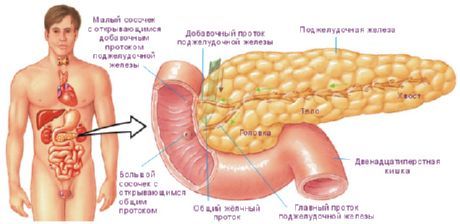
The pancreas is an elongated gland, gray-pink in color, and located retroperitoneally. The pancreas is a large digestive gland of mixed type. It has both an exocrine part with typical secretory sections, a duct apparatus, and an endocrine part. As an exocrine gland, it produces 500-700 ml of pancreatic juice daily, which enters the lumen of the duodenum. Pancreatic juice contains proteolytic enzymes, trypsin, chymotrypsin, and amylolytic enzymes (lipase, etc.). The endocrine part of the gland in the form of small cell clusters (pancreatic islets) produces hormones (insulin, glucagon, etc.) that regulate carbohydrate and fat metabolism.

The length of the pancreas in an adult is 14-18 cm, width - 6-9 cm, thickness - 2-3 cm, its weight is 85-95 g. The gland is covered with a thin connective tissue capsule. The gland is located transversely at the level of the I-II lumbar vertebrae. The tail of the gland lies slightly higher than its head.
Behind the pancreas are the spine, aorta, inferior vena cava and left renal vein. In front of the gland is the stomach. The pancreas has a head, body and tail.
The head of the pancreas (caput pancreatis) is surrounded by the duodenum from above on the right and below. The head is slightly flattened in the anteroposterior direction. At the border between the lower part of the head and the body there is a deep notch of the pancreas (incisura pancreatis), in which the superior mesenteric artery and vein pass. The posterior surface of the head of the pancreas is adjacent to the right renal vein, and closer to the median plane - to the initial part of the portal vein. The right part of the transverse colon is located in front of the head of the gland.
The body of the pancreas (corpus pancreatis) has a prismatic shape, it has anterior, posterior and inferior surfaces. The anterior surface (facies anterior) is covered by the parietal peritoneum. At the border of the body of the gland with its head there is a bulge forward - the so-called omental tubercle (tuber omentale). The posterior surface (facies posterior) is adjacent to the spine, large blood vessels (inferior vena cava and aorta), celiac plexus. The inferior surface (facies inferior) is narrow, partially covered by the peritoneum, separated from the anterior surface by the anterior edge of the gland. The splenic artery and vein are adjacent to the upper edge of the gland.
The tail of the pancreas (cauda pancreatis) is directed to the left, where it touches the visceral surface of the spleen, below its gate. Behind the tail of the gland are the left adrenal gland, the upper part of the left kidney.
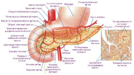
The parenchyma of the gland is divided into lobules by connective tissue interlobular septa (trabeculae) extending deep from the capsule of the organ. The lobules contain secretory sections resembling hollow sacs measuring 100-500 µm. Each secretory section, the pancreatic acinus (acinus pancreaticus), consists of 8-14 cells - exocrine pancreatocytes (acinocytes) that have a pyramidal shape. Secretory (acinous) cells are located on the basal membrane. Intercalated excretory ducts (diictuli intercalatus), lined with a single-layer flattened epithelium, begin from the cavity of the secretory section. Intercalated ducts give rise to the ductal apparatus of the gland. The intercalated ducts pass into the intralobular ducts (ductuli intralobulares), formed by a single-layer cuboidal epithelium, and then into the interlobular ducts (ductuli interlobulares), passing in the interlobular connective tissue septa. The walls of the interlobular ducts are formed by high prismatic epithelium and their own connective tissue plate. The interlobular ducts flow into the excretory duct of the pancreas.

The excretory duct (main) of the pancreas (ductus pancreaticus), or the duct of Wirsung, runs in the thickness of the gland, closer to its posterior surface. The duct begins in the area of the tail of the gland, passes through the body and head, and receives smaller interlobular excretory ducts along the way. The main duct of the pancreas flows into the lumen of the descending part of the duodenum, opens on its major papilla, having previously connected with the common bile duct. The wall of the final section of the pancreatic duct has a sphincter of the pancreatic duct (sphincter ductus pancriaticae), which is a thickening of the circular bundles of smooth muscles. Often, the pancreatic duct and the common bile duct flow into the duodenum separately at the apex of the major papilla of the duodenum. Other options for the entry of both ducts are possible.
In the area of the head of the pancreas, an independent accessory pancreatic duct (ductus pancreatis accesorius), or Santorini's duct, is formed. This duct opens into the lumen of the duodenum on its minor papilla. Sometimes both ducts (main and accessory) anastomose with each other.
The walls of the main and accessory ducts are lined with columnar epithelium. In the epithelium of the ductal apparatus of the pancreas there are goblet cells that produce mucus, as well as endocrinocytes. Endocrine cells of the ducts synthesize pancreozymin and cholecystokinin. In the proper plate of the mucous membrane of the interlobular ducts, accessory and main ducts there are multicellular mucous glands.
Endocrine pancreas
Endocrine pancreasformed by pancreatic islets (islets of Langerhans), which are clusters of endocrine cells. The islets are located mainly in the tail region, and there are fewer of them in the thickness of the body of the gland. Pancreatic islets have a round, oval, ribbon-shaped or star-shaped form. The total number of islets is 0.2-1.8 million, the diameter of the islet varies from 100 to 300 µm, the mass of all islets is 0.7-2.6 g. There are several types of endocrine cells that form islets.
Innervation of the pancreas
The pancreas is innervated by branches of the vagus nerves (mainly the right), sympathetic nerves from the celiac plexus.

Blood supply of the pancreas
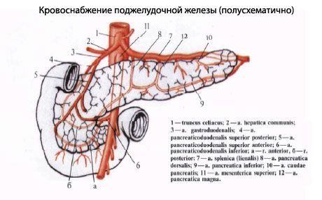
The pancreas is supplied with blood by the following vessels: the anterior and posterior superior pancreaticoduodenal arteries (from the gastroduodenal artery), the inferior pancreaticoduodenal artery (from the superior mesenteric artery). Venous outflow: into the pancreatic veins (tributaries of the superior mesenteric, splenic and other veins from the portal vein system).
Lymph drainage: into the pancreas: pancreatic-duodenal, pyloric and lumbar lymph nodes.
 [ 7 ], [ 8 ], [ 9 ], [ 10 ], [ 11 ], [ 12 ], [ 13 ], [ 14 ], [ 15 ], [ 16 ], [ 17 ]
Age-related features of the pancreas
The pancreas of a newborn is small. Its length is 4-5 cm, and its weight is 2-3 g. The gland is located slightly higher than in an adult. By 3-4 months of life, the gland's weight doubles, by 3 years it reaches 20 g, and at 10-12 years its weight is 30 g. Due to the lack of strong fixation to the back wall of the abdominal cavity, the pancreas of a newborn is relatively mobile. By 5-6 years, the gland takes on the appearance typical of the gland of an adult. The topographic relationships of the pancreas with neighboring organs, typical of an adult, are established by the end of the first year of life.

